Digital Poster
Cerebrovascular & Stroke Imaging II
ISMRM & ISMRT Annual Meeting & Exhibition • 03-08 June 2023 • Toronto, ON, Canada

Digital Poster
Cerebrovascular & Stroke Imaging II
| Computer # | |||
|---|---|---|---|
2095.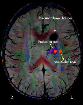 |
101 | Evaluation of the Glymphatic System Using the DTI-ALPS Index in Patients with Spontaneous Intracerebral Haemorrhage
Chao Zhang1, Jingyun Sha1, Lulu Cai1, Houliang Zhao1, Weiqiang Dou2, and Kai Xu1
1Department of Radiology, Affiliated Hospital of Xuzhou Medical University, Xuzhou, China, 2MR Research China, GE Healthcare, Beijing, China Keywords: Stroke, Diffusion Tensor Imaging, glymphatic system We evaluated the function of the human glymphatic system (GS) in patients with spontaneous intracerebral haemorrhage (sICH) using diffusion tensor imaging analysis along with the perivascular space (DTI-ALPS). Twenty patients with sICH and 31 healthy controls (HCs) were recruited for analysis. The results showed that DTI-ALPS index on the lesion side was significantly decreased, but not in the contralateral side in sICH or in HCs. And, the decreased DTI-ALPS index was significantly correlated with disease duration. This study confirmed the presence of GS dysfunction only ipsilateral to the lesion, indicating the GS may be a separate system in bilateral hemispheres. |
|
2096.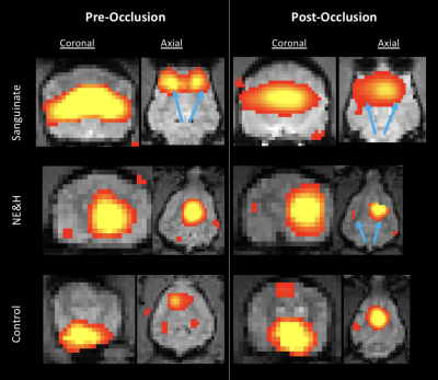 |
102 | Flow augmentation after occlusion maintains functional connectivity and mean diffusivity
Chisondi Simba Warioba1, Mira Liu1, Sean Foxley1, Julian Bertini1, Gregory Christoforidis2, and Timothy Carroll1
1Radiology, University of Chicago, Chicago, IL, United States, 2Mount Carmel, Columbus, OH, United States Keywords: Stroke, fMRI (resting state) Through the use of resting state functional MRI, diffusion tensor imaging (DTI), and a unique canine stroke model, our study explored the functional and structural effects of maintaining perfusion pressure as a means of extending the window of opportunity for thrombectomy. In our preliminary results, the employment of flow augmentation indicated a maintenance of functional connectivity, mean T2* signal intensity, and mean diffusivity in the ischemic region. Through this we have shown a potential method of extending the "door-to-needle time" in stroke treatment and possible therapeutic effects of flow augmentation therapy in acute stroke. |
|
2097.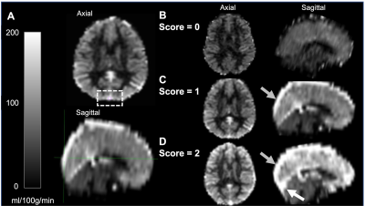 |
103 | Diffuse dural sinus hyperintensities on arterial spin labeling reflects reduced cerebral metabolic rate of oxygen in sickle cell anemia patients
Alexander K. Song1,2, Spencer L. Waddle3, Randall Sky Jones4, Niral J. Patel4, Samantha Davis4, Chelsea Custer1, Larry Taylor Davis3, Sumit Pruthi3, Jarrod J. Eisma1, Megan A. Aumann1, Lori C. Jordan4, and Manus J. Donahue1
1Neurology, Vanderbilt University Medical Center, Nashville, TN, United States, 2Vanderbilt Brain Institute, Vanderbilt University, Nashville, TN, United States, 3Radiology, Vanderbilt University Medical Center, Nashville, TN, United States, 4Pediatric Neurology, Vanderbilt University Medical Center, Nashville, TN, United States Keywords: Stroke, Stroke Sickle cell anemia (SCA) patients have elevated cerebral blood flow and flow velocities to partially compensate for reduced blood oxygen content; this phenomenon may result in inefficient oxygen delivery to tissue due to accelerated red cell capillary transit. Pseudo-continuous arterial spin labeling (pCASL) and T2-relaxation-under-sping-tagging (TRUST) MRI methods were used to assess arterial-to-venous transit artifacts and oxygen metabolism in 150 SCA participants. Patients with diffuse dural sinus hyperintensities on pCASL had 22.7% lower cerebral metabolic rates of oxygen compared to their counterparts; however, values remained within a normal range of 2.91±0.69 ml O2/100g/min and were not associated with infarct history. |
|
2098.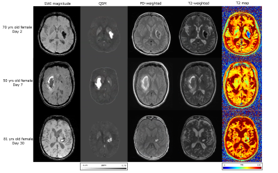 |
104 | T2 Mapping in Hemorrhagic and Ischemic Stroke using Standard Clinical Images
Ashmita De1, Jeff Snyder1, Mahesh Kate2, Derek J. Emery3, and Alan H. Wilman1
1Department of Biomedical Engineering, University of Alberta, Edmonton, AB, Canada, 2Division of Neurology, Department of Medicine, University of Alberta, Edmonton, AB, Canada, 3Department of Radiology and Diagnostic Imaging, University of Alberta, Edmonton, AB, Canada Keywords: Stroke, Brain T2 mapping can provide a route to monitor both iron changes in hemorrhage and water changes in ischemic stroke. Our aim is to investigate the value of T2 mapping from standard proton density and T2-weighted fast spin echo images for hemorrhagic and ischemic stroke. Rapid T2 maps using sequence modeling were reconstructed and compared with susceptibility and apparent diffusion coefficient for 13 hemorrhages and 14 ischemic stroke lesions respectively. Quantitative T2 is highly correlated with susceptibility for hemorrhage and diffusion for ischemic stroke, providing complementary measurements of water and iron changes. |
|
2099.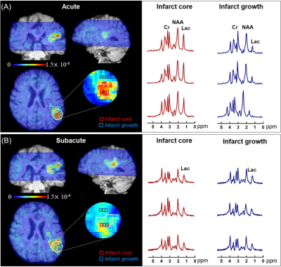 |
105 | Progressive Lactic Acidosis is Associated with Infarct Growth Volume in Acute Ischemic Stroke: A Longitudinal 3D MRSI Study
Bin Bo1, Tianyao Wang2, Ziyu Meng1, Yibo Zhao3,4, Yudu Li3,5, Rong Guo3,6, Wen Jin3,4, Xin Yu7, Zhi-Pei Liang3,4, and Yao Li1
1School of Biomedical Engineering, Shanghai Jiao Tong University, Shanghai, China, 2Radiology Department, The Fifth People's Hospital of Shanghai, Fudan University, Shanghai, China, 3Beckman Institute for Advanced Science and Technology, University of Illinois at Urbana-Champaign, Urbana, IL, United States, 4Department of Electrical and Computer Engineering, University of Illinois at Urbana-Champaign, Urbana, IL, United States, 5National Center for Supercomputing Applications, University of Illinois at Urbana-Champaign, Urbana, IL, United States, 6Siemens Medical Solutions USA, Inc., Urbana, IL, United States, 7Department of Biomedical Engineering, Case Western Reserve University, Cleveland, OH, United States Keywords: Stroke, Spectroscopy Imaging lactic acidosis is of significance for injury assessment in acute stroke. In this study, we investigated the progression of lactate, along with the concomitant changes in N-acetylaspartate and creatine concentrations, and its relationship to infarct growth using high-resolution MRSI data acquired from a longitudinal cohort of 42 ischemic stroke patients. The results showed that the progression in lactate level was predictive of infarct growth volume from acute to subacute stroke. Our study might provide a useful biomarker for lactic acidosis and tissue injury assessment in ischemic stroke. |
|
2100.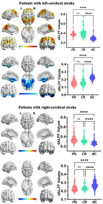 |
106 | Temporal dynamic change patterns of intrinsic brain activity in multidomain brain networks in subcortical stroke patients
Caihong Wang1, Jingchun Liu2, Jun Guo3, Peifang Miao1, Ying Wei1, Kaiyu Wang4, and Jingliang Cheng1
1The First Affiliated Hospital of Zhengzhou University, Zhengzhou, China, 2Tianjin Medical University General Hospital, Tianjin, China, 3Tianjin Huanhu Hospital, Tianjin, China, 4GE Healthcare MR Research China, Beijing, China Keywords: Stroke, fMRI, ischemic, cognitive, dynamic This study aimed to explore the temporal dynamic change patterns and the mechanisms of verbal memory deficit in chronic subcortical stroke patients with motor pathway based on the static and dynamic amplitude of low-frequency fluctuations. A total of 136 patients and 88 normal controls were included in the study. We found verbal memory deficits in the patients of chronic subcortical stroke involving the motor pathway, especially in patients with partial recovery. Moreover, subcortical stroke-induced functional deficits may not only occur in the motor system but also in the cognitive functional system. |
|
2101.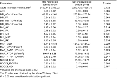 |
107 | Quantitative Parameters of Diffusion Spectrum Imaging: Prediction of Severe Neurologic Deficits at Discharge in Acute Ischemic Stroke Patients
Peirong Jiang1, Xiuzhu Xu1, Yanping Zheng1, Jialin Chen2, Peng Wu3, and Yunjing Xue1
1Radiology, Fujian Medical University Union Hospital, Fuzhou, China, 2Neurology, Fujian Medical University Union Hospital, Fuzhou, China, 3Philips Healthcare, Shanghai, China Keywords: Stroke, Ischemia, Neurologic deficits This study explores the value of quantitative parameters derived from diffusion spectrum imaging (DSI) in distinguishing between mild (NIHSS score ≤ 5) and severe (NIHSS score > 5) neurologic deficits at discharge in DWI positive acute ischemic stroke (AIS) patients. The method is to compare conventional diffusion and DSI parameters of acute infarction between the two groups. Our research shows DKI_AK, DKI_MKT, DKI_RK, MAP_RTPP and NODDI_ICVF can predict poor neural function outcome at discharge, with NODDI_ICVF demonstrates the highest diagnostic performance. DSI is a promising approach in detecting microstructural brain tissue changes in acute infarction lesion. |
|
2102.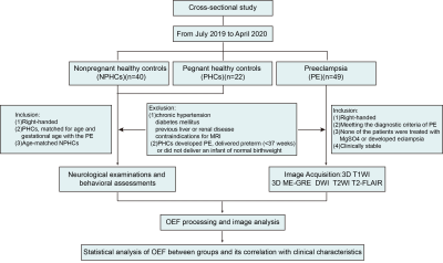 |
108 | Assessing cerebral oxygen metabolism changes in preeclampsia using voxel-based morphometry of oxygen extraction fraction (OEF) maps in MRI
Chaofan Sui1, Qihao Zhang2, Junghun Cho2, Linfeng Yang3, Tao Chen3, Bin Guo3, Kelly M. Gillen2, Jing Li4, Lingfei Guo1, and Yi Wang2
1Department of Radiology, Shandong Provincial Hospital Affiliated to Shandong First Medical University, Jinan, China, 2Department of Radiology, Weill Cornell Medical College, New York, NY, United States, 3Department of Radiology, Jinan Maternal and Child Care Hospital, Jinan, China, 4Department of Radiology, Beijing Friendship Hospital, Capital Medical University, Beijing, China Keywords: Stroke, Hypertension The objective of this study was to analyze the different brain oxygen metabolism statuses in preeclampsia. Furthermore, we also investigated the influencing factors that affect cerebral oxygen metabolism in preeclampsia. Forty-nine preeclampsia patients, forty nonpregnant healthy controls (NPHCs) and twenty-two pregnant healthy controls (PHCs) were included in this study. Brain OEF values were computed using quantitative susceptibility mapping (QSM) plus quantitative blood oxygen level‐dependent magnitude-based OEF mapping (QSM+qBOLD, or QQ). Voxel-based morphometry (VBM) was applied to investigate the differences of OEF values in brain regions among groups. |
|
2103.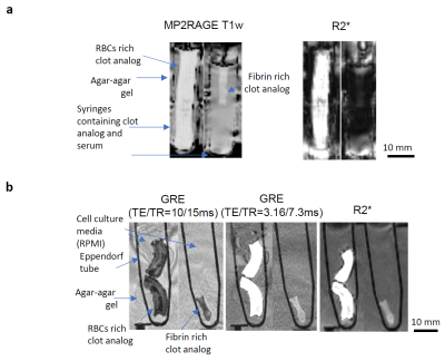 |
109 | Ultra-high field MRI sequences for clot characterization in acute ischemic stroke
Karl-Olof Lövblad1,2, Daniela Dumitriu LaGrange2, Lijing Xin3, Philippe Reymond2, Seàn Fitzgerald4, Karen M. Doyle4,5, Paolo Machi1,2, Maria Isabel Vargas1,2, François Lazeyras2,3, and Isabel Wanke6,7,8
1Division of Diagnostic and Interventional Neuroradiology, HUG Geneva University Hospitals, Geneva, Switzerland, 2Radiology and Medical Informatics, University of Geneva, Geneva, Switzerland, 3Ecole Polytechnique Fédérale de Lausanne (EPFL), Center for Biomedical Imaging (CIBM), Lausanne, Switzerland, 4Department of Physiology, University of Galway, Galway, Ireland, 5CURAM, Sciene Foundation Ireland (SFI) Centre for Research in Medical Devices, University of Galway, Galway, Ireland, 6Division of Neuroradiology, Klinik Hirslanden, Zurich, Switzerland, 7Swiss Neuroradiology Institute, Zurich, Switzerland, 8Division of Neuroradiology, University of Essen, Essen, Germany Keywords: Stroke, Stroke In acute ischemic stroke, the composition of the clot occluding the arteries is associated with the underlying pathophysiology and with the response to treatment. Innovative MRI sequences aim to improve the ability to recognize clot composition from neuroimaging signs. Using ultra-high field MR imaging of clot analogs, we demonstrate the ability of R2* map to distinguish between red blood cells-rich and fibrin-rich compositions. This technique can also be used for the volumetric analysis of formalin fixed clots. Further studies with intermediate clot compositions and improved phantoms will reveal the full potential of MRI sequences for depicting clot composition. |
|
2104. |
110 | The Relationship between MR Imaging Characteristics of Bilateral Carotid High-risk Plaques and Recurrence of Stroke
Ruijing Xin1, Maoxue Wang1, and Bing Zhang1
1Department of Radiology, The Affiliated Drum Tower Hospital of Nanjing University Medical School, Nanjing, China Keywords: Stroke, Atherosclerosis, High-risk plaque Carotid HRP is one of the major sources of ischemic stroke. Recent studies have focused on identifying characteristics of HRP which is defined as lesion with IPH, FCR, or LRNC. We compared the plaque characteristics of patients with bilateral carotid HRPs between patients with recurrent stroke and those without recurrent stroke using MR vessel wall imaging. We determined the association between HRP plaque features and recurrent stroke in patients with bilateral carotid HRPs. We found that those with larger plaque burden, more juxta luminal IPH and/or thrombus, and juxta luminal calcification in HRPs were more likely to have recurrent stroke. |
|
2105. |
111 | Quantitative susceptibility mapping reveals an association between iron load in gray matter nuclei and different cerebral perfusion patterns
Yu Guo1, Weiqiang Dou2, Huimin Mao1, and Xinyi Wang1
1Radiology, The First Affiliated Hospital of Shandong First Medical University&Shandong Provincial Qianfoshan Hospital, Jinan, China, 2MR Research, GE Healthcare, Beijing, China Keywords: Stroke, Perfusion The main purpose was to explore iron alterations in gray matter (GM) nucleus of patients with unilateral middle cerebral artery (MCA) stenosis or occlusion with varied cerebral perfusion states using quantitative susceptibility mapping (QSM). Sixty-one patients with unilateral MCA stenosis or occlusion were divided into three groups based on different cerebral perfusion patterns and examined with QSM. Iron-related susceptibility of GM nucleus subregions were assessed. The results showed that iron deposition in bilateral putamen and globus pallidus at the lesion side significantly increased in patients with extensive impaired but not hypo- or normal cerebral perfusion. |
|
2106.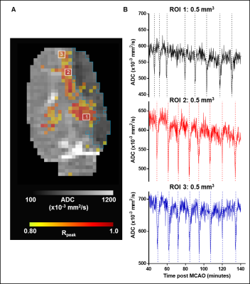 |
112 | Automated Analysis of Dynamic Diffusion Abnormalities and Infarct Growth Following Ischemic Injury
Mihika Gangolli1,2, Alexander Kharlamov3, David Y. Chung4, Fernando Boada5, Victor E. Yushmanov3, Dzung L. Pham1,6, John A. Butman1,7, and Stephen C. Jones3,8
1Center for Neuroscience and Regenerative Medicine, Bethesda, MD, United States, 2The Henry M. Jackson Foundation for the Advancement of Military Medicine, Inc., Bethesda, MD, United States, 3Allegheny-Singer Research Institute, Pittsburgh, PA, United States, 4Radiology, Harvard Medical School, Charlestown, MA, United States, 5Radiology, Stanford University School of Medicine, Palo Alto, CA, United States, 6Radiology and Radiological Sciences, Uniformed Services University, Bethesda, MD, United States, 7Radiology and Imaging Sciences, Clinical Center, National Institutes of Health, Bethesda, MD, United States, 8CerebroScope, Pittsburgh, PA, United States Keywords: Stroke, Diffusion/other diffusion imaging techniques, Peri-infarct depolarization, dynamic diffusion MRI An automated analysis pipeline was developed to detect and analyze the trajectories of dynamic diffusion abnormalities (DDAs) in a rat model of severe ischemic injury. DDAs were detected in both hemispheres of the brain and perilesional DDAs propagated along the infarct boundary. Infarct volume, quantified using apparent diffusion coefficient (ADC), increased during the recording period concomitantly with detected DDAs. Our approach could provide the means to explore the causal relationship between DDAs hypothesized to correspond with peri-infarct depolarizations and secondary expansion of infarcted tissue. |
|
2107.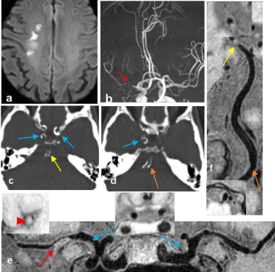 |
113 | Identification of intracranial atherosclerotic plaque features associated with recurrent stroke: a multi-modality imaging study
Lingling Wang1, Beibei Sun1, Maysam Orouskhani2, Mahmud Mossa-Basha2, Jianrong Xu1, Yan Zhou1, Chengcheng Zhu2, and Huilin Zhao1
1Radiology, Renji Hospital, Shanghai Jiao Tong University School of Medicine, Shanghai, China, 2University of Washington, Seattle, United States, Seattle, WA, United States Keywords: Stroke, Atherosclerosis, Calcification Intracranial atherosclerotic plaque is a major cause of stroke. Both CTA and vessel wall magnetic resonance imaging (VW-MRI) can identify high risk plaque features. We aim to investigate the intracranial atherosclerotic plaque features associated with recurrent stroke using a multi-modality imaging approach by combining CTA and whole brain VW-MR. We found that higher calcium burden quantified by volume and Agatston score of intracranial arteries, as well as whole-brain plaque number and culprit plaque burden were independently associated with recurrent acute stroke. |
|
2108.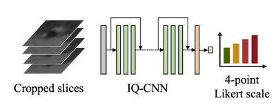 |
114 | Artificial Intelligence-Driven Image Quality Assessment for Intracranial Vessel Wall Magnetic Resonance Imaging
Wenjia Peng1, Haiyan Zhao1, Xuefeng Zhang1, Luguang Chen1, Hao Li2, Shuo Wang2, and Jianping Lu1
1The First Affiliated Hospital of Naval Medical University, Shanghai, China, 2Fudan University, Shanghai, China Keywords: Stroke, Stroke, vessel wall imaging Image quality control is a prerequisite for quantitative image analysis. We develop a convolutional neural network-based model for assessing the image quality of intracranial vessel wall MRI. Experimental results show that the model prediction is in good agreement with a senior radiologist, with a Cohen’s Kappa of 0.689. The model demonstrates real-time evaluation speed which is 500 times faster than the radiologist. It has the potential to be used in performing quality control on historical data for research purposes, and also can be used to examine the image quality immediately after the clinical MRI scan. |
|
2109.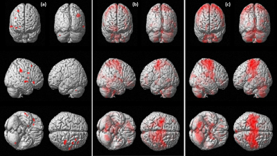 |
115 | Neural correlates of bi-cephalic transcranial direct current stimulation in upper limb hand function post stroke
Ashu Bhasin1, Rahul Sharma1, MV Padma Srivastava1, and S Senthil Kumaran2
1Department of Neurology, All India Institute of Medical Sciences, New Delhi, India, 2Department of NMR, All India Institute of Medical Sciences, New Delhi, India Keywords: Stroke, fMRI (task based) Non invasive brain stimulation holds great promise in post stroke recovery in upper limb hand function and inducing neural plasticity. Transcranial direct current stimulation (tDCS) enables the alteration of cortical excitability by passing direct currents causing hypo or hyperpolarization of neuronal resting membrane potentials. Bi-cephalic tDCS with anode applied on the affected cortex especially M1 and cathode over the non-affected cortex has been used to normalize excitatory and inhibitory corticospinal networks. |
|
2110.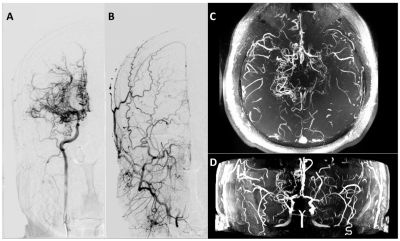 |
116 | Comparison of 7T MRA and DSA in assessment of the Suzuki staging system for Moyamoya disease
Qi Duan1, Jinhao Lyu1, Caohui Duan1, Xiangbing Bian1, Jianxun Qu2, and Xin Lou1
1Chinese PLA General Hospital, Beijing, China, 2MR Collaboration, Siemens Healthineers Ltd., Beijing, China Keywords: Stroke, High-Field MRI, Suzuki staging system Moyamoya disease (MMD) is an uncommon chronic cerebrovascular disease and a leading cause of stroke in pediatric and young patients. Suzuki staging system based on conventional digital subtraction angiography (DSA) is a useful and important index in clinical. However, DSA is invasive and unnecessary in some situations. 7T TOF-MRA enables the visualization of the "puff of smoke" collateral network in MMD compared to 1.5T and 3T. We tried to grade the Suzuki staging system by using 7T TOF-MRA in MMD patients. Our results indicated that 7T TOF-MRA showed excellent performance as DSA on grading the Suzuki staging system in MMD. |
|
2111.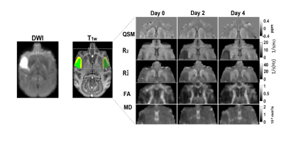 |
117 | Evaluation of progressive evolution of oxygen extraction fraction in the brain during acute stroke by using quantitative susceptibility mapping
Xiaodong Zhang1, Yuguang Meng1, and Chun-Xia Li1
1Emory University, Atlanta, GA, United States Keywords: Stroke, Brain, Oxygen extraction fraction Oxygen extraction fraction (OEF) has been suggested to be an effective measure to assess the oxygen metabolism and viability of tissue at risk. The preliminary results in a monkey model of stroke demonstrated progressive OEF reduction in both grey matter and white matter after pMCAO, in agreement with the neuron loss and fiber denegation as indicated by diffusion MRI indices following stroke. The findings suggest QSM derived OEF could provide additional information about the oxygen metabolism of the tissue and may be used to assess the ischemia-induced damage of the brain during acute stroke. |
|
2112.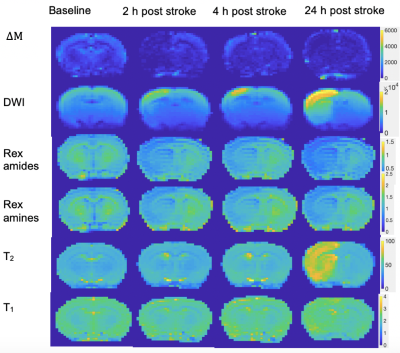 |
118 | Following evolving pH changes in the penumbra using a novel MRI method
Eleni Demetriou1,2, Valerie Taylor3, Mohamed Tachrount4, Kimmo Jokivarsi 5, Olli Grohn5, Rolf Jager6, Karin Shmueli7, and Xavier Golay8
1Advanced Imaging Research Center, University of Texas Southwestern Medical Center, Dallas, TX, United States, 2Brain repair and Rehabilitation, Institute of Neurology, Dallas, TX, United States, 3University College of London, Center for Advanced Biomedical Imaging, London, United Kingdom, 4Wellcome Centre for Integrative Neuroimaging, FMRIB, Nuffield Department of Clinical Neurosciences, University of Oxford, London, United Kingdom, 5A. I. Virtanen Institute for Molecular Sciences, University of Eastern Finland, Kuopio, Finland, Kuopio, Finland, 6Institute of Neurology, Brain repair and rehabilitation, London, United Kingdom, 7Department of Medical Physics & Bioengineering, University College London, UK, London, United Kingdom, 8Brain repair and rehabilitation, Institute of Neurology, London, United Kingdom Keywords: Stroke, Ischemia, ischemic penumbra Here, our goal was to visualise dynamic pH changes in a rat model of MCAO and their relation to other imaging techniques for identifying the penumbra zone which is used to guide therapeutic intervention. |
|
2113.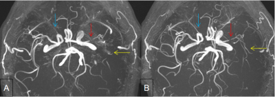 |
119 | A Preliminary study on evaluation of collateral compensatory by high-resolution CS TOF MRA and 4D flow:Case Series
Yue Ma1, Yueluan Jiang2, Dan Tong1, Zechen Yu3, and Xinpeng Liu1
1Radiology, First Hospital of Jilin University, Changchun of Jilin province, China, 2MR Scientific Marketing, Diagnostic Imaging, Siemens Healthineers Ltd., Beijing, China, 3Siemens Healthineers Digital Technology (Shanghai) Co., Ltd., Changchun of Jilin province, China Keywords: Stroke, Blood vessels Evaluation of hemodynamic and geometric changes of intracranial atherosclerosis and follow-up is of great clinical significance. A comprehensive quantification of the whole collateral recruitment in the circle of Willis (CW) is needed. In this work, we combined High-resolution CS TOF-MRA and 4D Flow MRI to investigate the situation of collateral recruitment. We were able to demonstrate a new way to visual and quantify the cerebrovascular impact of a significant intracranial stenosis and the compensatory mechanism of the collaterals. |
|
2114.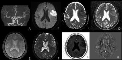 |
120 | The application of MTP synthetic sequence in the diagnosis of acute ischemic stroke
Peipei Chang1, xinyue liang2, Yanwei Miao1, and yongming dai3
1The First Affiliated Hospital of Dalian Medical University, Dalian, China, 2MR Collaboration,Central Research Institute, United Imaging Healthcare, Shanghai, China, 3MR Collaboration,Central Research Institute, United Imaging Healthcare, shanghai, China Keywords: Stroke, Ischemia, T2 Star Multiple parametric (MTP) is a novel quantitative MRI technique that allows the generation of quantitative maps as well as synthetic weightings from a single acquisition. Preliminary results show that MTP synthetic sequence can effectively distinguish infarcted brain from normal tissue with a good diagnostic performance. MTP imaging offers the potential to create a standardized brain imaging protocol providing multiple types of quantitative tissue property information and qualitative information in just a few minutes. The aim of this study is to evaluate the potential of MTP in the mapping-based identification of acute ischemic stroke. |
|
 Back to Meeting Home
 Back to the Program-at-a-Glance
The International Society for Magnetic Resonance in Medicine is accredited by the Accreditation Council for Continuing Medical Education to provide continuing medical education for physicians.
 Back to Meeting Home
 Back to the Program-at-a-Glance
 View Presentation Video