Digital Poster
Advanced Imaging in White Matter
ISMRM & ISMRT Annual Meeting & Exhibition • 03-08 June 2023 • Toronto, ON, Canada

Digital Poster
Advanced Imaging in White Matter
| Computer # | |||
|---|---|---|---|
2784. |
81 |
High Heterogeneity of White Matter Observed with Short TE
Magnetic Resonance Elastography
Samuel Patz1,2,
W. Scott Hoge3,4,
Bin Deng2,5,
Lauren O'Donnell1,2,
Fan Zhang1,2,
Yanmei Tie2,6,
Emma Tinney7,
Ralph Sinkus8,
and Katherine M. Breedlove1,2
1Radiology, Brigham and Women's Hospital, Boston, MA, United States, 2Harvard Medical School, Boston, MA, United States, 3SigProc Expert Solutions, Westwood, MA, United States, 4Imaginostics, Inc., Cambridge, MA, United States, 5Radiology, Massachusetts General Hospital, Charlestown, MA, United States, 6Neurosurgery, Brigham and Women's Hospital, Boston, MA, United States, 7Psychology, Northeastern University, Boston, MA, United States, 8INSERM, Paris, France Keywords: White Matter, Elastography, superficial white matter heterogeneity, myelin water Prior MRE work in the brain has utilized long TE pulse sequences (spiral and EPI) and therefore has necessarily not observed the signal from short T2 components. In particular, the signal from water compartmentalized in myelin membranes and that is a sensitive measure of white matter structural integrity, has been missed. Here we demonstrate high heterogeneity of the shear stiffness within the superficial white matter when utilizing MRE sequences with a short (10ms) TE. |
|
2785. |
82 |
Longitudinal change of white matter-specific brain age is
associated with Alzheimer's disease-related regional atrophy
Chang-Le Chen1,
Jinghang Li1,
Linghai Wang1,
Noah Schweitzer1,
Dana Tudorascu2,3,
Howard Aizenstein1,2,
and Minjie Wu1,2
1Department of Bioengineering, University of Pittsburgh, Pittsburgh, PA, United States, 2Department of Psychiatry, University of Pittsburgh, Pittsburgh, PA, United States, 3Department of Biostatistics, University of Pittsburgh, Pittsburgh, PA, United States Keywords: White Matter, Aging, brain age The white matter (WM) network integrity is assumed to associate with Alzheimer's disease (AD)-related gray matter (GM) atrophy. To investigate this hypothesis, we estimated WM-specific brain age to quantify WM integrity and calculated twelve AD-related GM signatures in a longitudinal cognitively normal cohort. We identified that the change rate of WM brain age was significantly correlated with the left hippocampal and amygdala volumetric changes; that is, the accelerated aging in WM was associated with the more atrophic GM volumes. This result suggested that changes of structural network characterized by brain age metrics can reflect the alteration of AD-related GM signatures. |
|
2786.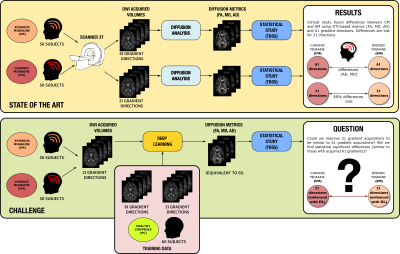 |
83 |
Validation of Deep Learning techniques for quality augmentation
in diffusion MRI for clinical studies
Santiago Aja-Fernandez1,
Carmen Martin-Martin1,
Tomasz Pieciak1,
Alvaro Planchuelo-Gomez1,2,
Abrar Faiyaz3,
Nasir Uddin3,
Abhishek Tiwari4,
Saurabh J. Shigwan4,
Tianshu Zheng5,
Zuozhen Cao5,
Stefano B. Blumberg6,
Snigdha Sen6,
Mehmet Yigit Avci7,
Zihan Li7,
Xinyi Wang8,
Zihao Tang8,
Amelie Rauland9,
Dorit Merhof10,
Renata Manzano Maria11,
Vinicius P. Campos11,
SeyyedKazem HashemizadehKolowri12,
Edward DiBella12,
Chenxu Peng13,
Zan Chen13,
Irfan Ullah14,
Merry Mani14,
Samuel Eckstrom15,
Steven H. Baete15,
Scifitto Scifitto3,
Rajeev Kumar Singh4,
Dan Wu5,
Tobias Goodwin-Allcock6,
Paddy J. Slator6,
Berkin Bilgic7,
Qiyuan Tian7,
Mariano Cabezas8,
Tales Santini11,
Marcelo Andrade da Costa Vieira11,
Zhimin Shen13,
Hesam Abdolmotalleby14,
Patryk Filipiak15,
Antonio Tristan-Vega1,
and Rodrigo de Luis-Garcia1
1Universidad de Valladolid, Valladolid, Spain, 2CUBRIC, Cardiff University, Cardiff, United Kingdom, 3University of Rochester, Rochester, NY, United States, 4Shiv Nadar University, Delhi NCR, India, 5Zhejiang University, Hangzhou, China, 6Centre for Medical Image Computing, University College London, London, United Kingdom, 7Athinoula A. Martinos Center for Biomedical Imaging, Charlestown, MA, United States, 8University of Sydney, Sidney, Australia, 9RWTH Aachen University, Aachen, Germany, 10University of Regensburg, Regensburg, Germany, 11University of São Paulo, São Paulo, Brazil, 12University of Utah, Salt Lake City, UT, United States, 13Zhejiang University of Technology, Hangzhou, China, 14University of Iowa, Iowa City, IA, United States, 15New York University, New York, NY, United States Keywords: White Matter, Brain, Migraine This work gathers the results of the QuadD22 challenge, held in MICCAI 2022. We evaluate whether Deep Learning (DL) Techniques are able to improve the quality of diffusion MRI data in clinical studies. To that end, we focused on a real study on migraine, where the differences between groups are drastically reduced when using 21 gradient directions instead of 61. Thus, we asked the participants to augment dMRI data acquired with only 21 directions to 61 via DL. The results were evaluated using a real clinical study with TBSS in which we statistically compared episodic migraine to chronic migraine. |
|
2787.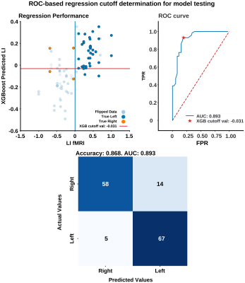 |
84 |
Machine-learning prediction of fMRI language laterality based on
morphological features of Arcuate fasciculi CSD tractograms
Ahmed Radwan1,
Robert Pretorius1,
and Stefan Sunaert1
1Imaging and pathology, Translational MRI, KU Leuven, Leuven, Belgium Keywords: White Matter, Machine Learning/Artificial Intelligence, Tractography & Fiber Modelling Shape features of the arcuate fasciculi (AF) can be used for predicting language laterality by a machine-learning algorithm as determined by language task-based functional MRI (tb-fMRI) laterality index (LI) relatively accurately (AUC = 0.893, accuracy = 0.868) in a sample of 60 clinical preoperative patients with variable pathology. Constrained spherical deconvolution (CSD) tractograms seemed to give the best outcome of model training regardless of additional streamline filtering or anatomical constraint. The best-performing model appeared to prioritise bundle curl, irregularity and span over the more conventional measures of surface-area and volume. |
|
2788. |
85 |
Association between arterial stiffness and white matter
integrity in older adults
Junko Kikuta1,
Koji Kamagata1,
Masahiro Abe1,
Andica Christina1,
Yuya Saito1,
Kaito Takabayashi1,
Wataru Uchida1,
Hitoshi Naito2,
Hiroki Tabata3,
Akihiko Wada1,
Yoshifumi Tamura2,3,
Ryuzo Kawamura2,3,
Hirotaka Watada2,3,
and Shigeki Aoki1
1Department of Radiology, Juntendo University Graduate School of Medicine, Bunkyo-ku, Japan, 2Department of Metabolism and Endocrinology, Juntendo University Graduate School of Medicine, Bunkyo-ku, Japan, 3Sportology Center, Juntendo University Graduate School of Medicine, Bunkyo-ku, Japan Keywords: White Matter, Brain This study aimed to use advanced MRI techniques to investigate the effect of arterial stiffness on the white matter (WM) microstructure among older adults. Arterial stiffness was examined using the cardio-ankle vascular elasticity index (CAVI). The high-CAVI group (mean CAVI ≥ 9 points) and the low-CAVI group (mean CAVI < 9 points) were used. The neuronal fiber integrity of the WM was assessed by neurite orientation dispersion, density imaging, and magnetization transfer-saturation imaging. Our results suggest that arterial stiffness may be related to demyelination rather than axonal degeneration.
|
|
2789. |
86 |
Serial Acquisition of radiofrequency pulse MOdes (SAMO) for
correction of transmit field inhomogeneities in ultra high field
(7T) diffusion MRI.
Bradford A Moffat1,
Robert A Williams2,
Rebecca A Glarin3,
Braden Thai3,
Jennie Ponsford4,
and Christopher C Rowe5
1Radiology, University of Melbourne, Parkville, Australia, 2University of Melbourne, Parkville, Australia, 3Radiology, Royal Melbourne Hospital, Parkville, Australia, 4Psychology, Monash University, Clayton, Australia, 5Department of Nuclear Medicine and Centre for PET,, Austin Health, Heidelberg,, Australia Keywords: White Matter, Diffusion Tensor Imaging, 7T DWI, CSD, tractography This study presents a simple and robust correction of B1+ inhomogeneities in high resolution multi-shell DWI at 7T. |
|
2790.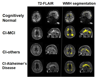 |
87 |
Brain WMH Load, Kinetics and regional Distribution with Aging: A
signature of Structural and Cognitive Health
Niraj Kumar Gupta1,
Neha Yadav1,
Aniket Aman1,
and Vivek Tiwari1
1Department of Biological Sciences, Indian Institute of Science Education and Research Berhampur, Berhampur, India Keywords: White Matter, Aging Discriminating Cognitive Impairment and Alzheimer's Disease is challenging due to overlapping subtle structural changes and cognitive variations. Load and distribution of white matter hyperintensity across brain regions may elicit structural atrophy and cognitive disabilities via continuous vascular insult. Here, we have investigated the kinetics and regional distribution of White matter hyperintensity with aging and its implications in brain structure and cognitive health using T2-FLAIR and T1w longitudinal MRI from the NACC cohort. |
|
2791.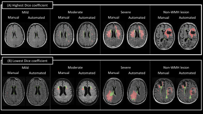 |
88 |
Deep learning-based quantification of white matter
hyperintensity applicable to real-world clinical FLAIR images
Kengo Onda1,
Jill Chotiyanonta1,
Yuto Uchida1,
Xin Li1,
Susumu Mori1,2,
and Kenichi Oishi1,2,3
1The Russell H. Morgan Department of Radiology and Radiological Science, The Johns Hopkins University School of Medicine, Baltimore, MD, United States, 2F. M. Kirby Research Center for Functional Brain Imaging, Kennedy Krieger Institute, Baltimore, MD, United States, 3The Richman Family Precision Medicine Center of Excellence in Alzheimer’s Disease, The Johns Hopkins University School of Medicine, Baltimore, MD, United States Keywords: White Matter, Neurodegeneration, White Matter Hyperintensity White matter hyperintensity (WMH) in the brain is known to correlate with cognitive prognosis in many diseases; automated quantification tools for WMH have been developed, but most have been used to quantify study data from specific diseases imaged with a single scanning protocol. The low accuracy of these tools when used for clinical data with diverse scan protocols and diseases has been a problem in clinical applications. To overcome this limitation, we developed a deep-learning-based WMH quantification model for real-world clinical FLAIR images with high heterogeneity. The results show the potential of this method as a clinical tool. |
|
2792. |
89 |
The value of MR diffusion tensor imaging in assessing white
matter changes in short-term methamphetamine withdrawal
Yanyao Du1,
Wenhan Yang1,
Sihong Huang1,
Fei Tang1,
Wei Zhao1,
Hu Guo2,
Huiting Zhang3,
and Jun Liu1
1The Second Xiangya Hospital, Central South University, Changsha, China, 2MR Application,Siemens Healthineers Ltd., Changsha, China, 3MR Scientific Marketing,Siemens Healthineers Ltd., Wuhan, China Keywords: White Matter, Diffusion/other diffusion imaging techniques This study aimed to explore the changes of brain white matter changes in short-term methamphetamine (MA) abstinence and to investigate potential imaging markers. Compared with healthy controls (HC), fractional anisotropy (FA), axial diffusivty (AD) and mean diffusivity (MD) values in short-term abstinence group were all increased, but there was no significant difference in the radial diffusivty value. The changes of FA, AD and MD value may be a new biomarker which is helpful to explore the potential mechanism of neurotoxicity damage. |
|
2793.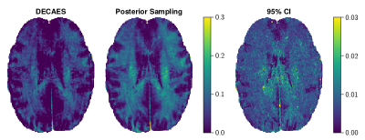 |
90 |
Improving T2 distribution estimation using deep generative
priors
Jonathan Doucette1,2,3,
Christian Kames1,2,3,
and Alexander Rauscher1,2,3,4
1UBC MRI Research Centre, Vancouver, BC, Canada, 2Physics and Astronomy, University of British Columbia, Vancouver, BC, Canada, 3Pediatrics, University of British Columbia, Vancouver, BC, Canada, 4Djavad Mowafaghian Centre for Brain Health, Vancouver, BC, Canada Keywords: White Matter, Relaxometry, Brain, Myelin Water Fraction $$$T_2$$$ distributions are typically computed using point estimates such as nonnegative least-squares (NNLS). This characterizes the most likely $$$T_2$$$-distribution arising from the data, but disregards other plausible solutions - of which there are many, due to the ill-posed nature of the inverse problem. Here, we instead propose to use Bayesian posterior sampling methods. To guide the difficult high-dimensional sampling problem, a data-driven domain transformation is learned alongside a deep generative prior. The resulting posterior samples produce more spatially consistent myelin water fraction (MWF) maps compared to NNLS, despite the purely voxelwise analysis, and additionally yields novel MWF uncertainty estimates. |
|
2794.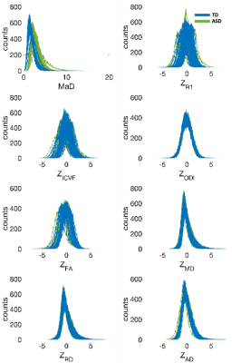 |
91 |
Individual-specific TBSS of diffusion and relaxometry measures
reveal differences of global white matter in autistic
individuals
Jose Guerrero-Gonzalez1,
Olivia Surgent2,
Nagesh Adluru3,
Gregory Kirk3,
Steven Kecskemeti3,
Douglas Dean III1,
Brittany Travers4,
and Andrew Alexander1
1Medical Physics, University of Wisconsin - Madison, Madison, WI, United States, 2Neuroscience Training Program, Waisman Center, University of Wisconsin - Madison, Madison, WI, United States, 3Waisman Center, University of Wisconsin - Madison, Madison, WI, United States, 4Kinesiology, University of Wisconsin - Madison, Madison, WI, United States Keywords: White Matter, Diffusion/other diffusion imaging techniques, Autism For neurological conditions that effect brain regions differently across individuals, quantitative neuroimaging-based metrics of individual deviation may be more informative in characterizing neuroanatomical variation including normal from abnormal variations than commonly used group-wise analyses. This work uses a joint analysis of iTBSS (individual-specific Tract-Based Spatial Statistics) of multiple neuroimaging measures from diffusion and relaxometry to better characterize differences between autistic and non-autistic individuals. |
|
2795.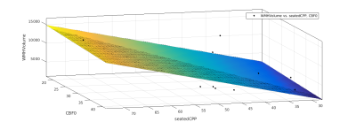 |
92 |
Measures of vascular function influence the number and volume of
white matter hyperintensities in older, hypertensive subjects
Andrew Crofts1,
Jessica Steventon2,
Joseph Whittaker3,
Marcello Venzi3,
Hannah Chandler4,
Michael Germuska4,
Mahfoudha Al Shezawi5,
Eric Stohr5,
Chris Pugh5,
Barry McDonnell5,
and Kevin Murphy3
1National Institute for Quantum and Radiological Science and Technology, Chiba, Japan, 2CUBRIC, School of Medicine, Cardiff University, Cardiff, United Kingdom, 3CUBRIC, School of Physica and Astronomy, Cardiff University, Cardiff, United Kingdom, 4CUBRIC, School of Psychology, Cardiff University, Cardiff, United Kingdom, 5Cardiff Metropolitan University, Cardiff, United Kingdom Keywords: White Matter, Aging Damage to the deep white matter of the brain has been shown to correlate with hypertension and advanced age. However, the changes in the cerebral microvasculature that cause white matter lesions are unclear. Increased blood pressure causes morphological changes in cerebral vessels, and impaired vascular function and neurovascular coupling is a potential factor. Here, we demonstrate that age, central pulse pressure, and dual-echo MRI measures of CBF, CVR, CMRO2 and OEF influence the number and volume of white matter hyperintensities. |
|
2796.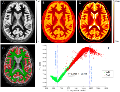 |
93 |
Fast T1 mapping of whole brain using Multiplied Added Subtracted
Divided Inversion Recovery (MASDIR)
Letizia Losa1,
Denis Peruzzo1,
Graeme Bydder2,
and Nivedita Agarwal3
1CESNE, IRCCS E. Medea, Bosisio Parini, Italy, 2Department of Radiology, University of California, San Diego, San Diego, CA, United States, 3Neuroradiology, IRCCS E. Medea, Bosisio Parini, Italy Keywords: White Matter, Quantitative Imaging, T1 mapping Whole brain T1 mapping has been performed using several different sequences but remains mostly confined to research applications. A clinically reliable and fast method to evaluate the white matter (WM) is important for accurate diagnosis, evaluating the severity of disease and monitoring disease progression over time. In this study we assessed Multiplied, Added, Subtracted and/or Divided Inversion Recovery (MASDIR) technique in nine healthy subjects and found evidence that such technique greatly improves the visualisation of tissue of interest and allows calculation of T1 values in a reliable, fast and clinically usable manner. |
|
2797.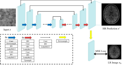 |
94 |
Unsupervised Super-Resolution of Magnetic Resonance Images Using
Deep Image Prior
Geng Chen#1,2,
Hao Yang#1,
Lemroussi Wissal1,
Yong Xia*1,
and Pew-Thian Yap*2
1Northwestern Polytechnical University, Xi'an, China, 2University of North Carolina at Chapel Hill, Chapel Hill, NC, United States Keywords: White Matter, Diffusion/other diffusion imaging techniques The anatomical resolution of MRI is typically limited by acquisition time constraints. While deep learning networks have shown great potential for post-acquisition MRI resolution enhancement, their training typically relies on low-high resolution image pairs, which are not always available in practice. Here, we propose using deep image prior (DIP) for unsupervised MRI resolution enhancement with network training relying only on low-resolution images. Experimental results indicate that our method super-resolve MR images effectively with realistic details. |
|
2798.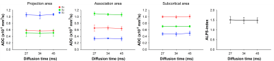 |
95 |
Reproducibility of diffusion tensor image analysis along the
perivascular space (DTI-ALPS) on variable diffusion times
Won Beom Jung1,
Jaekyun Ryu1,
Chuluunbaatar Otgonbaatar2,
Jeonghak Song3,
Juho Kim1,
and Hackjoon Shim1,3
1Medical Imaging AI Research Center, Canon Medical Systems Korea, Seoul, Korea, Republic of, 2College of Medicine, Seoul National University, Seoul, Korea, Republic of, 3Magnetic Resonance Business Unit, Canon Medical Systems Korea, Seoul, Korea, Republic of Keywords: White Matter, Diffusion/other diffusion imaging techniques Diffusion tensor imaging (DTI) allows noninvasively to investigate the function of glymphatic system with ALPS index by utilizing the behavior of water molecules along the perivascular space. The effects of diffusion time with fixed TE for the evaluation of glymphatic system using diffusion tensor imaging have not been investigated. Here, we evaluated the effects of DTI with different diffusion time to ALPS index in the glymphatic system. |
|
2799.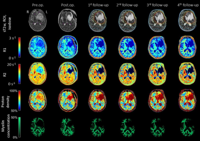 |
96 |
Radiation Dependent Demyelination in Normal Appearing White
Matter in Glioma Patients, Determined Using Quantitative MRI
Anna Ljusberg1,2,
Ida Blystad2,3,
Peter Lundberg1,2,
Emelie Adolfsson1,
and Anders Tisell1,2
1Department of Medical Radiation Physics, and Department of Health, Medicine and Caring Sciences, Linköping University, Linköping, Sweden, 2Center for Medical Image science and Visualization (CMIV), Linköping University, Linköping, Sweden, 3Department of Radiology in Linköping, and Department of Health, Medicine and Caring Sciences, Linköping University, Linköping, Sweden Keywords: White Matter, Radiotherapy Adding quantitative MRI to existing examinations for patients with malignant glioma is valuable for observing changes in tissue specific parameters such as R1, R2, proton density and myelin concentration. Changes observed in this study, with a decrease of myelin concentration and increase of proton density, in normal appearing white matter can be interpreted as a demyelination caused by radiation therapy. For low doses (<30 Gy), but not for high (>30 Gy) the myelin concentration returns to baseline. |
|
2800.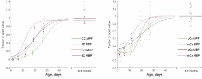 |
97 |
Macromolecular proton fraction as a biomarker of early
myelination: ultrastructural and immunochemical validation in a
rabbit model
Alexander Drobyshevsky1,
Daniil Aksenov1,
and Vasiliy Yarnykh2
1NorthShore University HealthSystems, Evanston, IL, United States, 2Radiology, University of Washington, Seattle, WA, United States Keywords: White Matter, Quantitative Imaging, myelin Ultrastructural and immunochemical methods of myelin quantification were applied to validate a model-based MRI techniques, macromolecular proton fraction mapping (MPF), during early development in major white matter tracts and cerebral cortex in rabbits. MPF trajectories were in general agreement with levels of myelination by histology and accurately reflected differential rate of development between white matter tracts and cortical regions. |
|
2801.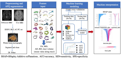 |
98 |
Comparison of machine learning models for discriminating
obsessive-compulsive disorder based on automated fiber
quantification
Suming Zhang1,
Xuan Bu2,
Lingxiao Cao1,
Hailong Li1,
Kaili Liang1,
Zilin Zhou1,
Yingxue Gao1,
Lianqing Zhang1,
Bin Li3,
and Xiaoqi Huang1
1Huaxi MR Research Center(HMRRC), Department of Radiology, West China Hospital of Sichuan University, Chengdu, China, 2State Key Laboratory of Cognitive Neuroscience and Learning, Beijing Normal University, Beijing, China, 3Mental Health Center, West China Hospital of Sichuan University, Chengdu, China Keywords: White Matter, Diffusion Tensor Imaging In this study, we compared the classification performance of different machine learning models for discriminating OCD patients based on DTI tractography. Firstly, we extracted DTI metrics and tract volumes as features. Following feature selection, four machine learning models were performed for classification. Finally, a novel SHapley Additive exPlanations (SHAP) analysis was used to intepret the value of importance for each feature. We found that XGBoost exhibited the best classification performance among the four models. The model explanation by SHAP suggested that the volume of callosal orbital frontal tract was the most important factor in differentiating OCD from healthy controls. |
|
2802. |
99 |
Modified cerebral small vessel disease total burden is useful to
screening study based on community people
Ronghua Mu1,
Wei Zheng1,2,
Xiaoyan Qin1,
Xin Li1,
Kan Deng3,
Fuzhen Liu1,
Zeyu Zhuang1,2,
Peng Yang1,
Jian Lv1,
and Xiqi Zhu1
1the Nanxishan Hospital, Guangxi Zhuang Autonomous Region, guilin, China, 2Graduate School of Guilin Medical University, guilin, China, 3Philips Healthcare, Guangzhou, China Keywords: White Matter, White Matter The objective of this study was to create a modified total load score suitable for community screening study by modifying mild WMH and EPVS. Our study found NCT-A (DST) variable abnormalities even in people with mild EPVS and WMH, suggesting that modified cerebral small vessel disease total burden is useful to screening study based on community people. |
|
2803.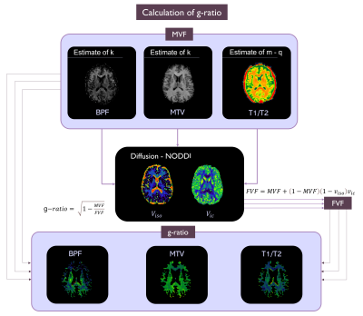 |
100 |
Myelin quantification: is it possible to obtain comparable
g-ratio maps from different acquisition techniques?
Marta Gaviraghi1,
Eleonora Lupi1,
Simone Rancati2,
Fulvia Palesi1,
Francesco Grussu3,4,
Marco Battiston3,
Carmen Tur3,4,
Alberto Calvi3,
Sara Collorone3,
Antonio Ricciardi3,
Ferran Prados3,5,6,
Baris Kanber3,7,
Egidio D'Angelo1,8,
Rebecca S. Samson3,
and Claudia A. M. Gandini Wheeler-Kingshott1,3,8
1Department of Brain and Behavioural Sciences, University of Pavia, Pavia, Italy, 2Department of Electrical, Computer and Biomedical Engineering, University of Pavia, Pavia, Italy, 3NMR Research Unit, Department of Neuroinflammation, Queen Square Multiple Sclerosis Centre, UCL Queen Square Institute of Neurology, University College London (UCL), London, United Kingdom, 4Neurology-Neuroimmunology Department Multiple Sclerosis Centre of Catalonia (Cemcat), Vall d’Hebron Barcelona Hospital Campus, Barcelona, Spain, 5Department of Medical Physics and Biomedical Engineering, Centre for Medical Image Computing (CMIC), University College London, London, United Kingdom, 6E-Health Center, Universitat Oberta de Catalunya, Barcelona, Spain, 7Department of Medical Physics and Biomedical Engineering, Centre for Medical Image Computing (CMIC), University College London, London, Italy, 8Brain Connectivity Centre, IRCCS Mondino Foundation, Pavia, Italy Keywords: White Matter, Multi-Contrast, Myelin Quantifying myelin in vivo with MRI is useful for studying multiple sclerosis by calculating the g-ratio. To estimate the g-ratio, special sequences often must be set. We have developed a preliminary method for calculating g-ratio from clinical images i.e. T1w and T2w. We then compared three methods to obtain the g-ratio: from quantitative Magnetization Transfer (qMT) (gold standard), from Macromolecular Tissue Volume (MTV) and from T1w/T2w. The MTV (g_MTV) and T1w/T2w (g_T1wT2w) g-ratios were different from the qMT g-ratio (g_qMT). g_MTV flattens differences between healthy and pathological subjects whereas g_T1wT2w has the same median trend as g_qMT. |
|
 Back to Meeting Home
 Back to the Program-at-a-Glance
The International Society for Magnetic Resonance in Medicine is accredited by the Accreditation Council for Continuing Medical Education to provide continuing medical education for physicians.
 Back to Meeting Home
 Back to the Program-at-a-Glance
 View Presentation Video