Digital Poster
Alzheimer's & Dementias I
ISMRM & ISMRT Annual Meeting & Exhibition • 03-08 June 2023 • Toronto, ON, Canada

| Computer # | |||
|---|---|---|---|
2646.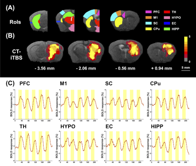 |
121 |
Central Thalamic Intermittent Theta-Burst Stimulation
Ameliorated Memory Impairment in Alzheimer's Disease Mouse Model
Yi-Chen Lin1,
Ssu-Ju Li1,
Yu-Chun Lo2,
Ting-Chieh Chen1,
Ching-Wen Chang1,
Tsai-Yu Cho1,
Mu-Hua Wang1,
Ching-Te Chen3,
Sheng-Huang Lin4,5,
and You-Yin Chen1,2
1Department of Biomedical Engineering, National Yang Ming Chiao Tung University, Taipei, Taiwan, 2PhD Program in Medical Neuroscience, Taipei Medical University, Taipei, Taiwan, 3Abbott Neuromodulation, Austin, TX, United States, 4Department of Neurology, Buddhist Tzu Chi Medical Foundation, Hualien, Taiwan, 5Department of Neurology, Tzu Chi University, Hualien, Taiwan Keywords: Alzheimer's Disease, fMRI, Intermittent theta-burst stimulation (iTBS) Intermittent theta-burst stimulation (iTBS) has been hypothesized to be a more effective paradigm for deep brain stimulation (DBS) in treating various neurological disorders by altering neuronal circuits. However, the effect of iTBS in improving memory deficits in AD patients remains unknown. Combining iTBS in central thalamus (CT-iTBS) with functional magnetic resonance imaging, our results demonstrated the restoration of brain functional connectivity (FC) in corticolimbic circuit of AD mice model, accompanied with enhancing memory cognitive function in novel object recognition test and T-maze test. These results revealed that CT-iTBS could be an effective treatment for improving symptoms of individuals with AD. |
|
2647.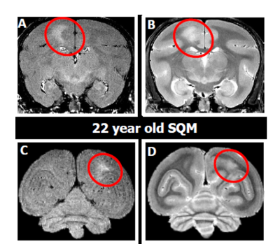 |
122 |
Multi-parametric MRI Characterization of the Aging Squirrel
Monkey as a Model of Sporadic Cerebral Amyloid Angiopathy
Youssef Z Wadghiri1,
Jelle Veerart1,
Sean Murray1,
Jakub Szabo1,
Carolyn Akers1,
Thomas Genovese1,
Hannah Goldman1,
Jonathan Leung1,
Charles V Kingsley 2,
Henry Rusinek1,
Stanton Gray2,
William Donald Hopkins2,
Thomas Wisniewski1,
and Henrieta Scholtzova1
1NYU Grossman School of Medicine, New York, NY, United States, 2UT - MD Anderson Cancer Center, Houston, TX, United States Keywords: Alzheimer's Disease, Aging, Non human primate The current study seeks to identify neuroimaging biomarkers in a unique squirrel monkey (SQM) as a spontaneous model of cerebral amyloid angiopathy both in vivo and ex vivo using conventional MRI metrics in young and geriatric SQM cohorts and demonstrate the utility of quantitative R2* to map age-related differences in SQM neuropathology cross-sectionally and longitudinally. |
|
2648.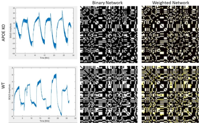 |
123 |
4D Oxy-wavelet MRI and Graph Theory in APOE KO mice: A new
perspective into the role of mitochondria and brain connectivity
Devin Raine Everalo Cortes1,2,3,4,
Margaret C. Stapleton2,4,
Samuel Wyman2,4,
Kristina E. Schwab5,6,
Noah W. Coulson2,7,
Dalton R West2,4,
Thomas Becker-Szurszewski5,6,
Sean Hartwick5,6,
Anthony G. Christodoulou8,
and Yijen L Wu2,3,4,9
1Department of Bioengineering, University of Pittsburgh, Pittsburgh, PA, United States, 2Department of Developmental Biology, School of Medicine, University of Pittsburgh, Pittsburgh, PA, United States, 3Department of Bioengineering, Swanson School of Engineering, University of Pittsburgh, Pittsburgh, PA, United States, 4Rangos Research Center Small Animal Imaging Core, Children's Hospital of Pittsburgh of UPMC, Pittsburgh, PA, United States, 5Rangos Research Center Animal Imaging Core, Children's Hospital of Pittsburgh of UPMC, Pittsburgh, PA, United States, 6Department of Pediatrics, University of Pittsburgh, School of Medicine, Pittsburgh, PA, United States, 7Rangos Research Center Small Animal Imaging Core, University of Pittsburgh, Pittsburgh, PA, United States, 8Biomedical Imaging Research Institue, Cedars-Sinai Medical Center, Los Angeles, CA, United States, 9Department of Pediatrics, School of Medicine, University of Pittsburgh, Pittsburgh, PA, United States Keywords: Alzheimer's Disease, Brain Connectivity, Metabolism, fMRI Apolipoprotein E (APOE) alleles are strong genetic risk factors for the Late-Onset-Alzheimer’s Disease (LOAD). In this study we utilize our novel 4D Oxy-wavelet functional MRI to study the bioenergetic footprint of APOE knockout (KO) mice compared to wild type (WT) mice. We have created neuronal networks in mice defined by bioenergetic dispersion throughout the whole brain, and demonstrated key differences between APOE KO and WT. We further validate our oxywavelet index for probing mitochondrial function non-invasively and present new signal processing methods for relevant biomarker extraction. |
|
2649.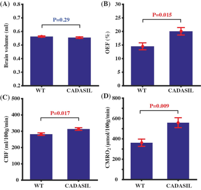 |
124 |
CADASIL mice are characterized by simultaneous metabolic and
vascular stress
Zhiliang Wei1,2,
Yuguo Li1,2,
Lin Chen3,
Hongshuai Liu4,
Minmin Yao4,
Jiadi Xu1,2,
Angeliki Louvi5,
Wenzhen Duan4,6,
and Hanzhang Lu1,2,7
1Russell H. Morgan Department of Radiology and Radiological Science, Johns Hopkins University School of Medicine, Baltimore, MD, United States, 2F. M. Kirby Research Center for Functional Brain Imaging, Kennedy Krieger Research Institute, Baltimore, MD, United States, 3Department of Electronic Science, Xiamen University, Xiamen, China, 4Department of Psychiatry and Behavioral Sciences, Johns Hopkins University School of Medicine, Baltimore, MD, United States, 5Departments of Neurosurgery and Neuroscience, Yale School of Medicine, New Haven, CT, United States, 6The Solomon H. Snyder Department of Neuroscience, Johns Hopkins University School of Medicine, Baltimore, MD, United States, 7Department of Biomedical Engineering, Johns Hopkins University School of Medicine, Baltimore, MD, United States Keywords: Dementia, Animals Vascular cognitive impairment and dementia (VCID) is the second leading cause of dementia and is often mixed with other pathologies. Related mouse models with relatively pure vascular pathologies are used for mechanistic studies or therapeutic trials. CADASIL (Cerebral autosomal dominant arteriopathy with subcortical infarcts and leukoencephalopathy) is a monogenic condition causing lacunar strokes and vascular dementia. Using the established mouse model of CADASIL, we aimed to investigate potential microvascular dysfunctions with advanced non-contrast MRI techniques. We found that CADASIL mice displayed elevated oxygen consumption and impaired cerebrovascular reactivity, suggesting simultaneous metabolic and vascular stress. |
|
2650.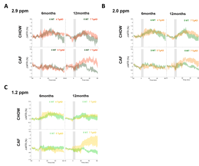 |
125 |
Metabolic Imaging using Chemical Exchange Saturation Transfer in
Rat Transgenic Model of Alzheimer’s Disease Comorbid with
Metabolic Syndrome
Dustin Loren Velasco Almanza1,2,
Wilfred Lam2,
Margaret Koletar2,
Mary Hill3,
JoAnne McLaurin3,4,
Greg Stanisz1,2,
and Bojana Stefanovic1,2
1Medical Biophysics, University of Toronto, Toronto, ON, Canada, 2Physical Sciences, Sunnybrook Research Institute, Toronto, ON, Canada, 3Biological Sciences, Sunnybrook Research Institute, Toronto, ON, Canada, 4Laboratory Medicine and Pathology, University of Toronto, Toronto, ON, Canada Keywords: Alzheimer's Disease, CEST & MT Our study characterized the hippocampal glucose uptake in an Alzheimer’s Disease rat model (TgF344AD) comorbid with metabolic syndrome modeled by cafeteria (CAF) diet. Glucose uptake was evaluated by detecting 2-deoxy-D-glucose (2DG) using chemical exchange saturation transfer (CEST) MRI in 6- and 12-month-old cohorts, reflecting pre-clinical and established disease stages, respectively. CAF diet attenuated glucose uptake of both non-transgenic and transgenic-AD at 6 months of age; whereas at 12 months of age, CAF diet enhanced the hippocampal glucose uptake of TgAD but attenuated it in chow-fed TgAD, when compared to that in correspondingly fed nTg littermates. |
|
2651.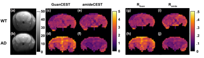 |
126 |
Decreased pH in the mouse brain of Alzheimer’s disease revealed
by guanidinium and amide CEST at 3T
Kexin Wang1,2,
Jianpan Huang3,
Ziqin Zhang1,2,
Kannie W. Y. Chan3,4,
and Jiadi Xu2,4
1Department of Biomedical Engineering, Johns Hopkins University, Baltimore, MD, United States, 2F.M. Kirby Research Center for Functional Brain Imaging, Kennedy Krieger Research Institute, Baltimore, MD, United States, 3Department of Biomedical Engineering, City University of Hong Kong, Hong Kong, China, 4Russell H. Morgan Department of Radiology and Radiological Science, Johns Hopkins University School of Medicine, Baltimore, MD, United States Keywords: Alzheimer's Disease, CEST & MT, pH mapping Guanidinium CEST (GuanCEST) and amideCEST are highly sensitive to pH change, while their correlations with pH are opposite. Thus taking (unitless) difference of GuanCEST and amideCEST provides a reliable indicator for pH. Polynomial and Lorentzian line-shape fitting (PLOF) method helps extract the two CEST signals precisely, and the (unitless) difference shows a significant increase (p=0.008, n=10) in Alzheimer’s disease (AD) mouse brain compared with that of wild type mouse at 3T. Our results light up a novel and promising method to detect the onset of AD noninvasively, bringing the recent breakthrough in AD mechanism to further clinical application. |
|
2652.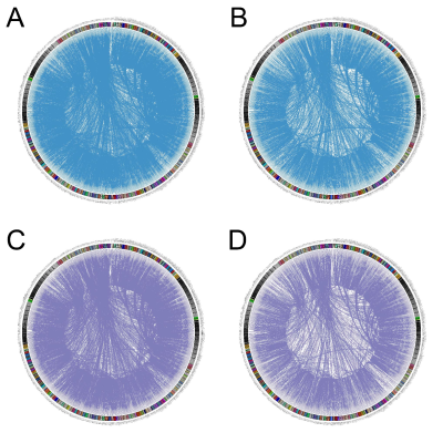 |
127 |
Effects of aquaporin-4 on neurovascular coupling and brain
network in Alzheimer’s disease mice model
Jing Li1,
Renyuan Liu1,
Pin Lv1,
and Bing Zhang1
1The Affiliated Drum Tower Hospital of Nanjing University Medical School, Nanjing, China Keywords: Alzheimer's Disease, fMRI, neurovascular coupling Neurovascular uncoupling can be found in the early stage of AD continuum. The effect of AQP4 content on neurovascular coupling (NVC) in AD mice is unclear. Adult 5xFAD mice and wild-type mice were employed in this study. Mice received TGN-020, an AQP4 inhibitor, intraperitoneally. The results showed that 20 min after the injection of TGN-020, reduced ReHo in the basal area of the frontal lobe, a trend of reduced functional connectivities and disordered network properties were found among the AQP4-inhibited WT and 5xFAD mice. Therefore, Inhibition of AQP4 may reduce the NVC, and disorder the functional network. |
|
2653.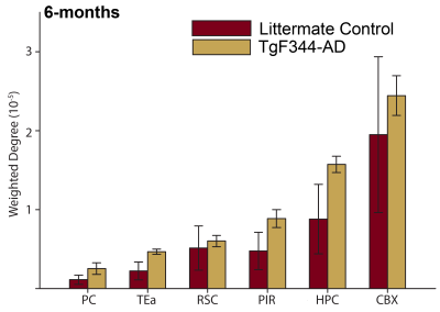 |
128 |
Functional and Structural Connectivity in the TgF344-AD
Transgenic Rat Model of Alzheimer's Disease using rs-fMRI & DTI
at 21.1 T
Jenna M. Radovich1,2,
Jordan Ogg3,
Zachary Baty1,2,
Aaron Wilber3,
and Samuel Colles Grant1,2
1National High Magnetic Field Laboratory, Florida State University, Tallahassee, FL, United States, 2Chemical & Biomedical Engineering, FAMU-FSU College of Engineering, Tallahassee, FL, United States, 3Psychology, Florida State University, Tallahassee, FL, United States Keywords: Alzheimer's Disease, Brain Connectivity, Graph theory, Progressive Degeneration A hallmark of Alzheimer's Disease (AD) is spatial disorientation, such as getting lost in new locations. A potential cause is disrupted exchange between egocentric and allocentric reference frames, in which the parietal and retrosplenial cortex have roles. This study aimed to examine rs-fMRI and DTI in relationship with coordination between reference frames in a transgenic AD rat model. Behavioral and DTI data suggest that both age and genotype lead to declines in action-orientation performance with alterations appearing at 5 mon, and at 6 mon structurally with profound changes developing longitudinally across multiple ROI. |
|
2654.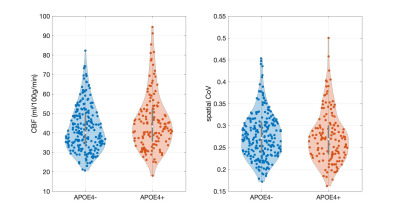 |
129 |
Hyperperfusion in middle-aged individuals with genetic risk of
sporadic Alzheimer’s disease
Maria-Eleni Dounavi1,
Elijah Mak1,
Audrey Low1,
Guy B Williams2,
Katie Wells3,
Graciela Muniz-Terrera3,4,
Brian Lawlor5,
Lorina Naci5,
Paresh Malhotra6,
Ivan Koychev7,
Karen Ritchie8,
Li Su1,9,
Craig W Ritchie3,
and John T O'Brien1
1Department of Psychiatry, University of Cambridge, Cambridge, United Kingdom, 2Department of Clinical Neurosciences and Wolfson Brain Imaging Centre, University of Cambridge, Cambridge, United Kingdom, 3Centre for Dementia Prevention, University of Edinburgh, Edinburgh, United Kingdom, 4Ohio University, Athens, OH, United States, 5Institute of Neuroscience, Trinity College Dublin, University of Dublin, Dublin, United Kingdom, 6Division of Brain Science, Imperial College Healthcare NHS Trust, London, United Kingdom, 7Department of Psychiatry, University of Oxford, Oxford, United Kingdom, 8INSERM, Montpellier, France, 9University of Sheffield, Sheffield, United Kingdom Keywords: Alzheimer's Disease, Arterial spin labelling Extensive brain hypoperfusion is well-established in people with Alzheimer’s disease (AD). However, mixed findings have emerged during the pre-symptomatic disease stage. In this study, we used arterial spin labelling (ASL) MRI in middle-aged, cognitively normal participants from the PREVENT-Dementia study to evaluate cerebral perfusion differences between carriers and non-carriers of the apolipoprotein ε4 (APOE4) allele. The ASL spatial coefficient of variation (CoV) was used as a proxy for arterial transit time (ATT) delays. We found hyperperfusion in APOE4 carriers which was not accompanied by increased spatial CoV, suggesting that the observed hyperperfusion is not driven by ATT delays. |
|
2655.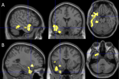 |
130 |
Cascade of Perfusion and Brain Atrophy in Alzheimer’s Disease: A
Longitudinal Study
Tony Zhou1,
Zongpai Zhang2,
Arvind Balachandrasekaran3,
Cyrus A. Raji4,
James T. Becker5,
Lewis H. Kuller6,
Yulin Ge7,
Oscar L. Lopez8,
Weiying Dai2,
and H. Michael Gach1,9
1Radiation Oncology, Washington University School of Medicine, Saint Louis, MO, United States, 2Computer Science, State University of New York at Binghamton, Binghamton, NY, United States, 3Boston Children's Hospital, Boston, MA, United States, 4Radiology and Neurology, Washington University School of Medicine, Saint Louis, MO, United States, 5Psychiatry, Psychology, and Neurology, University of Pittsburgh, Pittsburgh, PA, United States, 6Epidemiology, University of Pittsburgh, Pittsburgh, PA, United States, 7Radiology, New York University School of Medicine, New York City, NY, United States, 8Neurology and Psychiatry, University of Pittsburgh, Pittsburgh, PA, United States, 9Radiology and Biomedical Engineering, Washington University School of Medicine, Saint Louis, MO, United States Keywords: Alzheimer's Disease, Arterial spin labelling We conducted a longitudinal study to determine if reduced temporoparietal and frontal cerebral blood flow (CBF) in elderly population leads to reduced gray matter volumes (GMVs) in the temporal lobe, or vice versa. We observed smaller GMVs in the temporal pole (TP) region in Alzheimer’s disease (AD) patients. We also found associations of: (1) the TP GMVs with subsequent temporoparietal CBF declines; (2) the TP CBF with its own subsequent GMV changes; and (3) the hippocampal GMVs with longitudinal frontal CBF declines. Hypoperfusion in the temporal lobe may be an early event driving atrophy, followed by temporoparietal and frontal hypoperfusion. |
|
2656.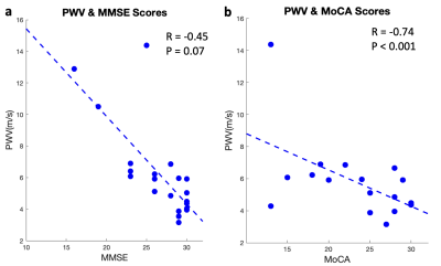 |
131 |
Elevated Carotid Pulse Wave Velocity is associated with
Cognitive Impairment and Brain Amyloid Deposition
Jianing Tang1,2,
Elizabeth Joe3,
Helena Chui3,
and Lirong Yan2,3
1Biomedical Engineering, Northwestern University, Evanston, IL, United States, 2Radiology, Northwestern University, Evanston, IL, United States, 3Department of Neurology, University of Southern California, Los Angeles, CA, United States Keywords: Alzheimer's Disease, Blood vessels Increasing evidence suggests that vascular compliance could offer valuable insights into the pathology of Alzheimer’s diseases. Recently, carotid pulse wave velocity (cPWV) between internal carotid artery and common carotid artery has been successfully measured by a fast single-slice oblique-sagittal PC-MRI technique. In this study, we evaluated the role of cPWV in brain amyloid deposition and cognitive decline in an aged cohort. The results showed that greater cPWV was associated with cognitive decline and amyloid pathology. Our findings suggest elevated cPWV may affect amyloid clearance in brain, leading to cognitive impairment. cPWV could be a potential sensitive imaging marker of AD. |
|
2657.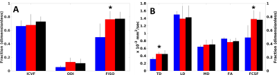 |
132 |
Microstructure of the Fornix Correlates with Cognitive Status in
Alzheimer’s Disease
Ken Sakaie1,
Katherine Koenig2,
Jian Lin2,
Alan Lerner3,
James Leverenz4,
and Mark J. Lowe2
1The Cleveland Clinic, Cleveland, OH, United States, 2Imaging Institute, The Cleveland Clinic, Cleveland, OH, United States, 3Brain Health and Memory Center, Neurological Institute, University Hospitals Cleveland Medical Center, Cleveland, OH, United States, 4Lou Ruvo Center for Brain Health, The Cleveland Clinic, Cleveland, OH, United States Keywords: Alzheimer's Disease, Dementia Imaging measures of tissue microstructure of the fornix are are potential biomarkers for cognitive decline in Alzheimer’s disease (AD). However, partial volume averaging (PVA) between the fornix and cerobrospinal fluid can be substantial. As a result, measured changes may reflect atrophy, not changes in tissue microstructure. Neurite Orientation Dispersion and Direction Imaging and Free Water Elimination diffusion tensor imaging account for PVA. We present results from a cohort of AD patients, patients with mild cognitive impairment (MCI) and cognitively normal (CN) subjects, showing that NODDI and DTI measures correlate with specific scores cognitive status. |
|
2658. |
133 |
Effects of Subcortical Atrophy and Alzheimer’s pathology on
Cognition in Elderly Type 2 Diabetes
Wen Zhang1 and
Bing Zhang2
1Radiology, The Affiliated Drum Tower Hospital of Nanjing University Medical School, Nanjing, China, 2The Affiliated Drum Tower Hospital of Nanjing University Medical School, Nanjing, China Keywords: Alzheimer's Disease, Diabetes Subcortical atrophy and increased cerebral β-amyloid and tau deposition are linked to cognitive decline in type 2 diabetes. However, whether and how subcortical atrophy is related to Alzheimer’s pathology in diabetes remains unclear. We investigated the subcortical structural alterations induced by diabetes and the relationship between subcortical alteration, Alzheimer’s pathology and cognition. Our results suggested that although both type 2 diabetes and AD are correlated with subcortical neurodegeneration, type 2 diabetes have no direct or indirect effect on the cerebral amyloid deposition and CSF p-tau. |
|
2659.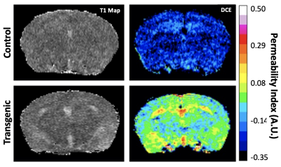 |
134 |
Detection of Capillary Leakage in a Transgenic Mouse Model of
Alzheimer’s Disease
Ning Hua1,
Olga Minaeva1,
Juliet Moncaster1,
Hernan Jara1,
and Lee Goldstein1
1Boston University, Boston, MA, United States Keywords: Alzheimer's Disease, DSC & DCE Perfusion The goal of the project is to explore the possibility of using dynamic contrast-enhanced (DCE)-MRI for detecting subtle blood-brain barrier leakage in a transgenic mouse model of Alzheimer’s disease. The washout slope of Gd concentration was used to assess microvascular integrity. We observed different washout patterns in transgenic relative to control mice. Our findings demonstrated that the washout slope could be used to assess status of blood-brain barrier permeability in AD mice. |
|
2660.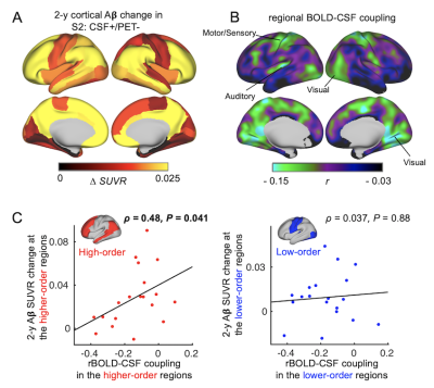 |
135 |
Resting-state global brain activity affects early β-amyloid
accumulation and hypoconnectivity in default mode network
Feng Han1,
Xufu Liu1,
Richard Mailman2,
Xuemei Huang2,
and Xiao Liu1
1the Pennsylvania State University, State College, PA, United States, 2Pennsylvania State University College of Medicine and Milton S. Hershey Medical Center, Hershey, PA, United States Keywords: Alzheimer's Disease, fMRI (resting state), cerebrospinal fluid The β-amyloid (Aβ) plaque, an Alzheimer’s disease (AD) hallmark, accumulates first in the default mode network (DMN) regions years before AD diagnosis. The underlying mechanisms remain elusive. Low-frequency (<0.1 Hz) resting-state global brain activity was recently found coupled by cerebrospinal fluid (CSF) movements, and this coupling has been linked to AD pathologies, including Aβ accumulations, presumably due to its role in glymphatic clearance. Here we showed the preferential Aβ accumulation in DMN was related to the reduced engagement of the global brain activity in these regions, partly explained by its failure to reach these regions as cortical propagating waves. |
|
2661.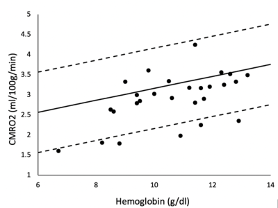 |
136 |
Iron deficiency anemia is associated with abnormal cerebral
metabolic rate, blood brain barrier permeability and cognitive
function.
Silvie Suriany1,
Clio Gonzalez-Zacarias2,
Chau Vu2,
Sharon ONeil3,
Gagan C Mathur4,
Dawn C Ward5,
Alyssa Ziman5,
Ellen B Klapper6,
Lefan Zhuang7,
and John C Wood1
1Pediatrics, Children's Hospital of Los Angeles-USC KSOM, Los Angeles, CA, United States, 2University of Southern California, Los Angeles, CA, United States, 3Neuropsychology, Children's Hospital of Los Angeles-USC KSOM, Los Angeles, CA, United States, 4Transfusion Medicine, Children's Hospital of Los Angeles-USC KSOM, Los Angeles, CA, United States, 5Transfusion Medicine, University of California, Los Angeles, Los Angeles, CA, United States, 6Transfusion Medicine, Cedar Sinai Medical Center, Los Angeles, CA, United States, 7Transfusion Medicine, City of Hope Medical Center, Duarte, CA, United States Keywords: Dementia, Metabolism, Iron Deficiency Iron deficiency is the predominant cause of anemia in adults, typically occurring in women with heavy menses, endometriosis or fibroids. We study cerebral metabolic rate of oxygen (CMRO2), blood brain barrier (BBB) permeability to water, and neurocognitive function in 34 women with iron deficiency anemia (IDA). We observed striking deficits in verbal and spatial memory, perceptual reasoning, and crystalized intelligence that were strongly correlated with hemoglobin level. CMRO2 was lower than predicted by anemia alone, correlated to BBB permeability to water, and associated with poorer neurocognitive performance, suggesting decreased brain capillary surface area for oxygen and water diffusion. |
|
2662.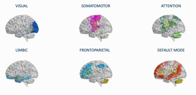 |
137 |
Personalized fingerprint of neurodegenerative phenotypes using
model-based simulations of brain networks
Anita Monteverdi1,
Fulvia Palesi2,
Michael Schirner3,4,5,6,7,
Francesca Argentino2,
Mariateresa Merante2,
Alberto Redolfi8,
Francesca Conca9,
Laura Mazzocchi10,
Matteo Cotta Ramusino2,11,
Alfredo Costa2,11,
Anna Pichiecchio2,10,
Lisa Maria Farina9,
Stefano Cappa9,12,
Viktor Jirsa13,
Petra Ritter3,4,5,6,7,
Claudia A.M. Gandini Wheeler-Kingshott1,2,14,
and Egidio D’Angelo1,2
1Brain Connectivity Center, IRCCS Mondino Foundation, Pavia, Italy, 2Dept of Brain and Behavioral Sciences, University of Pavia, Pavia, Italy, 3Berlin Institute of Health at Charité, Universitätsmedizin Berlin, Berlin, Germany, 4Department of Neurology with Experimental Neurology, Charité, Universitätsmedizin Berlin, Berlin, Germany, 5Bernstein Focus State Dependencies of Learning and Bernstein Center for Computational Neuroscience, Berlin, Germany, 6Einstein Center for Neuroscience Berlin, Berlin, Germany, 7Einstein Center Digital Future, Berlin, Germany, 8Laboratory of Neuroinformatics, IRCCS Istituto Centro San Giovanni di Dio Fatebenefratelli, Brescia, Italy, 9IRCCS Mondino Foundation, Pavia, Italy, 10Advanced Imaging and Radiomics Center, IRCCS Mondino Foundation, Pavia, Italy, 11Unit of Behavioral Neurology, IRCCS Mondino Foundation, Pavia, Italy, 12University Institute of Advanced Studies (IUSS), Pavia, Italy, 13Institut de Neurosciences des Systèmes, INSERM, INS, Aix Marseille University, Marseille, France, 14NMR Research Unit, Queen Square MS Centre, UCL Queen Square Institute of Neurology, Department of Neuroinflammation, University College London, London, United Kingdom Keywords: Alzheimer's Disease, Modelling, Frontotemporal Dementia This work provides the first personalized and non-invasive assessment of resting-state networks connectivity and excitatory/inhibitory balance in health and in neurodegenerative diseases (Alzheimer’s disease, Frontotemporal Dementia). Multiple networks were characterized at single-subject level performing brain dynamics simulations with The Virtual Brain (TVB). TVB-derived parameters identified specific network properties (at single network and inter-network level) and their disruption in neurodegeneration, underlined the relationship between neurophysiology and neuropsychology, and outlined a personalized fingerprint sensitive to clinical severity. This model-based simulation of brain networks functional dynamics lay the groundwork for customized biomarkers research and defines new trajectories for designing novel tailored interventional workflows. |
|
2663.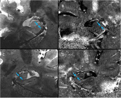 |
138 |
7T GRE Enables Detection of Subiculum Iron Deposits in AD
Mackenzie L Carlson1,
Phillip DiGiacomo1,
Brian Burns2,
Nicole Mouchawar1,
Julian Maclaren1,
Murat Aksoy1,
Pascal Spincemaille3,
Alexey Dimov3,
Yi Wang3,
Brian Rutt1,
and Michael Zeineh1
1Stanford University, Stanford, CA, United States, 2GE Healthcare, Seattle, WA, United States, 3Weill Cornell Medical College, New York, NY, United States Keywords: Alzheimer's Disease, Motion Correction Abnormal accumulation of iron has been found in the subiculum of Alzheimer’s Disease (AD) patients postmortem. This motivated us to push MRI resolution to observe iron accumulation in vivo in AD patients. Using optical prospective motion correction to enable higher resolution at 7T, we imaged 4 healthy controls, 3 mild cognitive impairment, and 1 Alzheimer’s Disease patient with a 60-minute research brain MRI protocol. Hippocampal iron deposits were detected by a neuroradiologist aware of diagnosis in 2 out of 4 MCI/AD subjects despite that mean R2* quantification in the subiculum was not significantly different between healthy control and MCI/AD subjects. |
|
2664.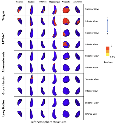 |
139 |
Associations of the shape of subcortical brain structures with
age-related neuropathologies in community-based older adults
Khalid Saifullah1,
Nazanin Makkinejad1,
Arnold M. Evia2,
David A. Bennett2,
Julie A. Schneider2,
and Konstantinos Arfanakis1,2
1Biomedical Engineering, Illinois Institute of Technology, Chicago, IL, United States, 2Rush Alzheimer's Disease Center, Rush University Medical Center, Chicago, IL, United States Keywords: Alzheimer's Disease, Neurodegeneration, Aging, Atherosclerosis Multiple age-related neuropathologies lead to atrophy of subcortical brain structures. However, definitive diagnosis of most of these pathologies is only possible at autopsy, complicating investigations into the effects of age-related neuropathologies on the shape of subcortical brain structures. The present work combined ex-vivo MRI and detailed pathologic assessment in a large number (N=842) of community-based older adults to study the relationship between age-related neuropathologies and the shape of subcortical brain structures. The resulting deformation patterns may be used as features in in-vivo classifiers of age-related neuropathologies. |
|
2665.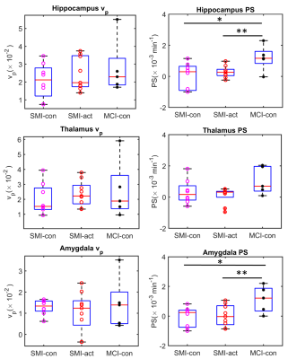 |
140 |
Improved DCE-MRI analyses of blood-brain barrier integrity in
subjective memory and mild cognitive impairment: New results
from the CANN trial
Rashed Sobhan1,
David R. Willis2,
Rachel Gillings2,
Michael J. Thrippleton3,
Joanna M. Wardlaw3,
Anne-Marie Minihane2,
Narelle M. Berry2,
and Donnie Cameron1,4
1Norwich Medical School, University of East Anglia, Norwich, United Kingdom, 2University of East Anglia, Norwich, United Kingdom, 3University of Edinburgh, Edinburgh, United Kingdom, 4Department of Radiology, C.J. Gorter Centre for High Field MRI, Leiden University Medical Centre, Leiden, Netherlands Keywords: Alzheimer's Disease, Permeability, DCE-MRI, Low-level Blood-brain-barrier Leakage, Mild cognitive impairment We implemented recent developments in DCE-MRI pre-processing and analysis techniques to compare local differences in blood brain barrier (BBB) integrity in subjective memory impairment (SMI) and mild cognitive impairment (MCI), two early indicators of Alzheimer’s disease. Also, as part of the CANN trial, we explored the impact of a combined flavanol and omega-3 fatty acid-based nutritional intervention on BBB leakage. The results show that BBB leakage in memory processing regions can distinguish between SMI and MCI, and thus can be used to quantitatively assess the progress of cSVD towards onset of MCI and/or early indication of Alzheimer’s disease. |
|
 Back to Meeting Home
 Back to the Program-at-a-Glance
The International Society for Magnetic Resonance in Medicine is accredited by the Accreditation Council for Continuing Medical Education to provide continuing medical education for physicians.
 Back to Meeting Home
 Back to the Program-at-a-Glance
 View Presentation Video