Digital Poster
Alzheimer's & Dementias II
ISMRM & ISMRT Annual Meeting & Exhibition • 03-08 June 2023 • Toronto, ON, Canada

| Computer # | |||
|---|---|---|---|
2804.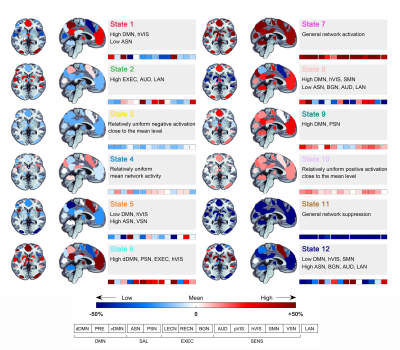 |
101 |
Reduced dynamism of brain activity underlying spatial deficits
in subjective cognitive decline
Qian Chen1,2 and
Bing Zhang1,2
1Department of Radiology, Drum Tower Hospital, Clinical College of Nanjing Medical University, Nanjing, China, 2Department of Radiology, The Affiliated Drum Tower Hospital of Nanjing University Medical School, Nanjing, China Keywords: Alzheimer's Disease, fMRI (resting state), subjective cognitive decline The alterations of brain dynamics and the associations with spatial navigation in individuals with subjective cognitive decline (SCD) remain unknown. In this study, 12 states with distinct brain activity were identified in a cohort of 80 SCD and 77 normal control (NC) participants using the hidden Markov model (HMM). The SCD group showed an inability to dynamically upregulate and downregulate the state with general network activation. Significant correlations between brain dynamics and spatial navigation were observed. The combined features of spatial navigation and brain dynamics showed an area under the curve of 0.854 in distinguishing between SCD and NC. |
|
2805.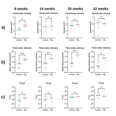 |
102 |
Non-invasive MRI of Blood-Cerebrospinal-Fluid-Barrier Function:
A Novel Functional Biomarker of Alzheimer's Disease Pathology
Charith Perera1,
Renata Cruz2,
Noam Shemesh2,
David L. Thomas3,4,5,
Jack Wells1,
and Andrada Ianus2
1UCL Centre for Advanced Biomedical Imaging, Division of Medicine, University College London, London, United Kingdom, 2Champalimaud Foundation, Lisbon, Portugal, 3Neuroradiological Academic Unit, Department of Brain Repair and Rehabilitation,, UCL Queen Square Institute of Neurology, London, United Kingdom, 4Dementia Research Centre, UCL Queen Square Institute of Neurology, London, United Kingdom, 5Wellcome Centre for Human Neuroimaging, UCL Queen Square Institute of Neurology, London, United Kingdom Keywords: Alzheimer's Disease, Arterial spin labelling Alzheimer’s disease (AD) is characterised by many pathophysiological changes, such as the accumulation of amyloid-β. The clearing of detrimental agents, including amyloid-β proteins, from brain tissue is linked to the function of choroid plexus (CP) or blood-cerebrospinal fluid barrier (BCSFB).This study investigates for the first time BCSFB function using non-invasive ASL-based methods in a mouse model of AD. Significantly higher values of total BCSFB-mediated water delivery in AD mice relative to controls were observed as early as 8 weeks of age, and a possible (though currently non-significant) correlation with behavioural tests was identified. |
|
2806.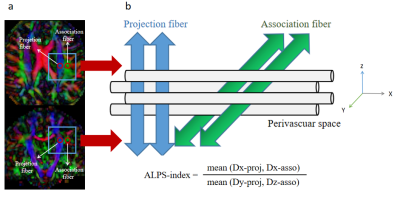 |
103 |
Glymphatic Dysfunction Mediates the Influence of White Matter
Hyperintensities on Episodic Memory in Cerebral Small Vessel
Disease
Zhihong Ke1,
Yuting Mo1,
and Yun Xu1
1Department of Neurology, Nanjing Drum Tower Hospital Clinical College of Nanjing Medical University, Nanjing 210008, China, nanjing, China Keywords: Dementia, Dementia In this study, we used the diffusion tensor image analysis along the perivascular space (ALPS)-index to evaluate glymphatic function and highlighted the reliability of using the ALPS-index in the early recognition of cognitive impairment (CI) in cerebral small vessel disease (CSVD) patients. We reported that 1) ALPS-index was a sensitive indicator to distinguish mild CI (MCI); 2) ALPS-index was an independent influencing factor of episodic memory in CSVD patients with MCI; 3) ALPS-index mediated the relationship between white matter hyperintensities and episodic memory in CSVD patients with MCI. |
|
2807.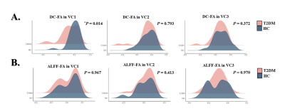 |
104 |
Functional-structural decoupling in visual network is associated
with cognitive decline in patients with type 2 diabetes
mellitus.
Minhua Ni1,
Zeyang Li1,
Ying Yu1,
Sining Li1,
Linfeng Yan1,
and Guangbin Cui1
1Tangdu Hospital, Xi`an, China Keywords: Dementia, fMRI (resting state) In this study, functional and structural changes in visual network (VN) of type 2 diabetes mellitus (T2DM) were investigated using multimodal magnetic resonance imaging. We explored degree centrality (DC), amplitude of low frequency fluctuation (ALFF), fractional anisotropy (FA), DC-FA and ALFF-FA in VN. Compared with healthy controls, deteriorated DC, ALFF and DC-FA coefficients in VN were observed in T2DM. These indicators showed positive correlations with cognitive function in T2DM, especially memory and executive function. Functional-structural decoupling may be a potential image biomarker of cognitive function change in T2DM. |
|
2808.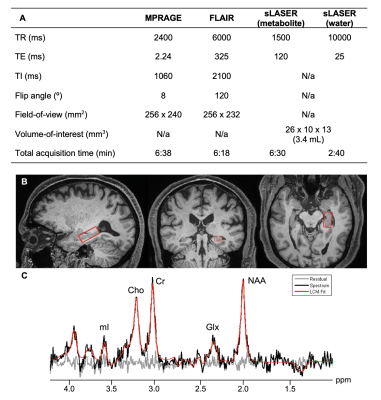 |
105 |
Hippocampal sLASER 1H-MRS in cognitively unimpaired elderly:
Associations with APOE4, CSF p-tau181 and MR morphometry
Anna Marie Chen1,
Martin Gajdošík1,2,
Rosemary Peralta1,
Helena Zheng1,
Mia Gajdošík1,
Mickael Tordjman1,3,
Julia Zabludovsky1,
Dishari Azad4,
Ajax George1,
Henry Rusinek1,4,
Yuxin Zhang5,
LianLian Chen5,
Guillaume Madelin1,
Christoph Juchem2,6,
Ricardo Osorio4,
and Ivan Kirov1,7,8
1Center for Biomedical Imaging, Department of Radiology, New York University Grossman School of Medicine, New York, NY, United States, 2Department of Biomedical Engineering, Columbia University, New York, NY, United States, 3Department of Radiology, Cochin Hospital, Paris, France, 4Department of Psychiatry, New York University Grossman School of Medicine, New York, NY, United States, 5Department of Biostatistics, New York University School of Global Public Health, New York, NY, United States, 6Department of Radiology, Columbia University, New York, NY, United States, 7Department of Neurology, New York University Grossman School of Medicine, New York, NY, United States, 8Center for Advanced Imaging Innovation and Research, Department of Radiology, New York University Grossman School of Medicine, New York, NY, United States Keywords: Alzheimer's Disease, Gray Matter, Hippocampus The hippocampus, a vulnerable structure in Alzheimer’s disease progression, is one of the most challenging brain structures for 1H-MRS applications. To examine whether hippocampal metabolic dysfunction in cognitively normal elderly may contribute to disease pathology, we used a validated long-TE sLASER sequence to minimize macromolecular signal contamination and chemical shift displacement errors. We tested whether, after adjusting for age, metabolites were associated with APOE4 genotype, a risk factor for amyloid accumulation; and CSF p-tau181 and left hippocampal volume, indicators of tau burden and neurodegeneration, respectively. The main result was a statistically significant direct correlation between Glx and CSF p-tau181. |
|
2809.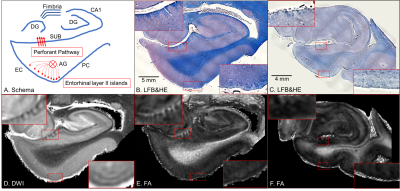 |
106 |
Microstructural neurodegeneration of the entorhinal-hippocampus
pathway in preclinical Alzheimer’s disease: ex vivo 11.7T MRI
and histology
Yuto Uchida1,
Kengo Onda1,
Jill Chotiyanonta1,
Zhipeng Hou1,
Juan Troncoso2,
Susumu Mori1,
and Kenichi Oishi1
1Department of Radiology, Johns Hopkins University School of Medicine, Baltimore, MD, United States, 2Department of Pathology, Johns Hopkins University School of Medicine, Baltimore, MD, United States Keywords: Alzheimer's Disease, Microstructure Conventional neuroimaging biomarkers for the neurodegeneration of Alzheimer’s disease (AD) are not sensitive enough to detect neurodegenerative alterations in preclinical AD individuals. We aimed to examine microstructural neurodegeneration of the entorhinal-hippocampus pathway during the pathological process of AD. Our ex vivo comparative study of 11.7T MRI and histology successfully visualized microstructural neurodegeneration of the entorhinal-hippocampus pathway in preclinical AD brain tissues. In a future study, we aim to translate the findings from ex vivo 11.7T MRI and histology into in vivo MRI clinical research for preclinical AD individuals. |
|
2810.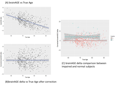 |
107 |
Brain Age Prediction using optimized MRI Brain Features: A novel
strategy to understand Aging and Aging Associated Pathologies
Arkaprava Majumdar1,
Neha Yadav1,
and Vivek Tiwari1
1Department of Biological Sciences, Indian Institute of Science Education and Research Berhampur, Berhampur, India Keywords: Dementia, Modelling, Brain Age A certain subset of the aging population maintains the cognitive abilities with minimal structural changes with aging while another subset of people transforms to mild cognitive impairment and dementia with manifold structural variations. Since a subset maintains the cognitive abilities, brain structural volume with aging while another subset undergoes cognitive-impairment and drastic brain structure alterations, we believe that the brain age pattern is distinct from chronological age. Here, we have used a set of MRI determined brain volumetry to determine the brain age as a measure of normal and pathological aging. |
|
2811.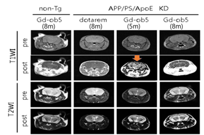 |
108 |
Validation Study of MRI Contrast Agent of Gd-DOTA Conjugated to
DNA Aptamer for Detecting Oligomeric Amyloid-beta
Geon-Ho Jahng1,
Sang-Tae Kim2,
Hyug-Gi Kim3,
Yu Mi Kim2,
and Jee-Hyun Cho4
1Radiology, Kyung Hee University Hospital at Gangdong, Seoul, Korea, Republic of, 2Neuroscience of Lab, Seoul National University College of Medicine, Seongnam city, Korea, Republic of, 3Radiology, Kyung Hee University Hospital, Seoul, Korea, Republic of, 4Korea Basic Science Institute, Cheongju-si, Korea, Republic of Keywords: Alzheimer's Disease, Molecular Imaging The oligomeric amyloid-b (oAβ) is a reliable feature for an early diagnosis of Alzheimer's disease (AD). Therefore, the objective of this study was to demonstrate imaging of oAβ deposits using our developed DNA aptamer called ob5 conjugated with gadolinium (Gd)- DOTA as a contrast agent for early diagnosis of AD using MRI. An oAβ-specific aptamer was developed by amide bond formation and conjugated to Gd-DOTA MRI contrast agent and/or cyanine5 (cy5). We verified the performance of our new contrast agent with AD model mice using In vivo and ex vivo fluorescent imaging and animal MRI experiments. |
|
2812.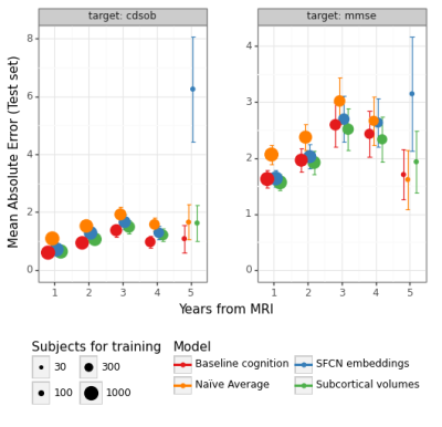 |
109 |
On the prediction of cognitive impairment trajectories from
anatomical MRI in ADNI: preliminary results
Bruno Hebling Vieira1,2,3 and
Nicolas Langer1,2,3
1Methods of Plasticity Research, Department of Psychology, University of Zurich, Zürich, Switzerland, 2Neuroscience Center Zurich (ZNZ), UZH & ETHZ, Zürich, Switzerland, 3University Research Priority Program (URPP) Dynamic of Healthy Aging, University of Zurich, Zürich, Switzerland Keywords: Alzheimer's Disease, Neurodegeneration We predict yearly differences in MMSE and CDR-SOB using data from the ADNI cohort ranging from five years before to five years after the imaging session. We show that models that use embeddings from a deep-learning model trained to predict brain-age or models that use bilateral hippocampi, amygdalae, and accumbens perform approximately the same as models that use baseline cognitive scores as inputs. This has potential ramifications for both clinical machine learning applications and the neurobiology of cognitive decline. In future work, we will finetune the deep-learning model and substantially increase the sample size. |
|
2813.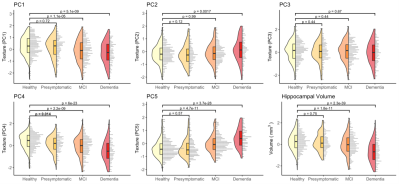 |
110 |
Longitudinal changes in hippocampal texture from healthy aging
to Alzheimer’s disease
Alfie Wearn1,
Lars Lau Raket2,3,
and Nathan Spreng1,4,5,6
1Montreal Neurological Institute, McGill University, Montreal, QC, Canada, 2Clinical Memory Research Unit, Lund University, Lund, Sweden, 3Novo Nordisk A/S, Søborg, Denmark, 4McConnell Brain Imaging Centre, McGill University, Montreal, QC, Canada, 5Departments of Psychology and Psychiatry, McGill University, Montreal, QC, Canada, 6Douglas Mental Health University Institute, McGill University, Montreal, QC, Canada Keywords: Alzheimer's Disease, Preclinical Microstructural brain changes caused by early Alzheimer’s disease neuropathology may cause subtle changes in MR signal that are quantifiable using texture analysis, a branch of radiomics. We used cross-sectional and longitudinal analysis techniques in the ADNI dataset to examine changes in texture across the disease continuum. We found that biomarker positive but cognitively healthy older adults had measurably different hippocampal texture than those without biomarker risk. Longitudinal modelling revealed progressive textural change with disease severity, but with high inter-subject variability. Nonetheless, hippocampal texture provided additional information to volume in predicting cognitive decline in older adults without a diagnosis of dementia. |
|
2814.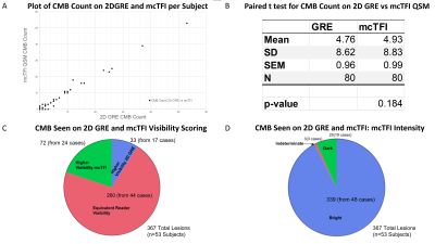 |
111 |
Comparability of mcTFI QSM and 2D GRE CMB Detection:
Implications for for Alzheimer’s Anti-Amyloid Therapy
Nikita Seth1,
Geunwon Kim2,
Magdy Selim3,
Ajith J Thomas4,
Aristotelis Filippidis5,
Yan Wen6,
Pascal Spincemaille7,
Yi Wang7,
and Salil Soman1
1Radiology, Beth Israel Deaconess Medical Center, Harvard Medical School, Boston, MA, United States, 2Atrius Health, Boston, MA, United States, 3Neurology, Beth Israel Deaconess Medical Center, Harvard Medical School, Boston, MA, United States, 4Department of Neurological Surgery, Cooper University Health Care, Cooper Medical School of Rowan University, Camden, NJ, United States, 5Neurosurgery, Beth Israel Deaconess Medical Center, Harvard Medical School, Boston, MA, United States, 6GE Healthcare, New York, NY, United States, 7Department of Radiology, Weill Cornell Medicine, New York, NY, United States Keywords: Alzheimer's Disease, Quantitative Susceptibility mapping, CMB, 2D GRE, mcTFI, Anti-amyloid therapy Cerebral microbleeds/microhemorrhages (CMB) are used for risk stratification, including for the hemorrhagic complication ARIA-H of Alzheimer’s anti-amyloid therapy. For AD, risk information is based on many trials using 2DGRE technique, which is MRI field and parameter dependent. The use of techniques that better distinguish CMB mimics, like calcifications, is limited by an unclear relationship to 2D GRE CMB depiction. In this study we found the number of CMB candidate lesions between 2D GRE and mcTFI QSM obtained during the same scan session highly correlated, suggesting mcTFI could be used in the management of pathologies evaluating presence and number of CMBs. |
|
2815.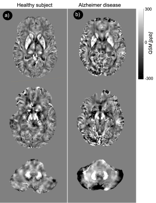 |
112 |
Ultra-High Resolution QSM of the Brain Iron Metabolism in the
Cognitively Declined Adults at 7T MRI
Akbar Alipour1,
Pinar S Ozbay2,
Ameen Alqadi1,
Mackenzie Langan1,
Mehmet Kurt3,
Trey Hedden 4,
Bradley N Delman1,
and Priti Balchandani1
1Radiology, Icahn School of Medicine at Mount Sinai, New York, NY, United States, 2Biomedical Engineering, Boğaziçi University, Istanbul, Turkey, 3Department of Mechanical Engineering, University of Washington, Seattle, WA, United States, 4Neurology, Icahn School of Medicine at Mount Sinai, New York, NY, United States Keywords: Alzheimer's Disease, Quantitative Susceptibility mapping Detection of iron aggregations associated with beta-amyloid in Alzheimer’s disease would help to understand the related pathophysiology in these cohorts. Aggregations of iron associated beta-amyloid should increase electron density and induce notable changes in local susceptibility value. With higher susceptibility at ultra-high field strengths, induced iron susceptibility is large enough to generate contrast relative to surrounding normal tissues that can be visualized by quantitative susceptibility techniques at 7 Tesla MRI. In this study, we used 7T MRI to analyze the iron-related pathologic markers in Alzheimer patients using the quantitative susceptibility mapping technique. |
|
2816.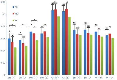 |
113 |
Investigation of iron content changes in Alzheimer's disease and
mild cognitive impairment using quantitative susceptibility
mapping
Chuan-Bin Huang1,
Yong Zhang2,
Ke-Xue Deng1,
and Chang Liu1
1Radiology, The First Affiliated Hospital of University of Science and Technology of China, Hefei, China, 2GE Healthcare, Shanghai, China Keywords: Alzheimer's Disease, Quantitative Susceptibility mapping This study investigated iron content changes using Quantitative Susceptibility Mapping (QSM) technique in AD and MCI patients, as compared with normal controls, and correlated iron deposit levels with cognitive scores to assess the clinical values of QSM in the diagnosis of AD and MCI. Bilateral caudate nucleus and right putamen showed significantly increased QSM values in AD and MCI patients as compared to healthy controls. The QSM values of right caudate nucleus correlated with the MMSE scores of AD patients. These results might indicate QSM as the potential biomarker for clinical diagnosis of AD and MCI. |
|
2817.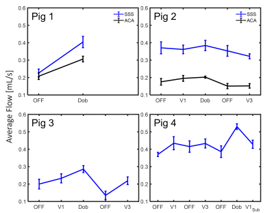 |
114 |
Evaluation of relationship between trigeminal nerve stimulation
and hemodynamics using 4D-Flow MRI: A pilot study in swine model
Mu-Lan Jen1,
Nishant Verma2,3,
Kevin P Cheng2,3,
Robert Moskwa1,
Grant S Roberts1,
Maria Laluzerne2,3,
Jennifer Frank2,3,
Justin Williams2,3,
Wally Block1,
Kip Ludwig2,3,4,
and Kevin M Johnson1,5
1Medical Physics, University of Wisconsin-Madison, Madison, WI, United States, 2Biomedical Engineering, University of Wisconsin-Madison, Madison, WI, United States, 3Wisconsin Institute for Translational Neuroengineering (WITNe) – Madison, Madison, WI, United States, 4Neurosurgery, University of Wisconsin-Madison, Madison, WI, United States, 5Radiology, University of Wisconsin-Madison, Madison, WI, United States Keywords: Alzheimer's Disease, Velocity & Flow This study utilizes 4D-Flow imaging to evaluate hemodynamic alterations during the period of nerve stimulation and investigates cerebral blood flow changes modulated by using 4D-Flow MRI in large animal models. Preliminary results showed cerebral blood flow increases while flow pulsatility decreases with the electrical stimulation of trigeminal nerves. This effect from neuromodulation is on the contrary to the hemodynamic alterations with aging and AD progression, which implies the potential of neuromodulation as an alternative treatment approach in repairing glymphatic system function. |
|
2818.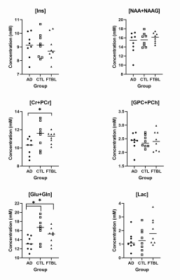 |
115 |
Comparison of MR Spectroscopy of Probable Alzheimer’s Disease
and Symptoms of Chronic Traumatic Encephalopathy
Jessica J. Chen1,
Michael L. Alosco2,
Huijun Liao1,
Inga K. Koerte3,4,
Martha E. Shenton3,
Robert A. Stern5,
and Alexander P. Lin1
1Center for Clinical Spectroscopy,Department of Radiology, Brigham and Women's Hospital, Harvard Medical School, Boston, MA, United States, 2Boston University School of Medicine, Department of Neurology, Boston University Alzheimer's Disease and CTE Centers, Boston, MA, United States, 3Psychiatry Neuroimaging Laboratory, Department of Psychiatry, Brigham and Women's Hospital, Harvard Medical School, Boston, MA, United States, 4Department of Child and Adolescent Psychiatry, Psychosomatic, and Psychotherapy, Ludwig-Maximilian-University, Munich, Germany, 5Neurosurgery, and Anatomy & Neurobiology, Departments of Neurology, Boston University Alzheimer’s Disease and CTE Center, Boston, MA, United States Keywords: Alzheimer's Disease, Spectroscopy, traumatic brain injury, chronic traumatic encephalopathy Chronic traumatic encephalopathy (CTE) is widely found in individuals exposed to repetitive head impacts (RHI) in organized contact sports, such as football. Previous findings have determined that there is a distinct deposition of phosphorylated tau (p-tau) in its neuropathology compared to other tauopathies, including Alzheimer’s disease. However, there is a lack of diagnostic criteria that examine later-life neurochemical changes due to long-term neurologic consequences to RHI. This study uses magnetic resonance spectroscopy (MRS) to compare neurochemical markers between participants with probable Alzheimer’s disease and symptomatic chronic traumatic encephalopathy in relation to healthy controls. |
|
2819.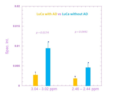 |
116 |
Discovering the Metabolomic Relationships between Lung Cancer
and Alzheimer’s Disease using Nuclear Magnetic Resonance
Spectroscopy
Leo Ling Cheng1,2,
Zuzanna Kobus1,2,3,
Marta Kobus1,2,4,
Li Su5,
and David C. Christiani5
1Department of Radiology, Massachusetts General Hospital, Boston, MA, United States, 2Harvard Medical School, Boston, MA, United States, 3Charité - Universitätsmedizin Berlin, Berlin, Germany, 4Department of Radiation Oncology, Charité - Universitätsmedizin Berlin, Berlin, Germany, 5Department of Environmental Health, Harvard T.H. Chan School of Public Health, Boston, MA, United States Keywords: Alzheimer's Disease, Spectroscopy, Metabolomics Herein, we report our preliminary data on metabolomic associations between human lung cancer (LuCa) and Alzheimer’s disease (AD) measured from serum samples using high resolution magic angle spinning (HRMAS) MRS. The current project’s objective is to establish MRS-based tissue pathology-guided serum metabolomic profiles for matched LuCa patients, with and without AD, by comparing serum profiles measured from matched healthy controls. Initial results demonstrate the feasibility of HRMAS MRS for LuCa-AD metabolomic mechanisms investigation. Our findings serve as a foundation for innovative future diagnostic and treatment studies. |
|
2820.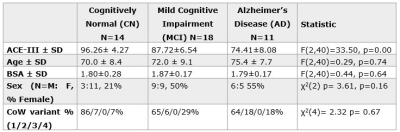 |
117 |
4D flow MRI reveals cerebrovascular changes in early Alzheimer’s
disease
Ayah Elsayed1,
Tracy Melzer2,3,4,
Lynette Tippett5,
and Catherine Morgan5,6
1Faculty of Health & Environmental Studies, Auckland University of Technology, Auckland, New Zealand, 2Department of Medicine, University of Otago, Christchurch, New Zealand, 3NZ Brain Research Institute, Christchurch, New Zealand, 4School of Psychology, Speech and Hearing, University of Canterbury, Christchurch, New Zealand, 5Department of Psychology, University of Auckland, Auckland, New Zealand, 6Centre for Advanced MRI, University of Auckland, Auckland, New Zealand Keywords: Alzheimer's Disease, Velocity & Flow Currently 50 million people suffer from Alzheimer’s disease (AD), the most common form of dementia, yet treatment options are limited. A growing focus on the contribution of cerebrovascular (CV) health presents new possible targets for treatment. We investigated novel CV imaging markers using 4D flow MRI in a total of 43 participants with mild cognitive impairment, early AD and controls. Both the MCI and AD group had significantly reduced mean blood flow in the larger vessels of the Circle of Willis. A significantly increased pulsatility index (indicative of poorer vessel compliance) was found in the AD group compared to controls. |
|
2821. |
118 |
Identifying fiber tracts strategic for cognitive impairments in
cerebral small vessel disease with harmonized diffusion MRI
Peiyu Huang1,
Carolina Canelas Valentim2,
Geert-Jan Biessels2,
Christopher Chen3,
Anna Dewenter4,
Marco Duering4,5,
Saima Hilal3,
Huiberdina L Koek6,
Anna Kopczak4,
Bonnie Yin Ka Lam7,
Alexander Leemans8,
Vincent Mok7,
Laurien Onkenhout2,
Hilde van den Brink2,
and Alberto De Luca9
1Department of Radiology, Zheijang University School of Medicine,, Hangzhou, China, 2UMC Utrecht Brain Center, University Medical Center Utrecht, Utrecht, Netherlands, 3Aging and Cognition Center, Yong Loo Lin School of Medicine, National University of Singapore, Singapore, Singapore, 4Institute for Stroke and Dementia Research, LMU Munich, Munich, Germany, 5Medical Image Analysis Center, University of Basel, Basel, Switzerland, 6Department of Geriatric Medicine, University Medical Center Utrecht, Utrecht, Netherlands, 7Division Neurology, The Chinese University of Hong Kong, Hong Kong, China, 8Image Sciences Institute, University Medical Center Utrecht, Utrecht, Netherlands, 9Division Imaging and Oncology, University Medical Center Utrecht, Utrecht, Netherlands Keywords: Dementia, White Matter Small vessel disease (SVD) is a worldwide leading cause of dementia. Damage to white matter tracts is a key mechanism through which SVD impacts cognition. In this work, we leverage a large multicenter dataset of patients with SVD to investigate which white matter tracts are strategic in SVD, that is, robustly associated with cognitive decline. Our early results show that the corpus callosum, superior longitudinal fasciculus and thalamo parietal tracts were the most associated to processing speed, verbal memory and executive function, respectively. Tract-based measures explained additional variance (2-5%) as compared to whole brain measures. |
|
2822. |
119 |
Relationship between white matter hyperintensity load and
cognitive decline in patients with carotid artery stenosis
Wen Zhang1,
Xiance Zhao2,
and Bing Zhang1
1Radiology, The Affiliated Drum Tower Hospital of Nanjing University Medical School, Nanjing, China, 2Philips Healthcare, Shanghai, China Keywords: Dementia, White Matter, carotid artery stenosis Patients with carotid artery stenosis (CAS) have a high prevalence of cognitive impairment. White matter hyperintensity (WMH) is one of the most common imaging signs of chronic brain injury in the elderly. We investigated the relationship between WMH load and cognitive decline in CAS patients. Our results suggested that Fazekas score and WMH volume quantification can be a potential simple biomarker for predicting cognitive function in CAS patients. |
|
| 2823. | 120 |
Functional connectivity changes of olfactory neural circuits in
Subjective Cognitive Decline under task fMRI
Yajing Zhu1,
Xin Zhang1,
and Bing Zhang1
1Department of Radiology, Nanjing Drum Tower Hospital Clinical College of Nanjing Medical University, Nanjing, China Keywords: Alzheimer's Disease, Alzheimer's Disease, fMRI Analysis of functional connectivity changes of olfactory neural circuits in subjective cognitive decline(SCD)under specific odor stimuli using generalized physiological and psychological interactions. |
|
 Back to Meeting Home
 Back to the Program-at-a-Glance
The International Society for Magnetic Resonance in Medicine is accredited by the Accreditation Council for Continuing Medical Education to provide continuing medical education for physicians.
 Back to Meeting Home
 Back to the Program-at-a-Glance
 View Presentation Video