Digital Poster
Alzheimer's & Dementias III
ISMRM & ISMRT Annual Meeting & Exhibition • 03-08 June 2023 • Toronto, ON, Canada

| Computer # | |||
|---|---|---|---|
3038. |
161 |
Comparison of High-Frequency Conductivity in the Brains of
Alzheimer’s Disease Patients and Cognitively Normal Elderly
Controls
Geon-Ho Jahng1,
Soonchan Park1,
Sue Min Jung2,
Mun Bae Lee3,
Hak Young Rhee4,
Chang-Woo Ryu1,
A- Rang Cho5,
and Oh In Kwon3
1Radiology, Kyung Hee University Hospital at Gangdong, Seoul, Korea, Republic of, 2Biomedical Engineering, Kyung Hee University, Yongin-si, Korea, Republic of, 3Mathematics, Konkuk University, Seoul, Korea, Republic of, 4Neurology, Kyung Hee University Hospital at Gangdong, Seoul, Korea, Republic of, 5Psychiatry, Kyung Hee University Hospital at Gangdong, Seoul, Korea, Republic of Keywords: Alzheimer's Disease, Electromagnetic Tissue Properties The objective of this study was to investigate high-frequency conductivity (HFC) obtained using magnetic resonance electrical property tomography (MREPT) in participants with Alzheimer’s disease (AD), amnestic mild cognitive impairment (MCI), and cognitively normal (CN) elderly controls. High-frequency conductivity (HFC) values in the brain are significantly increased in Alzheimer’s disease (AD) patients compared to cognitively normal (CN) elderly people, are negatively associated with Mini-Mental State Examination (MMSE) scores, and therefore can be used as an imaging biomarker to improve the differentiation of AD from CN. |
|
3039.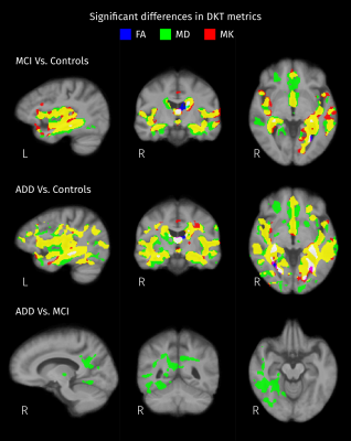 |
162 |
Comparison of diffusion kurtosis tensor imaging and multi-tissue
CSD for the investigation of group differences in Alzheimer’s
disease
Diana L. Giraldo1,2,3,
Robert E. Smith4,
Hanne Struyfs5,
Ellis Niemantsverdriet5,
Ellen De Roeck5,
Sebastiaan Engelborghs5,
Eduardo Romero2,
Jan Sijbers1,3,
and Ben Jeurissen1,3,6
1imec-Vision Lab, University of Antwerp, Antwerp, Belgium, 2Computer Imaging and Medical Applications Laboratory - Cim@Lab, Universidad Nacional de Colombia, Bogota, Colombia, 3µNEURO Research Center of Excellence, University of Antwerp, Antwerp, Belgium, 4The Florey Institute of Neuroscience and Mental Health, Melbourne, Australia, 5Reference Center for Biological Markers of Dementia (BIODEM), University of Antwerp, Antwerp, Belgium, 6Lab for Equilibrium Investigations and Aerospace, University of Antwerp, Antwerp, Belgium Keywords: Alzheimer's Disease, Data Analysis In this work, we compare the results of analyzing group differences in Alzheimer’s Disease (AD) with two different models for multi-shell diffusion MRI: the Diffusion Kurtosis Tensor (DKT) and Multi-Tissue Constrained Spherical Deconvolution (MT-CSD). Separate analysis for DKT metrics and measures derived from MT-CSD were performed to investigate differences between control subjects and patients with mild cognitive impairment and dementia due to AD. Statistical analyses included strong family-wise error correction for multiple comparisons. Results indicate that analyses with MT-CSD capture the differences that are detectable with DKT while offering more specific details that facilitate the interpretation of those differences. |
|
3040.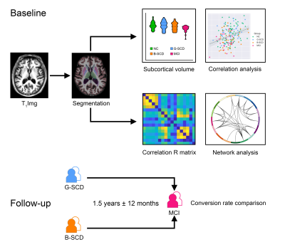 |
163 |
Spatial navigation reveals subcortical structural covariance and
progression risk in subjective cognitive decline
Qian Chen1,2 and
Bing Zhang1,2
1Department of Radiology, Drum Tower Hospital, Clinical College of Nanjing Medical University, Nanjing, China, 2Department of Radiology, The Affiliated Drum Tower Hospital of Nanjing University Medical School, Nanjing, China Keywords: Alzheimer's Disease, Gray Matter, structural covariance network We aimed to investigate whether spatial navigation could reveal subcortical structural alterations and the risk of clinical progression in subjective cognitive decline (SCD). SCD subjects were divided into SCD-good (G-SCD) and SCD-bad (B-SCD) groups according to their navigation performance. The B-SCD group showed decreased volumes in the basal forebrain and had a larger shortest path length in the subcortical structural covariance network than the G-SCD group. Follow-up data suggested that the B-SCD group had a higher conversion rate to MCI than the G-SCD group. This study may provide new insights for the risk assessment and early intervention for SCD subjects. |
|
3041.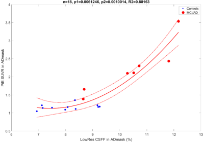 |
164 |
MR-based Parenchymal CSF water fraction (CSFF) is a marker of
glymphatic clearance function and relates to beta-amyloid
deposition on 11C-PiB PET
Liangdong Zhou1,
Thanh Nguyen1,
Xiuyuan H Wang1,
Gloria C Chiang1,
Mony de Leon1,
and Yi Li1
1Weill Cornell Medicine, New York, NY, United States Keywords: Alzheimer's Disease, Neurofluids Glymphatic clearance has recently gained increasing attention as a major driver of neurodegenerative diseases like Alzheimer’s disease. Perivascular spaces is believed to reflect glymphatic clearance such that enlargement of the PVS is believed to reflect blocked glymphatic clearance. We present preliminary work on a MR-based FAST-T2 CSF water fraction map to study glymphatic clearance. Our results show a quadratic relationship between CSFF and PiB PET SUVR in AD-specific regions. This indicates that CSFF could be a biomarker of dysfunctional glymphatic clearance, leading to early beta amyloid accumulation, since early on, CSFF increases without a significant change in PiB PET SUVR. |
|
3042.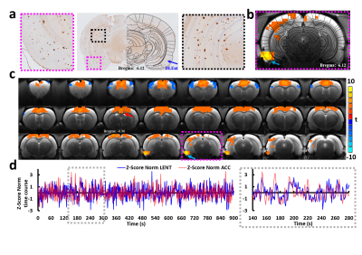 |
165 |
Functional connectivity to entorhinal cortex in hypertensive
Alzheimer’s disease rat models
Yi Chen1,2,
Zachary Fernandez3,4,
Norman Scheel3,
Mahsa Gifani5,
Chunqi Qian3,
Anne M. Dorrance6,
Scott E. Counts4,5,7,8,
and David C. Zhu3
1Max Planck Institute for Biological Cybernetics, Tuebingen, Germany, 2Department of Radiology, Michigan State University, East Lansing, MI, United States, 3Department of Radiology and Cognitive Imaging Research Center, Michigan State University, East Lansing, MI, United States, 4Neuroscience Program, Michigan State University, East Lansing, MI, United States, 5Department of Translational Neuroscience, Michigan State University, Grand Rapids, MI, United States, 6Department of Pharmacology and Toxicology, Michigan State University, East Lansing, MI, United States, 7Department of Family Medicine, Michigan State University, Grand Rapids, MI, United States, 8Hauenstein Neurosciences Center, Mercy Health Saint Mary’s Hospital, Grand Rapids, MI, United States Keywords: Alzheimer's Disease, Animals Extensive studies have revealed that the entorhinal cortex (EC) plays a critical role in Alzheimer’s disease (AD) development. However, EC functional connectivity and its associated network abnormalities are understudied, especially in rodent models. To the best of our knowledge, this is the first study to report disrupted EC functional connectivity in hypertensive AD rodents using resting-state fMRI. Our results may provide new insights into the impaired EC connectivity patterns and enable the search for novel preclinical EC-based fMRI biomarkers for AD studies. In addition, our novel animal model provides new information to understand the link between hypertension and AD. |
|
3043.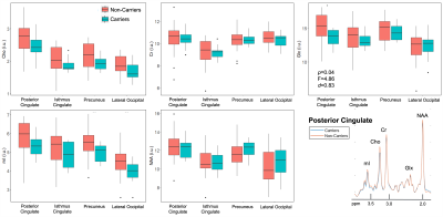 |
166 |
1H-MRSI of cortical and subcortical gray matter in cognitively
unimpaired elderly: Associations with APOE4, CSF p-tau181 and MR
morphometry
Anna Marie Chen1,
Martin Gajdošík1,
Rosemary Peralta1,
Dishari Azad2,
Helena Zheng1,
Mia Gajdošík1,
Ajax George1,
Sinyeob Ahn3,
Mickael Tordjman1,4,
Julia Zabludovsky1,
Yuxin Zhang5,
LianLian Chen5,
Henry Rusinek1,2,
Guillaume Madelin1,
Ricardo Osorio2,
and Ivan Kirov1,6,7
1Center for Biomedical Imaging, Department of Radiology, New York University Grossman School of Medicine, New York, NY, United States, 2Department of Psychiatry, New York University Grossman School of Medicine, New York, NY, United States, 3Siemens Medical Solutions USA Inc., Malvern, PA, United States, 4Department of Radiology, Cochin Hospital, Paris, France, 5Department of Biostatistics, New York University School of Global Public Health, New York, NY, United States, 6Department of Neurology, New York University Grossman School of Medicine, New York, NY, United States, 7Center for Advanced Imaging Innovation and Research, Department of Radiology, New York University Grossman School of Medicine, New York, NY, United States Keywords: Alzheimer's Disease, Gray Matter 1H-MRSI can examine spatiotemporal characteristics of metabolic dysfunction in multiple brain regions in early Alzheimer’s disease (AD) pathology. Since both cortical and subcortical gray matter structures have shown vulnerability to AD neurodegeneration, we tested whether regional gray matter metabolic abnormalities were associated with (i) APOE4 genotype, a risk factor for amyloid burden, (ii) CSF p-tau181, an indicator of tau burden, and (iii) morphometry metrics (volume, cortical thickness) indicative of neurodegeneration, in cognitively unimpaired elderly. We found lower caudate Cho and lower posterior cingulate Glx in APOE4 carriers compared to non-carriers. There were no metabolite relationships with CSF p-tau181 or morphometry. |
|
3044.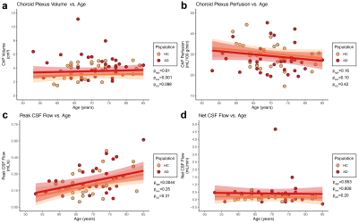 |
167 |
Quantitative choroid plexus and bulk cerebrospinal fluid metrics
in older adults with and without Alzheimer’s disease
Jarrod J. Eisma1,
Colin D. McKnight2,
Ciaran M. Considine1,
Kilian Hett1,
Alexander K. Song1,
Caleb J. Han1,
Jason Elenberger1,
Daniel O. Claassen1,
and Manus J. Donahue1,3
1Department of Neurology, Vanderbilt University Medical Center, Nashville, TN, United States, 2Department of Radiology and Radiological Sciences, Vanderbilt University Medical Center, Nashville, TN, United States, 3Department of Psychiatry and Behavioral Sciences, Vanderbilt University Medical Center, Nashville, TN, United States Keywords: Alzheimer's Disease, Neurofluids, Aging, Perfusion, Arterial spin labelling The goal of this work is to characterize changes in the choroid plexus (ChP) and aqueductal cerebrospinal fluid dynamics in patients with Alzheimer’s disease utilizing arterial spin labeling (ASL) and phase contrast (PC), respectively. We observed that across all participants, ChP hypertrophy was associated with reduced ChP perfusion. However, we did not observe differences in ChP perfusion or net CSF flow between AD and healthy cohorts. The observed increase in peak CSF flow with age, despite the general decrease in ChP perfusion with age, suggests that the ChP performs functions beyond simply increasing or decreasing CSF production. |
|
3045.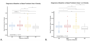 |
168 |
Decreased cholinergic nucleus 4 basal forebrain density is
associated with disease progression in Alzheimer’s Disease.
Coreylyn A. deBettencourt1,
Kirubel Kentiba1,
Alexander Atalay2,
Benjamin T. Newman3,
John D. Van Horn4,
and T. Jason Druzgal3
1University of Virginia School of Medicine, Charlottesville, VA, United States, 2University of Virginia, Charlottesville, VA, United States, 3Radiology and Medical Imaging, University of Virginia, Charlottesville, VA, United States, 4Department of Psychology, University of Virginia, Charlottesville, VA, United States Keywords: Alzheimer's Disease, Alzheimer's Disease Density of the cholinergic nuclei of the basal forebrain has been shown to be associated with disease and symptom progression in Parkinson’s Disease. This project was aimed at determining if there is a similar effect of these brain regions in predicting cognitive impairment and disease progression in Alzheimer’s Disease. By using a longitudinal multicenter dataset of Alzheimer's patients in the Alzheimer’s Disease Neuroimaging Initiative (ADNI), we analyzed the relationship between grey matter density of cholinergic basal forebrain and disease progression. We demonstrated that patients with increased disease severity had significantly lower density in the cholinergic nuclei of the basal forebrain. |
|
3046.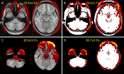 |
169 |
When texture does not matter: Misinterpretation of deep
learning-based Alzheimer's disease classification
Christian Tinauer1,
Stefan Ropele1,
and Christian Langkammer1
1Medical University of Graz, Graz, Austria Keywords: Alzheimer's Disease, Machine Learning/Artificial Intelligence Recent studies have shown Clever Hans effects in e.g. a widely used MRI tumor dataset. Clever Hans was a horse in the early 20th century that could supposedly do some arithmetic, although it only reacted to unintended cues of its owner. Inspired by these insights, we used T1w images from the ADNI database and removed the texture using threshold binarization. By using a standard deep neural network we separated images from Alzheimer's patients (n=404) from normal controls (n=905). This study revealed that volumetric measures are overwhelmingly relevant for the classification, while textures (T1w-contrast variations) are neglectable. |
|
3047.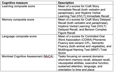 |
170 |
Predicting brain-behavior relationships using Voxel-based
Predictive Modeling in ASL data of older subjects
Scott Peltier1,
Michelle Karker2,
Luis Hernandez1,
Henry Paulson3,
Bruno Giordani4,
Benjamin M Hampstead4,
and Doug Noll2
1Functional MRI Laboratory, University of Michigan, Ann Arbor, MI, United States, 2Biomedical Engineering, University of Michigan, Ann Arbor, MI, United States, 3Neurology, University of Michigan, Ann Arbor, MI, United States, 4Psychiatry, University of Michigan, Ann Arbor, MI, United States Keywords: Alzheimer's Disease, Data Analysis A recent technique, Connectome-based Predictive Modeling (CPM), has shown promise in relating imaging-derived measures to clinical/behavioral observations. In this work, we adapt this method to relate volumetric CBF data to continuous composite measures of memory, learning, and language in subjects with MCI and DAT, along with healthy subjects. Models using the learning and memory composite scores had the largest effect sizes and statistical significance using permutation testing. Regions in the feature masks indicate involvement of brain regions that may be impacted in DAT. This demonstrates the utility of ASL-based perfusion measurement as a predictor of cognitive status in older subjects. |
|
3048.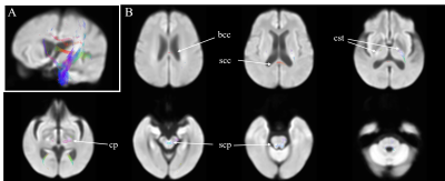 |
171 |
White matter damage in end-stage kidney disease patients with
cognitive impairment: a fixel-based analysis
Chih-Chien Tsai1,
YI-Chou Hou2,
Yao-Liang Chen3,
and Jiun-Jie Wang4
1Healthy Aging Research Center, Chang Gung University, Taoyuan, Taiwan, 2Department of Nephrology, Cardinal Tien Hospital at Xindian, New Taipei City, Taiwan, 3Department of Diagnostic Radiology, Chang Gung Memorial Hospital at Keelung, Keelung, Taiwan, 4Department of Medical Imaging and Radiological Sciences, Chang Gung University, Taoyuan, Taiwan Keywords: Dementia, Kidney Patients with end-stage kidney disease are vulnerable to cognitive impairment or dementia. Previous studies indicated that end-stage kidney disease subjects manifest prominent white matter hyperintensities. We use fixel-based analysis to examine the tract-specific differences in fixel-based metrics between end-stage kidney disease patients and normal participants. The results demonstrate the pattern of white matter degeneration in end-stage kidney disease patients. Our findings suggest that end-stage kidney disease patients with cognitive impairment exhibit white matter abnormalities. In clinical, the fixel-based analysis may serve as a potential biomarker for monitoring white matter change caused by disease. |
|
3049.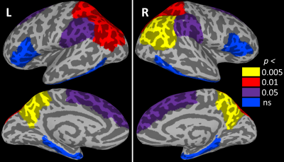 |
172 |
Cortical thickness related to future score on the Dementia
Questionnaire for People with Learning Disabilities in adults
with Down syndrome
Katherine Anne Koenig1,
Stephen Ruedrich2,
Se-Hong Oh1,
Mark J Lowe1,
and James B Leverenz1
1The Cleveland Clinic, Cleveland, OH, United States, 2University Hospitals, Cleveland, OH, United States Keywords: Alzheimer's Disease, Genetics, Down Syndrome Down syndrome (DS) is a genetic condition that is associated with early onset Alzheimer’s disease (AD). Individuals with DS show changes in neuroanatomy, including regional increases in cortical thickness. Here, we assess cortical thickness in ten non-demented adults with DS. We focus on regions that show cortical thinning in AD and relate cortical thickness to scores on a measure of dementia. We find that regional thickness measures are related to the 4.6 year change in a measure of dementia, suggesting that the magnitude of early DS-related neuroanatomical differences may relate to aspects of dementia onset. |
|
3050.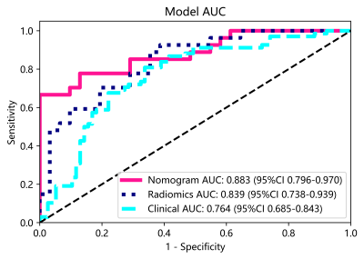 |
173 |
Prediction of amnestic mild cognitive impairment using
radiomics-clinical model based on 3D-T1WI imaging
Wei Zheng1,2,
Ronghua Mu1,
Xiaoyan Qin1,
Xin Li1,
Fuzhen Liu1,
Zeyu Zhuang1,2,
Peng Yang1,
Jian Lv1,
Xiqi Zhu1,
and Kan Deng3
1the Nanxishan Hospital, Guangxi Zhuang Autonomous Region, guilin, China, 2Graduate School of Guilin Medical University, guilin, China, 3Philips Healthcare, Guangzhou, China Keywords: Alzheimer's Disease, Radiomics Mild cognitive impairment (MCI) is a transitional state between normal aging and dementia disorders, especially Alzheimer's disease (AD). In this study, we aimed to develop a radiomics model based on 3D-T1W imaging to distinguish amnestic mild cognitive impairment (aMCI) patients from the normal elderly population by measuring changes in the frontal lobe. And the result shows that a quantitative nomogram based on clinical feature and radiomic features of MRI imaging can be used to distinguish aMCI and CN with excellent predictive ability, which can be served as a potential decision support tool to assist clinicians in Screening community aMCI population.
|
|
3051. |
174 |
Treatment response prediction of major depressive disorder using
brain magnetic resonance imaging
Fenghua Long1,
Qian Zhang1,
Yaxuan Wang1,
Qian Li1,
Youjin Zhao1,
and Fei Li1
1Huaxi MR Research Center (HMRRC), Department of Radiology, West China Hospital of Sichuan University, Chengdu 610041, Sichuan, P.R. China, Chengdu, China Keywords: Dementia, MR Value We performed a meta-analysis by including published studies using machine learning for unrestricted modalities and interventions on magnetic resonance imaging (MRI) data to predict the effectiveness of treatment for patients with major depressive disorder (MDD). The results showed that the resting-state functional MRI (rs-fMRI) had higher predictive performance in the modality subgroups, suggesting brain rs-fMRI may have an advantage in prediction performance. Using machine-learning analysis to predict treatment effectiveness is promising, but should not yet be implemented into clinical practice. |
|
3052. |
175 |
Studying the Impact of Diabetes on the Pathogenesis of
Alzheimer’s Disease
Thomas Jue1,
James Graham2,
Usman Rehman1,
Keshav Datta3,
Shie-Chau Liu3,
Jane Chen4,
Ralph Hurd3,
and Daniel Spielman3
1Biochemistry and Molecular Medicine, University of California, Davis, CA, United States, 2Nutrition, University of California, Davis, CA, United States, 3Radiology, Stanford University, Stanford, CA, United States, 4Center for Genomic Pathology Laboratory, University of California, Davis, CA, United States Keywords: Alzheimer's Disease, Neurodegeneration, Hyperpolarized C13 MRI/MRS Metabolism How type 2 diabetes (T2D) increases the risk of Alzheimer’s disease (AD) frames the central research question. The study has developed a rat model that exhibits both T2D and AD (T2D-AD +/-). These hemizygous animals contain the genes APPswe and PS1ΔE9 and display amyloid burden as early as 6 mos of age. Standard Tg344-AD +/- model shows amyloid plaque at 9 mos of age. AD impairs cognitive performance but brain metabolism as observed with Barnes Maze test and dynamic nuclear polarization experiments. MRS/MRI and physiological studies can now use the animal model to interrogate how insulin insensitivity accelerates AD onset/progression. |
|
3053. |
176 |
Multimodal deep learning for Alzheimer’s disease classification
and clinical score prediction
Vaibhavi Sanjeet Itkyal1,
Ishaan Batta2,
Anees Abrol3,
and Vince Calhoun2
1Neuroscience, Emory University, Decatur, GA, United States, 2Georgia Tech, Decatur, GA, United States, 3Georgia State University, Decatur, GA, United States Keywords: Alzheimer's Disease, Multimodal, Neurodegeneration, Deep Learning In this project, we use different neuroimaging modalities in the ADNI data, including structural magnetic resonance imaging (sMRI) and temporal transformations of resting-state functional magnetic resonance imaging (rs-fMRI) data, for several classification (e.g., diagnosis) and regression (e.g., age and clinical assessments) objectives. |
|
3054.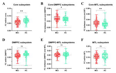 |
177 |
Disrupted subsystem interactions of default mode network in mild
cognitive impairment: A Resting-State Functional Connectivity
MRI Study
Sirong Piao1,
Keliang Chen1,
Na Wang1,
Yong Zhang2,
and Yuxin Li1
1Huashan Hospital, Fudan University, Shanghai, China, 2GE Healthcare, Shanghai, China Keywords: Alzheimer's Disease, fMRI (resting state) The default mode network (DMN) could be further divided into three subsystems, and each subsystem of DMN serves different cognitive functions. In this study, we revealed that cognitive decline is driven by weakened functional connectivity within the core subsystems, but enhanced functional connectivity between the core and MTL subsystems as well as the core and DMPFC subsystems. This dissociation may reflect abnormal core-centered connection abnormalities, along with the disruption of the MTL subsystem and the DMPFC subsystem to some degree. Our findings provided a promising imaging biomarker for the early diagnosis of AD. |
|
3055.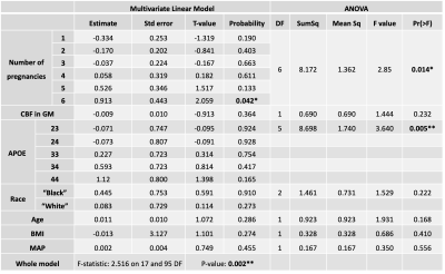 |
178 |
Investigating Alzheimer’s disease in women: is pregnancy a risk
factor?
Joana Pinto1,
Sierra Sparks1,
Genevieve Hayes1,
and Daniel P. Bulte1
1Institute of Biomedical Engineering, Department of Engineering Science, University of Oxford, Oxford, United Kingdom Keywords: Alzheimer's Disease, Alzheimer's Disease The influence of biological sex on clinical outcomes has been often neglected. Nevertheless, it is known that women have higher risk of dementia. Pregnancy might play an important role in this discrepancy, and studies have shown that this event is related to an increased risk of cerebrovascular impairment later in life. In this work we investigated the relation between the pregnancy related metrics and cognitive status and cerebral blood flow. Our results confirm that the number of pregnancies might be an important factor to consider when studying AD. Baseline CBF is not related to cognitive status or number of pregnancies. |
|
3056. |
179 |
Multi-shell DTI diffusion MRI Based Graph Theory Measures for
Prediction of Cognitive Impairment in the Alzheimer’s Disease
Connectome Project
Timothy Jaehyun Choi1,
Nagesh Adluru1,2,
Veena Nair1,
Daniel Chu1,
Camille Garcia-Ramos3,4,
Anusha Adluru1,
Shi-Jiang Li5,
Andrew L Alexander2,4,
Barbara Bendlin6,
and Vivek Prabhakaran1
1Department of Radiology, University of Wisconsin-Madison, Madison, WI, United States, 2Waisman Center, University of Wisconsin-Madison, Madison, WI, United States, 3Department of Neurology, University of Wisconsin-Madison, Madison, WI, United States, 4Department of Medical Physics, University of Wisconsin-Madison, Madison, WI, United States, 5Medical College of Wisconsin, Milwaukee, WI, United States, 6Department of Medicine, University of Wisconsin-Madison, Madison, WI, United States Keywords: Alzheimer's Disease, Brain Connectivity The prevalence of Alzheimer’s Dementia (AD) is on the rise, with an estimated economic burden to the US rivaling that of diabetes. Graph theory (GT) offers topological measures that can be used to detect connectivity changes due to aging and AD related processes. We aim to investigate the utility of GT measures based on multi-shell diffusion MRI (ms-dMRI) in discriminating aging individuals with cognitive impairment (CI) from healthy controls, and to find any intrinsic differences within the cognitively impaired subjects by clustering on the GT measures. |
|
 Back to Meeting Home
 Back to the Program-at-a-Glance
The International Society for Magnetic Resonance in Medicine is accredited by the Accreditation Council for Continuing Medical Education to provide continuing medical education for physicians.
 Back to Meeting Home
 Back to the Program-at-a-Glance
 View Presentation Video