Digital Poster
Brain Connectivity in Health I
ISMRM & ISMRT Annual Meeting & Exhibition • 03-08 June 2023 • Toronto, ON, Canada

| Computer # | |||
|---|---|---|---|
2666.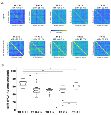 |
141 |
TR(acking) individuals down: Exploring the effect of temporal
resolution in resting-state fingerprinting
Barbara Cassone1,
Francesca Saviola1,
Stefano Tambalo1,
Enrico Amico2,3,
Dimitri Van De Ville3,4,
and Jorge Jovicich1
1CIMeC, University of Trento, Trento, Italy, 2Institute of Bioengineering, Center for Neuroprosthetics, EPFL, Geneva, Switzerland, 3Department of Radiology and Medical Informatics, University of Geneva, Geneva, Switzerland, 4Institute of Bioengineering, EPFL, Lausanne, Switzerland Keywords: Brain Connectivity, fMRI (resting state) Brain fingerprints reside in timescale-specific functional connectivity of specific regions. However, the effect of acquisition’s temporal resolution on fingerprinting is unknown. Here, we manipulated repetition time (TR) in resting-state fMRI acquisitions, and observed that subject identifiability was maximized when using fast (TR = 0.5 s) or slow (TR = 3 s) protocols, and decreased with TR = 0.7, 1 or 2 s. Moreover, while high-level association areas gave the highest contribution to individual fingerprinting regardless of TR, low-level sensorimotor areas were mostly involved in discriminating between acquisitions with different TRs. |
|
2667.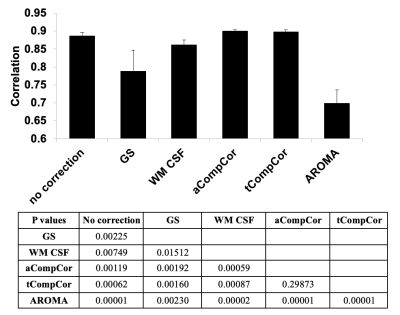 |
142 |
Comparing the efficacy of data-driven noise regression
techniques in preserving age-related resting-state connectivity
information
Ali M Golestani1 and
J Jean Chen2,3
1University of Calgary, Calgary, AB, Canada, 2Rotman Research Institute at Baycrest, Toronto, ON, Canada, 3Department of Biophysics, University of Toronto, Toronto, ON, Canada Keywords: Brain Connectivity, Artifacts Data-driven denoising methods (global-signal regression (GS), white matter and CSF (cerebrospinal fluid) regression, anatomical and temporal CompCor, ICA AROMA) have been shown to remove cardiac and respiratory contributions from the fMRI signal. In this study, we compared the effectiveness of these methods in preserving the signals associated with age-related brain connectivity changes. We show that GS and AROMA resulted in diminished age-related brain connectivity differences, aCompCor and tCompCor retained the most connectivity differences while denoising effectively. |
|
2668.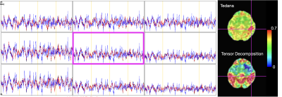 |
143 |
Exploring Tensor Decomposition as an Alternative to ICA for
Denoising Multi-Echo fMRI data
Eneko Uruñuela1,
Miguel Ánguel Veganzones2,
and César Caballero-Gaudes1
1Basque Center on Cognition, Brain and Language, Donostia - San Sebastián, Spain, 2University of Deusto, Donostia - San Sebastián, Spain Keywords: Brain Connectivity, fMRI, Tensor Decomposition Denoising of the blood oxygen level-dependent signal is critical for the study of brain dynamics with functional MRI data. However, disentangling neurobiological signals from non-neurobiological ones such as head motion-related artifacts, and cardiac-related and respiration-related fluctuations. Multi-echo ICA approaches are often used to denoise the data by exploiting the echo-time dependence of the BOLD signal. Nevertheless, these rely on the optimally combined data and do not employ the information contained in the different echo-time signals. Here, we explore the potential of tensor decomposition techniques, which can simultaneously consider all the information available, as a way to process multi-echo fMRI data. |
|
2669.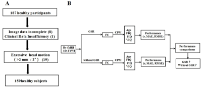 |
144 |
Global signal regression improves model performance of
connectome-based predictive modeling
Dafa Shi1,
Haoran Zhang1,
Guangsong Wang1,
and Ke Ren1
1Department of Radiology, Xiang’an Hospital of Xiamen Uneversity,School of Medicine, Xiamen University, Xiamen, China Keywords: Brain Connectivity, fMRI (resting state), connectome-based predictive modeling Physiologic significance of the global signal and the use (or omission) global signal regression (GSR) in fMRI data preprocessing remain controversial. Connectome-based predictive modeling(CPM) is one of the most commonly used machine-learning models. The effect of GSR on the performance of the CPM model is not well understood. We performed two preprocessing procedures for fMRI data: GSR and without GSR, and we used different brain atlases to construct CPM models to predict age, full-scale, performance and verbal IQ. We found that GSR can improve the predictive performance of CPM, at least for age, full-scale, performance and verbal IQ . |
|
2670.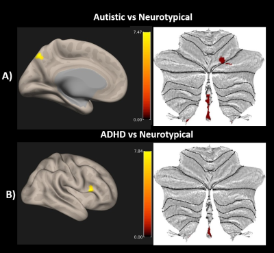 |
145 |
Whole Brain Multivoxel Pattern Analysis of resting fMRI in
Autism and Attention Deficit Hyperactivity Disorder
Smitha Karavallil Achuthan1,
Despina Stavrinos2,
Haley B Holm3,
Sheeba Arnold Anteraper4,
and Rajesh K Kana1
1Department of Psychology & The Center for Innovative Research in Autism., University of Alabama, Tuscaloosa, AL, United States, 2University of Alabama at Birmingham, Birmingham, AL, United States, 3Children’s Healthcare of Atlanta, Atlanta, GA, United States, 4Carle Illinois Advanced Imaging Center, Urbana, IL, United States Keywords: Brain Connectivity, Brain Connectivity, Multivoxel Pattern Analysis This study examined whole-brain resting state fMRI connectivity patterns in autistic and attention deficit hyperactivity disorder (ADHD) adults in comparison with neurotypical adults using multivoxel pattern analysis. Results highlight alterations in cerebellar-cortical functional connectivity (FC) in autistic participants and the involvement of cerebellum and inferior frontal gyrus in ADHD. |
|
2671.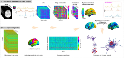 |
146 |
Uncovering the biological mechanism of depression based on
edge-centric functional connectivity: a correlation study
Fanghui Dong1,
Tongpeng Chu1,
Ning Mao1,
and Haizhu Xie1
1Yantai Yuhuangding Hospital, Yantai, China Keywords: Brain Connectivity, fMRI (resting state), Major depressive disorder, edge-centric functional connectivity, gene expression This study aims to use the combination of the edge-centric functional network (eFC) model with transcribed gene datasets to explore the molecular basis of depression. In our study, we for the first time reported the correlation between eFC and transcriptional profiles in MDD patients. These findings revealed eFC phenotypes in MDD and bridged the gap between transcriptome and neuroimaging. It advanced our understanding of the neurobiological mechanism underlying depression and provided potential biomarkers for the evaluation of MDD treatment. |
|
2672.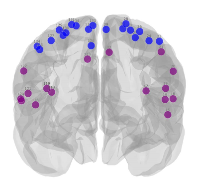 |
147 |
Characterization of cortical reorganization in persons with
chronic spinal cord injury using mesoscale graph measures
Farzad V Farahani1,
Cristina Sadowsky2,3,
James J Pekar4,5,
Martin Lindquist1,
and Ann S Choe4,5
1Department of Biostatistics, Johns Hopkins Bloomberg School of Public Health, Baltimore, MD, United States, 2International Center for Spinal Cord Injury, Kennedy Krieger Institute, Baltimore, MD, United States, 3Department of Physical Medicine and Rehabilitation, Johns Hopkins University School of Medicine, Baltimore, MD, United States, 4F.M. Kirby Research Center for Functional Brain Imaging, Kennedy Krieger Institute, Baltimore, MD, United States, 5Radiology and Radiological Science, Johns Hopkins University School of Medicine, Baltimore, MD, United States Keywords: Brain Connectivity, Spinal Cord, graph theory, spinal cord injury, plasticity, reorganization Cortical plasticity contributes to neurological recovery in persons with spinal cord injury (SCI) and can be studied using resting-state fMRI. Global graph measures offer insufficient detail to investigate local changes. Here, we investigated whether mesoscale graph measures provide additional insight. Changes in sensorimotor, visual, and ventral attention networks were revealed. Notably, decreased communication between lower body SMN with the rest of the functional networks across the brain and increased communication within the upper body SMN were observed. This suggests a potential for the mesoscale graph measures' utility in understanding the complex brain functional reorganizations in persons with chronic SCI. |
|
2673.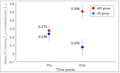 |
148 |
Neural perspective on observational drawing: A longitudinal
resting state functional connectivity study using 7T MRI
Apoorva Safai1,
Jeffrey Katz2,3,4,5,
Barbara Bondy6,
and Gopikrishna Deshpande2,3,4,5,7,8,9,10
1Symbiosis Centre for Medical Image Analysis, Symbiosis International University, Pune, India, 2Department of Psychological Sciences, Auburn University, Auburn, AL, United States, 3Department of Electrical & Computer Engineering, Auburn University, Auburn, AL, United States, 4Alabama Advanced Imaging Consortium, Birmingham, AL, United States, 5Center for Neuroscience, Auburn University, Auburn, AL, United States, 6Department of Art and Art History, Auburn University, Auburn, AL, United States, 7Key Laboratory for Learning and Cognition, Capital Normal University, Beijing, China, 8Department of Psychiatry, National Institute of Mental Health and Neurosciences, Bangalore, India, 9Centre for Brain Research, Indian Institute of Science, Bangalore, India, 10Department of Heritage Science and Technology, Indian Institute of Technology, Hyderabad, India Keywords: Brain Connectivity, Neuroscience, Neuroplasticity Observational drawing is representation of observed 3D real-life objects into 2D drawings on paper, which involves engaging complex skills like visual perception, spatial encoding, memory and decision making. This study investigates longitudinal functional connectivity (FC) in art students who underwent a 16-week long drawing course, in comparison to students who took unrelated courses. The art group showed improved drawing skills and demonstrated enhanced FC between DMN and salience network and in cerebellar regions post-art class, in comparison, with healthy non-art students. These findings imply positive changes in brain functioning on learning and practicing art, thereby highlighting its potential therapeutic applications. |
|
2674.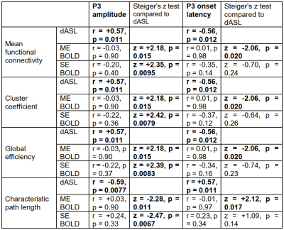 |
149 |
Association of Attention with Brain Global Efficiencies at Rest:
a Dynamic ASL and BOLD fMRI Comparison Study
Yakun Zhang1,
Shichun Chen1,
Zongpai Zhang1,
Wenna Duan1,
George Weinschenk1,
Li Zhao2,
Brandon Gibb3,
Adam Anderson4,
Wenming Luh5,
and Weiying Dai1
1Department of Computer Science, State University of New York at Binghamton, Binghamton, NY, United States, 2College of Biomedical Engineering & Instrument Science, Zhejiang University, Hangzhou, China, 3Department of Psychology, State University of New York at Binghamton, Binghamton, NY, United States, 4Department of Psychology, Cornell University, Ithaca, NY, United States, 5Cornell MRI Facility, Cornell University, Ithaca, NY, United States Keywords: Brain Connectivity, Arterial spin labelling We evaluated the relationship between attention levels and brain global efficiencies. This relationship was compared with brain global efficiencies measured with dynamic ASL (dASL) and with multi-echo (ME) BOLD fMRI. We found significantly greater correlation between attention levels (reflected by P3 properties when performing an attention task) and brain global efficiencies based on rsFC using dASL than those using ME BOLD fMRI, indicating that dASL can offer more accurate global neural signal fluctuations. |
|
2675.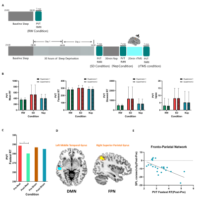 |
150 |
Task functional magnetic resonance imaging-guided individualized
repetitive transcranial magnetic stimulation after a brief nap
attenuates the sustained attention performance deterioration
induced by sleep deprivation
Yuanqiang Zhu1,
Fan Guo1,
and Yingjuan Chang1
1Air Force Medical University, Xi'an, China Keywords: Data Analysis, fMRI (task based) Our pilot study validated the effectiveness of rTMS after a brief nap in terms of improving sustained attention in the context of SD. An investigation of the dynamic changes of PVT task-related cerebral responses across the three conditions showed that the middle frontal gyrus recovered least after the nap and was selected as the stimulation target. Through modulating the functional connectivity within the FPN and DMN, individualized, 10-Hz rTMS showed promise in terms of improving the vigilance of military academy cadets accepting real stimulation. |
|
2676.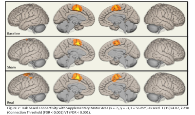 |
151 |
Exploring effect of rTMS at SMA in Parkinson’s Disease- An
fMRI-MVPA study.
Priyanka Bhat1,
S Senthil Kumaran2,
Vinay Goyal3,
Achal K Srivastava4,
S N Dwivedi5,
and Madhuri Behari6
1Dept. of Neurology, AIIMS, Delhi, India, 2Dept of Nuc. Mag. Res, AIIMS, Delhi, India, 3Medanta, The Medicity, Delhi, India, 4Dept of Neurology, AIIMS, Delhi, India, 5Dept. of Biostatistics, AIIMS, Delhi, India, 6Dept. of Neurology, Fortis Hospitals, Vasant Kunj, Delhi, India Keywords: Brain Connectivity, fMRI (task based), Parkinson's Disease, Supplementary Motor Area, Transcranial Magnetic Stimulation Transcranial Magnetic stimulation alters cortical connectivity in Parkinson’s disease (PD) imparting clinical benefits. The mechanisms by which this occurs remain unexplored. In this study, Multivariate pattern analyses of task-based connectivity was carried out to understand the effects of rTMS in PD. Visuospatial task based functional MRI was acquired before and after stimulation sessions. Correlation matrices revealed an increased connectivity between left supplementary motor area (SMA) and left primary motor area (M1), suggesting an improvement in the motor network. |
|
2677.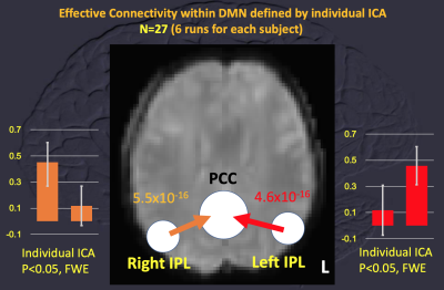 |
152 |
ROI-dependence in clarifying metabolic connectivity mapping
within default-mode network using simultaneous FDG PET and
BOLD-based fMRI
Tzu-chen Yeh1,2,
Chou-ming Cheng3,
and Chi-che Chou3
1Department of Radiology, Taipei Veterans General Hospital, Taipei, Taiwan, 2Institute of Brain Science, National Yang Ming Chiao Tung University, Taipei, Taiwan, 3Department of Medical Research, Taipei Veterans General Hospital, Taipei, Taiwan Keywords: Brain Connectivity, PET/MR, metabolic connectivity mapping Metabolic connectivity mapping (MCM) is a unique application of simultaneous PET/fMRI for solving the effective connectivity (EC) of brain circuits. Reproducibility of MCM was critical after the initial report. By applying the released database of MONASH (Melbourne, Australia), consistent EC among bilateral inferior parietal lobules (IPL) and posterior cingulate cortices (PCC), as nodes within default mode network (DMN), was identified using MCM with the statistic-based approach. One-way EC using dedicated ROI (regions of interest) derived from grouped ICA (independent component analysis) outperformed the atlas-based ROIs of DMN. |
|
2678. |
153 |
Multimodal neuroimaging data reveal intelligence-specific
neurobiological correlates
Dafa Shi1,
Haoran Zhang1,
Guangsong Wang1,
and Ke Ren1
1Department of Radiology, Xiang’an Hospital of Xiamen Uneversity,School of Medicine, Xiamen University, Xiamen, China Keywords: Brain Connectivity, fMRI (resting state) We used Brainnetome 246 atlas to construct FC matrices and extract image features from ALFF, DC, ReHo and VMHC maps.The classical CPM method using FC data and the combination model using multimodal data were constructed to predict FIQ scores. We found the combination model outperformed either classical models. Similar results were found with the Shen 268 atlas. The functional networks and regions related to intelligence mainly included intrafrontal, frontoparietal, frontotemporal and temporoparietal networks.The models constructed with the intelligence-related networks and regions could predict PIQ, VIQ, and CRT scores well, but not BDI, SAT, and TAT scores. |
|
2679.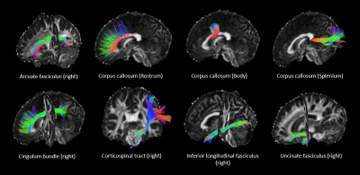 |
154 |
Assessing inter-tract independence across microstructural
metrics
Carolyn B McNabb1,
Eirini Messaritaki1,
Kristin Koller1,
and Derek K Jones1
1Psychology, Cardiff University, Cardiff, United Kingdom Keywords: Brain Connectivity, Brain Connectivity We aimed to identify potential redundancies in microstructural measures between major white matter tracts and asked which microstructural metrics correlate most within a tract. Using a combination of microstructural imaging techniques, including diffusion imaging, relaxometry and quantitative magnetisation transfer imaging, we identified strong correlations between homologous left and right fasciculi in the cingulum bundles, inferior longitudinal fasciculi, uncinate fasciculi and arcuate fasciculi as well as similar patterns of tissue microstructure between heterologous tracts. However, corticospinal tracts showed weak correlations with other tracts and a unique pattern of inter-metric correlations, suggesting that summarising metrics across all tracts would be inappropriate. |
|
2680.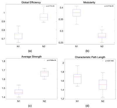 |
155 |
Measuring structural connectivity in migraine: the impact of
correcting for region volumes
Ana Matoso1,
Ana R Fouto1,
A. Ruiz-Tagle1,
I. Esteves1,
Gina Caetano1,
Nuno A. Silva2,
Pedro Vilela3,
Raquel Gil-Gouveia4,5,
Patrícia Figueiredo1,
and Rita G Nunes1
1Institute for Systems and Robotics- Lisboa and Department of Bioengineering, Instituto Superior Técnico, Universidade de Lisboa, Lisbon, Portugal, 2Learning Health, Hospital da Luz, Lisbon, Portugal, 3Imaging Department, Hospital da Luz, Lisbon, Portugal, 4Neurology Department, Hospital da Luz, Lisbon, Portugal, 5Center for Interdisciplinary Research in Health, Universidade Católica Portuguesa, Lisbon, Portugal Keywords: Brain Connectivity, Data Analysis, Migraine When studying structural brain connectivity, there are many normalization strategies that can be employed, however, there is still no consensus on which one is best to use. Hence, in this study, we investigated the impact of normalizing by the region volumes in the context of migraine as this is sometimes done to avoid bias towards higher connectivity in bigger regions. Several metrics showed significant differences between normalization strategies and, more critically, nodal metrics displayed different behaviours when compared between controls and migraineurs. |
|
2681. |
156 |
Brain connectome-based imaging markers for identifiable
signature of migraine with and without aura
Tong Fu1,
Yujia Gao1,
Xiaobin Huang1,
Di Zhang1,
Lindong Liu1,
Xindao Yin1,
Xinying Wu 1,
Hai Lin2,
and Yongming Dai2
1Department of Radiology, Nanjing first hospital,Nanjing Medical University, Nanjing, China, 2Central Research Institute, United Imaging Healthcare, Shanghai, China Keywords: Brain Connectivity, Machine Learning/Artificial Intelligence In comparison with migraine without aura (MwoA), migraine with aura (MwA) has its own characteristics in symptom, pathological mechanism, treatment and prognosis. In this study, we conducted connectome-based analysis to capture brain connectivity markers that would show identifiable signature of MwA and MwoA, using diffusion tensor imaging and resting-state functional MRI. We found that the alterations of structural and functional connectivity strength contributed to migraine patient subtyping. The whole brain connectome-based imaging markers might provide possible evidence in understanding the heterogeneity of migraine with aura and help for patient-specific decision. |
|
2682.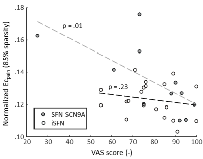 |
157 |
DTI reveals altered structural connectivity of pain-associated
regions in a genetic variant of small fiber neuropathy
Gerhard Drenthen1,
Amir Far2,
Catharina Faber2,
Jaymin Upadhyay3,
David Linden4,
Raquel van Gool4,
Walter Backes1,
Janneke Hoeijmakers2,
and Jacobus Jansen1
1Department of Radiology & Nuclear Medicine, School for Mental Health and Neuroscience, Maastricht University Medical Center, Maastricht, Netherlands, 2Department of Neurology, Maastricht University Medical Center, Maastricht, Netherlands, 3Boston Children's Hospital, Boston, MA, United States, 4School for Mental Health & Neuroscience, Maastricht University Medical Center, Maastricht, Netherlands Keywords: Brain Connectivity, Diffusion Tensor Imaging, Chornic pain Patients with small fiber neuropathy (SFN) suffer from chronic pain, which may lead to cerebral changes. Here, we studied structural network changes in idiopathic- and genetic-SFN compared to controls using diffusion-MRI. We found that for the genetic-SFN group pain-associated regions take a more prominent place in the network (in terms of nodal importance). Furthermore, in the genetic-SFN group, a higher nodal importance of pain-associated regions related to lower self-reported pain. This shows that genetic-SFN has a distinct structural pain pathway, which may be indicative of a compensatory mechanism where the structural organization is altered to inhibit the response to pain. |
|
2683.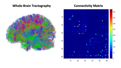 |
158 |
Graph theory-based analysis of brain diffusion data reveals
network alterations in World Trade Center first responders with
chronic PTSD
Daniel Suite1,
Thomas Hagan1,
Chuan Huang1,2,
Minos Kritikos3,
Sean Clouston3,
Megan Horton4,
Roman Kotov5,
Roberto G Lucchini6,7,
Evelyn J Bromet5,
and Benjamin Luft8
1Department of Biomedical Engineering, Stony Brook University, Stony Brook, NY, United States, 2Department of Radiology, Stony Brook Medicine, Stony Brook, NY, United States, 3Program in Public Health and Department of Family, Population, and Preventative Medicine, Stony Brook Medicine, Stony Brook, NY, United States, 4Environmental Medicine and Public Health, Icahn School of Medicine at Mount Sinai, New York City, NY, United States, 5Department of Psychiatry, Stony Brook Medicine, Stony Brook, NY, United States, 6Environmental Health Sciences, School of Public Health, Florida International University, Miami, FL, United States, 7Occupational Medicine, Department of Medical and Surgical Specialties, Radiological Sciences and Public Health, University of Brescia, Brescia, Italy, 8Department of Medicine, Stony Brook Medicine, Stony Brook, NY, United States Keywords: Brain Connectivity, Psychiatric Disorders Many World Trade Center (WTC) responders continue to suffer from chronic Post-Traumatic Stress Disorder (PTSD). Multimodal imaging techniques have shown potential as putative markers for PTSD but still lie in the developmental stages. Network connectivity techniques are showing promise for investigating neuropathology influencing PTSD symptom maintenance and course. This work utilizes a graph theory approach with brain diffusion images to probe the network alterations in WTC responders with PTSD. We identified a significant difference in Characteristic Path Length (CPL) between responders with and without chronic PTSD. |
|
2684.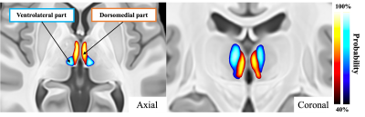 |
159 |
Identify the paraventricular thalamic nucleus in humans using a
structural connectivity approach
Koji Kamagata1,
Wataru Uchida1,
Christina Andica1,2,
Yasuhito Nagai3,
Masaki Nishioka3,
Mana Owaki1,4,
Yuya Saito1,
Kaito Takabayashi1,
Akifumi Hagiwara1,
Akihiko Wada1,
Toshiaki Akashi1,
Shigeki Aoki1,
and Tadafumi Kato3
1Department of Radiology, Juntendo University Graduate School of Medicine, Tokyo, Japan, 2Faculty of Health Science, Juntendo University, Chiba, Japan, 3Department of Psychiatry, Juntendo University, Tokyo, Japan, 4Department of Radiological Sciences, Graduate School of Human Health Sciences, Tokyo Metropolitan University, Tokyo, Japan Keywords: Brain Connectivity, Brain Connectivity We attempted to identify the paraventricular thalamic nucleus (PVT), which is known to regulate emotion, motivation, stress, and drug- and alcohol-related behaviors in humans. We used data-driven connectivity profiles obtained using probabilistic tractography and a k-means clustering method with diffusion-weighted imaging data. We consistently identified an anatomical connectivity-based parcellation of the PVT in two independent cohorts that included 601 healthy subjects. Furthermore, we discerned the specific structural pattern of the PVT, which agreed with findings from animal studies. Finally, we noted significant correlations between PVT structural and functional connectivity with the limbic structures and drug-, nicotine-, or alcohol-related scores.
|
|
2685.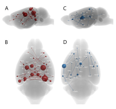 |
160 |
Application of Diffusion MRI to Characterize Connectome Changes
Associated with EED Ablation
Majd Alkhalily1,
Laura Currey2,
Michael Piper2,
and Nyoman D Kurniawan1
1Centre for Advanced Imaging, The University of Queensland, Brisbane, Australia, 2The School of Biomedical Sciences, The University of Queensland, Brisbane, Australia Keywords: Brain Connectivity, Genetic Diseases, Tractography, Connectome, EED, Neurodevelopment, MRI, HARDI Mutation in embryonic ectoderm development (EED) gene in mice have been shown to exhibit microcephaly at birth, however, the adult phenotype is unknown. This study investigates brain connectivity changes arising from EED ablation in adult mice using high angular resolution diffusion imaging. Three groups of adult mice (homozygotes, heterozygotes, controls) were scanned using 16.4 T to acquire diffusion data. Data was then used to compute iFOD2 maps, probabilistic whole brain tractograms, and the connectomes. Connectomic were compared both connectivity- and network-wise. Homozygotes had abnormal connectivity and network metrics suggesting brain under-development, highlighting the importance of EED’s role in brain development. |
|
 Back to Meeting Home
 Back to the Program-at-a-Glance
The International Society for Magnetic Resonance in Medicine is accredited by the Accreditation Council for Continuing Medical Education to provide continuing medical education for physicians.
 Back to Meeting Home
 Back to the Program-at-a-Glance
 View Presentation Video