Digital Poster
Tumors: The Importance of Diffusion & Perfusion I
ISMRM & ISMRT Annual Meeting & Exhibition • 03-08 June 2023 • Toronto, ON, Canada

Digital Poster
Tumors: The Importance of Diffusion & Perfusion I
| Computer # | |||
|---|---|---|---|
1565.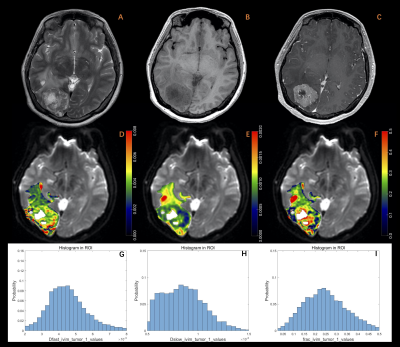 |
101 | Histogram analysis of Intravoxel incoherent motion (IVIM) in differentiating high-grade glioma and solitary brain metastasis
Yifei Su1, Jinxia Guo2, Junhao Wang1, Rui Cheng3, Chunhong Wang3, Liangliang Hao4, Hongming Ji3, Yexin He4, and Cheng Xu4
1The Fifth Clinical Medical College of Shanxi Medical University, Taiyuan, China, 2GE Healthcare, MR Research, Beijing, China, 3Department of Neurosurgery, Fifth Hospital of Shanxi Medical University, Taiyuan, China, 4Department of Radiology, Fifth Hospital of Shanxi Medical University, Taiyuan, China Keywords: Tumors, Cancer, high grade glioma, solitary brain metastasis We aim to explore the value of intravoxel incoherent motion (IVIM) in differentiating high-grade glioma (HGG) and solitary brain metastasis (SBM) by using histogram and first-order texture analysis in combination with the structural lesion feature. Results showed there were significant higher skewness (Dfast, f) and kurtosis (Dslow, f), and significant lower entropy (Dslow, f) and mean (f) in HGG solid tumor region as well as significant lower mean (f) in HGG peritumoral edema zone. Clear tumor margin in T2-weighted imaging help improve the differentiation when integrated with the IVIM parametric values. |
|
1566.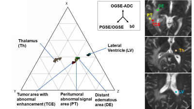 |
102 | Ternary plot method for presenting information acquired by OGSE and PGSE sequences: Evaluation of the peritumoral area in malignant glioma
Toshiaki Taoka1,2, Rintaro Ito1,2, Rei Nakamichi2, Toshiki Nakane2, Kazushige Ichikawa3, Mayuko Sakai4, Nobuyasu Ichinose4, Masanori Ozaki4, Takaya Mori4, Yoshiki Tanaka5, and Shinji Naganawa2
1Department of Innovative Biomedical Visualization (iBMV), Nagoya University, Nagoya, Japan, 2Radiology, Nagoya University, Nagoya, Japan, 3Division of Radiology, Nagoya University Hospital, Nagoya, Japan, 4Canon Medical Systems Corporation, Otawara, Japan, 5SORD Corporation, Chiba, Japan Keywords: Tumors, Diffusion/other diffusion imaging techniques, OGSE The characteristics of tissue structures can be estimated by comparing images with different diffusion times acquired using the OGSE and PGSE methods. However, for multiple images, it is difficult to make pixel-by-pixel comparison. We attempted a method for integrating the pixel values of these images using ternary plotting for malignant glioma cases. Tumor areas with abnormal enhancement, peritumoral abnormal signal areas, and distant edematous areas were evaluated. The ternary plot provided immediate visual information. It also highlighted similarities in the tissue characteristics of the tumor, with enhancement to the peritumoral area showing abnormal signals. |
|
1567.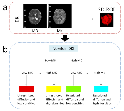 |
103 | Habitat analysis based on diffusion kurtosis imaging to predict adult-type diffuse gliomas grade
Peng Wang1,2, Yanhao Liu1, Qiong Wu1, Shaoyu Wang3, Huapeng Zhang3, Yang Song3, Xu Yan3, and Yang Gao1
1Affiliated Hospital of Inner Mongolia Medical University, Hohhot, China, 2Inner Mongolia Medical University, Hohhot, China, 3Siemens Healthineers, Shanghai, China Keywords: Tumors, Diffusion/other diffusion imaging techniques Glioma composition is complicated, making whole-tumor investigation with traditional pathological analysis typically challenging. It is possible to demonstrate intratumor heterogeneity using habitat analysis in radiography, a technique analogous to biological environmental analysis with individualized descriptions and quantitative expression of subregions inside the region of interest. Therefore, our goal was to create an integrated model for the habitat analysis-based diffusion kurtosis imaging diagnosis of adult-type diffuse glioma grade. According to the findings, diffusion kurtosis imaging based on habitat analysis accurately predicted adult-type diffuse gliomas grade, which may help to resolve some "intermediate situations" that are challenging to diagnose clinically. |
|
1568.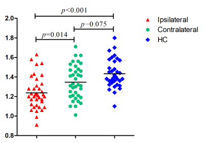 |
104 | Evaluation of the Glymphatic System Using the DTI-ALPS Index in Patients with Glioma
Chao Zhang1, Ruohan Li1, Jingyun Sha1, Lulu Cai1, Peng Wu2, and Kai Xu1
1Department of Radiology, Affiliated Hospital of Xuzhou Medical University, Xuzhou, China, 2Philips Healthcare, Shanghai, China Keywords: Tumors, Diffusion Tensor Imaging, edema We evaluated the function of the human glymphatic system (GS) in patients with glioma using diffusion tensor imaging analysis along with the perivascular space (DTI-ALPS). Thirty-seven patients with glioma and 34 healthy controls (HCs) were recruited for analysis. We found that DTI-ALPS index on the ipsilateral lesion side was significantly decreased, but not on the contralateral side in glioma when compared with HCs. The decreased DTI-ALPS index was significantly correlated with the volume of peritumoral edema (PTBE), but not the volume of tumor. This study suggested the PTBE may be an independent factor that affects the GS in glioma patients. |
|
1569.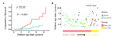 |
105 | The Value of Diffusion Weighted Imaging in Predicting the Growth Pattern of Meningioma: A Retrospective Study
Hui Zheng1, Zongmeng Wang1, Lingmin Zheng1, Rufei Zhang1, Yang Song2, and Lin Lin1
1Fujian Medical University Union Hospital, Fuzhou, China, 2MR Scientific Marketing, Siemens, Healthineers Ltd., Shanghai, China Keywords: Tumors, Diffusion/other diffusion imaging techniques The natural history of meningioma remains unclear and a simple, practical method is needed to identify fast growth tumors. This study evaluated correlations between clinical parameters, tumor growth rate (TGR), tumor volume doubling time (VDT), Ki-67 and relative apparent diffusion coefficient (rADC) derived from diffusion weighted imaging (DWI). The results showed that rADC was an independent predictive parameter of meningioma growth and baseline rADC had a good predictive ability for differentiating slow growth from fast growth meningioma. This suggests that in asymptomatic meningiomas, DWI might be a valuable predictive imaging method. |
|
1570.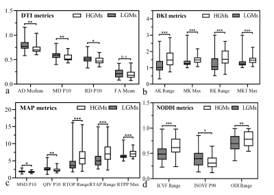 |
106 | Whole-Tumour Histogram Analysis of Multiple Diffusion Metrics for Meningiomas Grade
Dejun She1,2,3, Hao Huang1,2, Xiance Zhao4, and Dairong Cao1,2,3,5
1Department of Radiology, First Affiliated Hospital of Fujian Medical University, Fuzhou, China, 2Department of Radiology, National Regional Medical Center, Binhai Campus of the First Affiliated Hospital, Fujian Medical University, Fuzhou, China, 3Key Laboratory of Radiation Biology of Fujian higher education institutions, First Affiliated Hospital, Fujian Medical University, Fuzhou, China, 4Philips Healthcare, Shanghai, China, 5Department of Radiology, Fujian Key Laboratory of Precision Medicine for Cancer, First Affiliated Hospital of Fujian Medical University, Fuzhou, China Keywords: Tumors, Diffusion/other diffusion imaging techniques, Meningiomas; Diffusion-weighted MRI An accurate assessment of the World Health Organization grade is vital in meningiomas. While many studies have investigated the usefulness of conventional DWI and DTI for noninvasive grading intracranial meningiomas, there are neither any studies comparing three advanced diffusion model including DKI, MAP and NODDI with DTI for predicting meningioma grade. Thus, it is vital to evaluate whether these advanced models derived from diffusion spectrum imaging can also be beneficial in grading meningiomas. Our results suggested that whole tumour histogram analyses of the diffusion metrics from multiple diffusion models are promising methods in grading meningiomas. |
|
1571.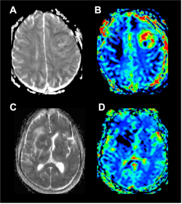 |
107 | Relative CBV Combined with Mean ADC in Assessing the Differential Between Glioblastoma and Primary Cerebral Lymphoma
Qi Lin1, Langlang Tang1, Yongzhou Xu2, and Peng Wu3
1Department of Radiology, Longyan First Hospital, Longyan, China, 2Philips Healthcare, Guangzhou, China, 3Philips Healthcare, Shanghai, China Keywords: Tumors, Cancer To further evaluate the efficacy of MRI in the differential diagnosis of glioblastoma (GBM) and primary cerebral lymphoma (PCL), this study retrospectively analysed rCBV and mADC-related data from patients with GBM and PCL and performed a quantitative comparison. The rCBV and mADC values were found to be significantly higher in the GBM group than in the PCL group. We diagnosed GBM when rCBV >23.2900 ml/100g or mADC >0.6550×10⁻³mm2/s, and the corresponding sensitivity, specificity, and predictive values were all high. This demonstrates the important clinical value of the combined application of mADC and rCBV for the definitive diagnosis of brain tumours. |
|
1572.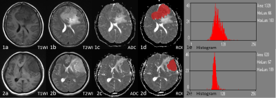 |
108 | The value of an apparent diffusion coefficient histogram model in predicting meningioma recurrence
Tao Han1, Junlin Zhou1, and Xianwang Liu1
1Lanzhou University Second Hospital, LanZhou, China Keywords: Tumors, Diffusion/other diffusion imaging techniques This study evaluated the feasibility of the model combining conventional MRI features and apparent diffusion coefficient (ADC) histogram parameters for predicting recurrent meningioma. The results showed that tumor shape and ADCp1 were independent risk factors for predicting meningioma recurrence after removing confounding factors by single and multiple logistic regression analysis. The logistic regression model nomogram constructed based on the above two risk factors for predicting meningioma recurrence, which had a higher predictive effect than a single risk factor. In conclusion, logistic regression model nomogram have important value in predicting meningioma recurrence and helping patients achieve the greatest survival benefit. |
|
1573.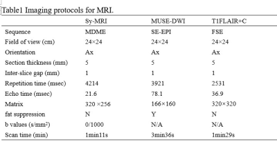 |
109 | Differentiation of progressive disease from pseudo-progression using Synthetic MRI and MUSE-DWI in patients with glioblastoma.
Ruirui Lv1, Wenfu Ma1, Yuhui Xiong2, Peng Yong1, Jiarui Zheng1, Xuhong Yang1, and Xiaodong Wang3
1Ningxia Medical University, Yinchuan, China, 2GE Healthcare MR Research, Beijing, China, 3General Hospital of Ningxia Medical University, Yinchuan, China Keywords: Tumors, Brain This work sought to investigate the performance of synthetic MRI(Sy-MRI) and MUSE-DWI in differentiating progressive disease (PD) from pseudo-progression (PsP). It was concluded that the pre-contrast T1(T1-pre) and post-contrast T1(T1-Gd) from synthetic MRI can be used as novel quantitative imaging biomarkers for discriminating PD from PsP. The combination of T1-pre, T1-Gd and ADC may explore as an effective strategy to improve the ability for discriminating PD from PsP, and provide a basis for clinical follow-up diagnosis and treatment. |
|
1574.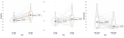 |
110 | Pre-treatment Turbo Spin-Echo (TSE) intravoxel incoherent motion (IVIM) predicts treatment outcome in nasopharyngeal carcinoma
Lijuan Qian1, Jie Yu2, Yuan Liu3, Peng Sun4, Manman Chen5, Xin Li5, and Fan Yang5
1Radiology, Union Hospital Tongji Medical College Huazhong University of Science and Technology, Wuhan, China, 2Union Hospital Tongji Medical College Huazhong University of Science and Technology, Wuhan, China, Wuhan, China, 3Union Hospital Tongji Medical College Huazhong University of Science and Technology, Wu, China, 4Philips healthcare,Beijing, Beijing, China, 5Union Hospital Tongji Medical College Huazhong University of Science and Technology, Wuhan, China Keywords: Tumors, Head & Neck/ENT TSE-DWI-IVIM as a new and feasible method for predicting the early response to Induction Chemotherapy(IC) for NPC patients.Diffusion-related IVIM parameters (pre-D) might be and perfusion-related parameters (pre-f) can be helpful as potential imaging biomarkers of the therapeutic response to IC in NPC patients.
|
|
1575.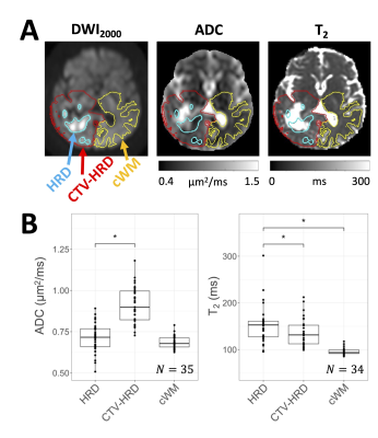 |
111 | High b-value diffusion-weighted imaging of glioblastoma on an MR-Linac
Liam S. P. Lawrence1, Rachel W. Chan1, James Stewart2, Mark Ruschin2, Aimee Theriault2, Sten Myrehaug2, Jay Detsky2, Pejman J. Maralani3, Chia-Lin Tseng2, Hany Soliman2, Mary Jane Lim-Fat4, Sunit Das5, Greg J. Stanisz1,6, Arjun Sahgal2, and Angus Z. Lau1
1Physical Sciences, Sunnybrook Research Institute, Toronto, ON, Canada, 2Department of Radiation Oncology, Sunnybrook Health Sciences Centre, Toronto, ON, Canada, 3Medical Imaging, Sunnybrook Health Sciences Centre, Toronto, ON, Canada, 4Division of Neurology, Department of Medicine, Sunnybrook Health Sciences Centre, Toronto, ON, Canada, 5Department of Surgery, St. Michael's Hospital, Toronto, ON, Canada, 6Department of Neurosurgery and Paediatric Neurosurgery, Medical University, Lublin, Poland Keywords: Tumors, Radiotherapy Hyperintense regions on high b-value DWI (HRDs) may reflect hypercellular tumour and are of interest for radiotherapy dose escalation. However, the extent to which diffusion restriction versus prolonged T2 creates these hyperintensities is not known. Additionally, the dynamics of HRDs during radiotherapy are not fully characterized. In 35 glioblastoma patients treated on a 1.5T MR-Linac, we found HRDs shrank during treatment and extended beyond the gross tumour volume. Apparent diffusion coefficient was reduced and T2 was elevated in HRD compared to the remainder of the clinical target volume, implying that both diffusion restriction and prolonged relaxation are responsible for HRDs. |
|
1576.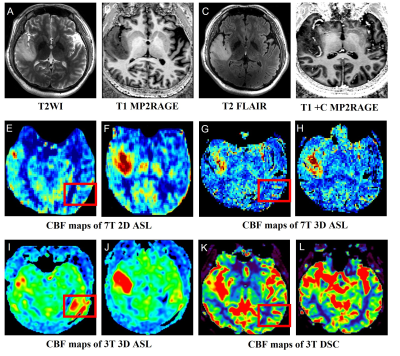 |
112 | The feasibility of arterial spin labeling imaging of intracranial tumors at 7T
Xiaoxiao Ma1, Kun Cheng1, Chenyang Zhao2, Jianxun Qu3, Chenxi Li1, Runze Li1, Caohui Duan1, Xiangbing Bian1, Danny JJ Wang2,4, and Xin Lou1
1Department of Radiology, Chinese PLA General Hospital, Beijing, China, 2Mark & Mary Stevens Neuroimaging and Informatics Institute, Keck School of Medicine, University of Southern California, Los Angeles, CA, United States, 3MR Collaboration, Siemens Healthineers Ltd., Beijing, China, 4Department of Neurology, Keck School of Medicine, University of Southern California, Los Angeles, CA, United States Keywords: Tumors, Perfusion The goal of this study was to assess the feasibility of pseudo-continuous arterial spin labeling (pCASL) at 7T on patients with intracranial tumors. We found that 7T ASL imaging was feasible and had a higher resolution than 3T ASL imaging and therefore, may enable in vivo assessment of subtle changes and provides a valuable tool for early detection and mechanism investigation of neurovascular function impairments in patients with intracranial tumors. |
|
1577.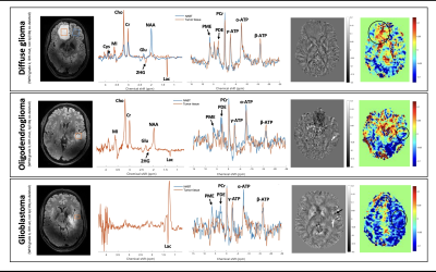 |
113 | 7T metabolic MRI in three types of diffuse glioma
Sarah M Jacobs1, Evita C Wiegers1, Zahra Shams1, Jannie P Wijnen1, Peter W.A. Willems2, Pierre A Robe2, Angelika Mühlebner3,4, Wim Van Hecke3, Maeike Zijlmans2,5, Dennis WJ Klomp1, and Anja G van der Kolk1,6
1Department of Radiology and Nuclear Medicine, University Medical Center Utrecht, Utrecht, Netherlands, 2UMC Utrecht Brain Center, Department of Neurology and Neurosurgery, University Medical Center Utrecht, Utrecht, Netherlands, 3Department of Pathology, University Medical Center Utrecht, Utrecht, Netherlands, 4Department of Pathology, Amsterdam University Medical Center, location AMC, Amsterdam, Netherlands, 5Stichting Epilepsie Instellingen Nederland (SEIN), Heemstede, Netherlands, 6Department of Medical Imaging, Radboud University Medical Center, Nijmegen, Netherlands Keywords: Tumors, Tissue Characterization We combined metabolic magnetic resonance imaging (MRI) sequences at 7 Tesla (7T) to characterize glioma tissue and discover metabolic MRI profiles that could help delineate the extent of disease spread and determine molecular tumor subtypes preoperatively. Here we show the first preliminary results of three different diffuse glioma subtypes imaged with 1H MR spectroscopy (MRS), 31P 3D chemical shift imaging (CSI), glutamate chemical exchange saturation transfer (GluCEST) and quantitative susceptibility mapping (QSM). |
|
1578.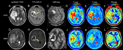 |
114 | Differentiation of High-Grade Gliomas from Solitary Brain Metastases in the Peritumoral Edema: using Combined Synthetic MRI and 3D-pCASL
Xin Ge1,2, Yuhui Xiong3, Min Li4, Xiaodong Wang5, and Jing Zhang2
1Second Clinical School, Lanzhou University, Lanzhou, China, Lanzhou, China, 2Department of Magnetic Resonance, Lanzhou University Second Hospital, Lanzhou, China, Lanzhou, China, 3GE Healthcare MR Research, Beijing, China, Beijing, China, 4GE Healthcare MR Enhancement Application, Beijing, China, Beijing, China, 5Department of Radiology, General Hospital of Ningxia Medical University, Yinchuan, China, Yinchuan, China Keywords: Tumors, Tumor The main work of our research was to investigate the difference of quantitative MR metrics from synthetic MRI and 3D-pCASL including T1, T2, PD, CBF, and the multiparametric strategies that contained more than one of the metrics between high-grade gliomas (HGGs) from solitary brain metastases (SBMs) in the peritumoral edema. We found that the T1, T2, and CBF were useful metrics for differentiating HGGs from SBMs which were easily confounded with HGGs in daily diagnosis. Furthermore, combining T1, T2, and CBF further improved their diagnostic performance. |
|
1579.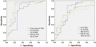 |
115 | Detecting microstructural features in gliomas with differences in malignancy and IDH-1 mutation status with a three-component diffusion model
Mengqiu Cao1, Xiaoqing Wang2, Fang Liu2, Ke Xue3, and Yongming Dai3
1Department of Radiology, Renji Hospital, School of Medicine, Shanghai Jiao Tong University, Shanghai, China, 2Renji Hospital, School of Medicine, Shanghai Jiao Tong University, Shanghai, China, 3MR Collaboration, Central Research Institute, United Imaging Healthcare, Shanghai, China Keywords: Tumors, Diffusion/other diffusion imaging techniques Evaluation of the degree of malignancy of gliomas, including the histopathological and molecular features, is of great importance for neurosurgeons in making individualized operation plans before surgery. In our study, the clinical value of a three-component diffusion model in characterizing gliomas malignancy and IDH-1 mutation status was evaluated. Our results showed that the three-component diffusion model exhibited great potential in evaluating the phenotype and genotype of gliomas by providing more complex microstructural information and showed significantly optimal diagnostic performance than the mono-exponential, bi-exponential and DKI models. |
|
1580.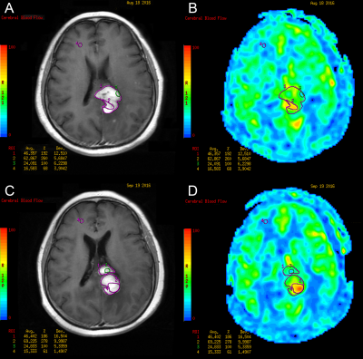 |
116 | Arterial spin labeling MRI can predict prognosis of patients with newly-diagnosed primary CNS lymphoma
Hao Wu1, Zhenwei Yao1, Bobin Chen2, Hui Kang2, Huaping Sun1, Yan Ren1, Shuguang Chu3, Na Lu1, Xianjing Zhao1, Jie Wu1, Yan Ma2, Yan Yuan2, Tianling Ding2, Zhiguang Lin2, Jingjing Ma2, Qing Li2, Xianjin Zhou4, Yiqian Zhu5, Tianyong Xu6, and Yong Zhang6
1Department of Radiology, Huashan Hospital, Fudan University, Shanghai, China, 2Department of Hematology, Huashan Hospital, Fudan University, Shanghai, China, 3Department of Radiology, Shanghai East Hospital, Tongji University School of Medicine, Shanghai, China, 4Eye Institute, Eye and ENT Hospital, College of Medicine, Fudan University, Shanghai, China, 5Department of Neurosurgery, Huashan Hospital, Fudan University, Shanghai, China, 6GE Healthcare, Shanghai, China Keywords: Tumors, Arterial spin labelling, primary CNS lymphoma, cerebral blood flow, prognostic factor PCNSL is a heterogeneous and aggressive non-Hodgkin lymphoma with poor prognosis. The present study revealed that 3D-ASL MR perfusion imaging-derived rCBF values pre- and posttreatment can be used as predictors for patients with newly diagnosed PCNSL. Furthermore, this is the first study identifying rCBFmean as an independent predictor for overall survival. Different from the traditional prognostic scoring system, the study provided a noninvasive MRI technique that requires no contrast agent or radiation exposure. Inclusion of the 3D-ASL sequence in routine MRI protocols is highly recommended to help to optimize individualized treatment and improve prognosis. |
|
1581.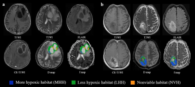 |
117 | Spatially Explicit Analysis of Tumor Hypoxia Heterogeneity from IVIM MRI Predicted Survival of Higher-Grade Glioma
Xiaoqing Wang1, Mengqiu Cao1, Yang Song2, Guang Yang3, and Yan Zhou1
1Renji Hospital, Shanghai Jiaotong University School of Medicine, Shanghai, China, 2MR Scientific Marketing, Siemens Healthineers Ltd. Shanghai, China, Shanghai, China, 3Shanghai Key Laboratory of Magnetic Resonance, East China Normal University, Shanghai, China Keywords: Tumors, Brain, Diffusion weighted MRI This study investigated the feasibility of spatially explicit analysis based on IVIM MRI to identify hypoxia-related habitats with higher-grade glioma, and to predict progress-free survival in patients. The results showed that spatial explicit analysis on IVIM MRI could be an appropriate method for distinguishing different hypoxic subregions in glioma. In addition, the volume percentage of less hypoxic habitats can effectively predict progress-free survival in higher-grade glioma. |
|
1582.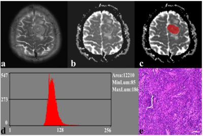 |
118 | Differentiation of intracranial solitary fbrous tumor from transitional meningioma using apparent difusion coefcient histogram analysis
Xianwang Liu1,2,3, Tao Han1,2,3, Hong Liu1,2,3, and Junlin Zhou1,2,3
1Department of Radiology, Lanzhou University Second Hospital, lanzhou, China, 2Gansu International Scientific and Technological Cooperation Base of Medical Imaging Artificial Intelligence, lanzhou, China, 3Key Laboratory of Medical Imaging of Gansu Province, lanzhou, China Keywords: Tumors, Quantitative Imaging, Solitary fibrous tumor, Transitional meningioma, Magnetic resonance imaging, Apparent diffusion coefficient, Histogram analysis This study investigates the value of whole volume apparent diffusion coefficient (ADC) histogram analysis in distinguishing intracranial solitary fibrous tumor (SFT) and transitional meningioma (TM). The method was to compare the differences between ADC histogram parameters in them and explore the relationships between these parameters and Ki-67 expression. Our research demonstrates that SFT with higher variance, lower AP1, and AP10. Significant correlations were observed between these parameters and Ki-67 expression. The best diagnostic performance was obtained by variance. Therefore, we consider that the whole volumetric ADC histogram analysis is a feasible tool for non-invasive distinguishing between SFT and TM. |
|
1583.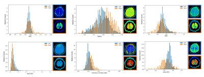 |
119 | Determination of Meningiomas Consistency by Histogram Analysis of Different Models of Diffusion-Weighted MR Imaging
Lingmin Zheng1, Zongmeng Wang1, Danjie Lin1, Yang Song2, Lin Lin1, and Yunjing Xue1
1Fujian Medical University Union Hospital, Fuzhou, China, 2MR Scientific Marketing, Siemens, Healthineers Ltd, Shanghai, China Keywords: Tumors, Diffusion/other diffusion imaging techniques The consistency of Intracranial tumors is crucial to determine the required surgical instruments as well as affecting the outcome of surgery, but no specific feature of conventional MRI is reliable in predicting the consistency of tumors. Histogram analysis of diffusion parameters has been successfully used in predicting the grade, subtype, and proliferative activity of meningiomas. This study prospectively evaluated and compared the potential of various diffusion metrics obtained from the mono-exponential model (MEM), bi-exponential model (BEM), and stretched exponential model (SEM)-based diffusion-weighted imaging (DWI) in predicting consistency of meningiomas. It was found that different models of DWI (MEM, BEM, and SEM) are useful in the differentiation between soft and hard meningiomas. However, Alpha obtained from SEM and fast-ADC from BEM are more promising diffusion parameters for predicting the consistency of meningiomas. |
|
1584.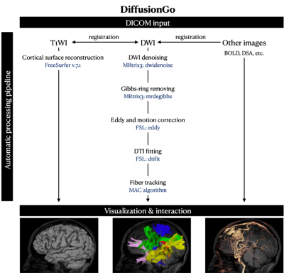 |
120 | Automatic bundle-specific white matter fiber tracking tool for language related glioma resection
Yifan Yuan1, Shin Tai Chong2, Sanford Pin-Chuan Hsu3, Ying-Hua Chu4, Yi-Cheng Hsu5, Yu-Ting Ko2, Kuan-Tsen Kuo2, Ching-Po Lin2,6, and Jianping Song1
1Department of neurosurgery, Huashan Hospital Fudan University, Shanghai, China, 2Institute of Neuroscience, National Yang Ming Chiao Tung University, Hsinchu, Taiwan, Hsinchu, Taiwan, 3Department of neurosurgery, Taipei Veterans General Hospital, Taipei, Taiwan, 4MR Collaboration, Siemens Healthineers Ltd, Shanghai, China, 5MR Collaboration, Siemens Healthineers Ltd, shanghai, China, 6Institute of Science and Technology for Brain-Inspired Intelligence, Fudan University, shanghai, China Keywords: Tumors, Diffusion Tensor Imaging Novel fiber tracking technology, based on diffusion imaging, can objectively reveal and visualize three-dimensional white matter tracts; through the cooperation of intraoperative navigation, it can help achieve maximum resection under the premise of ensuring function. We used an in-house developed software (DiffusionGo) specially designed for neurosurgeons. The fiber tracking result using DiffusionGo showed robust consistency with the surgical findings. We believe that this fully automatic processing pipeline provides the neurosurgeon with a solution that may reduce time costs and operating errors and improve care and surgical procedure quality across different neurosurgical centers. |
|
 Back to Meeting Home
 Back to the Program-at-a-Glance
The International Society for Magnetic Resonance in Medicine is accredited by the Accreditation Council for Continuing Medical Education to provide continuing medical education for physicians.
 Back to Meeting Home
 Back to the Program-at-a-Glance
 View Presentation Video