Digital Poster
Tumors: The Importance of Diffusion & Perfusion II
ISMRM & ISMRT Annual Meeting & Exhibition • 03-08 June 2023 • Toronto, ON, Canada

Digital Poster
Tumors: The Importance of Diffusion & Perfusion II
| Computer # | |||
|---|---|---|---|
1740.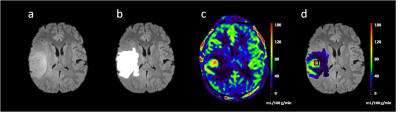 |
101 | 3D mTI-ASL perfusion for differentiating 1p/19q status in IDH1-mutant lower grade gliomas
Rui Wang1, Xianchang Zhang2, and Shaowu Li1
1Beijing Neurosurgical Institute/Beijing Tiantan Hospital, Capital Medical University, Beijing, China, 2MR Collaboration, Siemens Healthineers Ltd., Beijing, China Keywords: Tumors, Arterial spin labelling This study investigated the feasibility of using local maximum cerebral blood flow (maxCBF) in tumor area measured with multi-inversion-time ASL (mTI-ASL) to preoperatively predict the 1p/19q status in patients with IDH1-mutant lower-grade glioma (LGG). Mann-Whitney U test showed that maxCBF in the tumor region was significantly higher in 1p/19q Non-Codeleted group than Codeleted group. ROC showed maxCBF achieved high sensitivity (0.7) and specificity (0.737) in differentiation of IDH1-mutant-LGGs with distinct 1p/19q status. This finding suggested that ASL-derived maxCBF has the potential as a tool for the noninvasive preoperative characterization of 1p/19q status, thus providing evidence for individualized treatment planning. |
|
1741.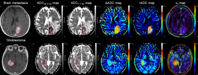 |
102 | Time-dependent diffusion-weighted imaging for differentiation between glioblastoma and brain metastasis
Kiyohisa Kamimura1, Tsubasa Nakano1, Tomohito Hasegawa1, Masanori Nakajo1, Hiroyuki Uchida2, Takashi Iwanaga3, Hiroshi Imai4, Thorsten Feiweier5, and Takashi Yoshiura1
1Radiology, Kagoshima University, Kagoshima, Japan, 2Neurosurgery, Kagoshima University, Kagoshima, Japan, 3Radiological Technology, Kagoshima University Hospital, Kagoshima, Japan, 4Siemens Healthcare K.K., Tokyo, Japan, 5Siemens Healthcare GmbH, Erlangen, Germany Keywords: Tumors, Diffusion/other diffusion imaging techniques To investigate the utility of time-dependent DWI for differentiating between glioblastoma and brain metastasis, 65 patients with glioblastoma and 27 patients with brain metastasis were examined. ADC was not significantly different between the two tumor types neither at a short (7.1ms) nor at a long (44.5ms) diffusion time, whereas the ADC difference (ΔADCmean, ΔADC5, ΔADC95) and ADC change ratio (rADCmean, rADC5, rADC95) were significantly higher in brain metastasis than in glioblastoma. The ΔADCmean showed the best diagnostic performance. The ΔADCmean and rADCmean showed a significant negative correlation with ADC44.5msmean, but not with extracellular extravascular space volume fraction derived from DCE-MRI. |
|
1742.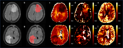 |
103 | NODDI maximum abnormal signal area histogram analysis for differentiation between glioblastoma Multiforme and solitary brain metastasis
Jinbo Qi1, Xiaoyue Ma1, Peipei Wang1, Eryuan Gao1, Guohua Zhao1, Kai Zhao1, Yang Song2, Huiting Zhang3, Ankang Gao1, Jie Bai1, Yong Zhang1, and Jingliang Cheng1
1Department of MRI, The First Affiliated Hospital of Zhengzhou University, Zhengzhou, China, 2Magnetic Resonance Scientific Marketing, Siemens Healthineers Ltd., Shanghai, China, 3Magnetic Resonance Scientific Marketing, Siemens Healthineers Ltd., Wuhan, China Keywords: Tumors, Diffusion/other diffusion imaging techniques, Neurite orientation dispersion and density imaging We aim to explore the value of neurite orientation dispersion and density imaging (NODDI) maximum abnormal signal area histogram analysis for differentiation between glioblastoma Multiforme (GBM) and solitary brain metastasis (SBM). 50 patients with GBM and 50 patients with SBM confirmed by surgical pathology were enrolled and underwent MR examination. Result showed that multiple histogram parameters can significantly distinguish GBM from SBM, and the performance of logistic regression model is better than that of optimal single parameter. |
|
1743.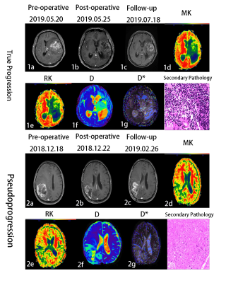 |
104 | Combination of DKI and IVIM for differentiating True Progression from Pseudoprogression of Glioma with Postoperative Radiation Therapy
Pei Dang1, Xueying Huang2, Zhihua Yang3, Aijun Wang2, Lidong Wang4, Yuhui Xiong5, Xuhong Yang6, Minglei Wang2, and Xiaodong Wang7
1Radiology, General Hospital of Ningxia Medical University, yinchuan, China, 2General Hospital of Ningxia Medical University, Yinchuan, China, 3Radiotherapy, General Hospital of Ningxia Medical University, Yinchuan, China, 4Radiology, Yinchuan Hospital of Traditional Chinese Medicine, Yinchuan, China, 5GE Healthcare MR Research, Beijing, China, 6Ningxia Medical University, Yinchuan, China, 7Radiology, General Hospital of Ningxia Medical University, Yinchuan, China Keywords: Tumors, Cancer, Glioma;True Progression;Pseudoprogression;DKI;IVIM The differentiation of pseudoprogression from true progression remains a crucial diagnostic dilemma in glioma with postoperative radiation therapy.This work to explore the role of DKI combined IVIM in differentiating true progression and pseudoprogression in glioma. It was concluded that the MK and RK from DKI、D and D* from IVIM can be used as novel imaging biomarkers for Clinical and radiologists in differentiating glioma true progression from pseudoprogression. Combining MK, RK, D and D* may explore as an effective strategy to improve the ability for discriminating glioma true progression and pseudoprogression. |
|
1744.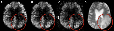 |
105 | Effects of peritumoral T2 hyperintensities in treated gliomas on the quantification of tumour blood flow with arterial spin labeling.
Magdalena Sokolska1, James K Ruffle2, Meetakshi Gupta3, Julia Markus4, Michael Kosmin3, Hans Rolf Jäger 2, Parashkev Nachev2, and Harpreet Harpreet4
1Medical Physics and Biomedical Engineering, University College London Hospitals, London, United Kingdom, 2Queen Square Institute of Neurology, University College London, London, United Kingdom, 3Radiotherapy, University College London Hospitals, London, United Kingdom, 4Imaging, University College London Hospitals, London, United Kingdom Keywords: Tumors, Arterial spin labelling Elevated tumour blood flow in treated gliomas t can be indicative of disease recurrence. TBF can be visualised and quantified with arterial spin labelling with quantification performed by scaling a perfusion-weighted image by a proton density image and a brain-blood partition coefficient, assuming mapping to a fully relaxed blood magnetisation signal. However, this assumption might not hold in T2/FLAIR hyperintense areas surrounding enhancing tumour due to much higher water content. Therefore, this work investigates blood flow quantification in three tumour compartments, enabled by recent developments in AI-generated labels of tumour components. |
|
1745.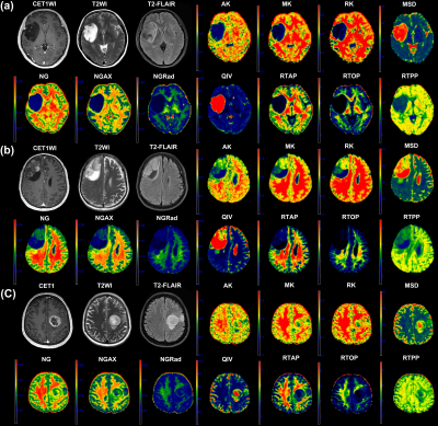 |
106 | Tumor habitat-derived diffusion spectrum imaging features in evaluating gliomas-A comparative study with DKI
Shanmei Zeng1, Zuliwei Ma1, Hui Ma2, Dingxiang Xie2, Yingqian Huang2, Mengzhu Wang3, Wenting Zeng1, Fangzeng Lin1, Zhiyun Yang1, Jianping Chu1, and Jing Zhao1
1Department of Radiology, The First Affiliated Hospital, Sun Yat-Sen University, Guangzhou, Guangdong, China, 2Department of Radiology, The First Affiliated Hospital, Sun Yat-Sen University, Guangzhou, China, 3MR Scientific Marketing, Siemens Healthineers Ltd., Guangzhou, Guangdong, China Keywords: Tumors, Tumor Currently, in diffuse glioma, in addition to histopathology, the molecular features have an important role in glioma diagnosis. As a new computing framework for diffusion spectrum imaging (DSI), mean apparent propagator (MAP) could investigate more subtle information of tumor microstructures than the Gaussian diffusion models, while MAP has not been studied much in glioma. Besides, the analysis of tumor habitat could reflect the glioma heterogeneity. Hence, we aim to analyze the tumor habitat-derived MAP metrics and further explore their clinical value in glioma evaluation, meanwhile, we introduce Diffusion-kurtosis imaging (DKI) as a reference to compare the diagnostic efficiency of DSI. |
|
1746. |
107 | Pretreatment Apparent Diffusion Coefficient Value for Prediction of Relapsed and Refractory Primary Central Nervous System Lymphoma
Ching-Chung Ko1,2, Lee-Ren Yeh3, and Jeon-Hor Chen4
1Chi Mei Medical Center, Tainan, Taiwan, 2Department of Health and Nutrition, Chia Nan University of Pharmacy and Science, Tainan, Taiwan, 3Department of Radiology, E-DA Hospital, I-Shou University, Kaohsiung, Taiwan, 4Department of Radiological Sciences, University of California, Irvine, CA, United States Keywords: Tumors, Cancer A subset of primary central nervous system lymphoma (PCNSL) may show early relapsed/refractory (R/R) disease after treatments. This study investigated the role of pretreatment apparent diffusion coefficient (ADC) values for the prediction of R/R in PCNSL. Total 52 patients with pathologically confirmed PCNSL were included, and 24 (46.2%) patients developed R/R (median time to relapse, 13 months) after treatment. The results showed female sex, complete response to first-line chemotherapy, and ADC value/ratio were significant predictors of R/R in PCNSL. Pretreatment ADC values/ratios offer objective and valuable evaluation for the treatment planning in PCNSL. |
|
1747.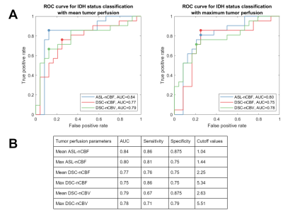 |
108 | Classification of IDH mutation with Arterial Spin Labeling and Dynamic Susceptibility Contrast MRI in adult gliomas
Yeva Prysiazhniuk1,2, Andres Server3, Øystein Bech-Aase3, Eirik Helseth4, David Kala1,5, Roelant Sjouke Eijgelaar6, Elies Fuster-García7, Petter Brandal8,9, Atle Bjørnerud3, Jakub Otáhal1,2, Jan Petr10,11, and Wibeke Nordhøy3
1Department of Pathophysiology, the Second Faculty of Medicine, Charles University, Prague, Czech Republic, 2Laboratory of Developmental Epileptology, Institute of Physiology, Czech Academy of Sciences, Prague, Czech Republic, 3Department of Physics and Computational Radiology, Division of Radiology and Nuclear Medicine, Oslo University Hospital, Oslo, Norway, 4Oslo University Hospital, Department of Neurosurgery, Oslo, Norway, 5Department of Circuit Theory, Faculty of Electrical Engineering, Czech Technical University, Prague, Czech Republic, 6Department of Neurosurgery, Amsterdam University Medical Center, Amsterdam, Netherlands, 7Instituto Universitario de Tecnologías de la Información y Comunicaciones, Universitat Politècnica de València, Valencia, Spain, 8Department of Oncology, Oslo University Hospital, Oslo, Norway, 9Institute for Cancer Genetics and Informatics, Oslo University Hospital, Oslo, Norway, 10Institute of Radiopharmaceutical Cancer Research, Helmholtz-Zentrum Dresden-Rossendorf, Dresden, Germany, 11Department of Radiology and Nuclear Medicine, Cancer Center Amsterdam, Amsterdam University Medical Center, Amsterdam, Netherlands Keywords: Tumors, Perfusion, Genetics IDH genotype status is an important marker in glioma diagnostics. Given that IDH mutation affects the tumor vascularization pattern, perfusion imaging has the potential to become a non-invasive tumor histopathology assessor. In this study, two methods of perfusion MRI – Dynamic Susceptibility Contrast (DSC) and Arterial Spin Labeling (ASL) – were compared in their ability to assess IDH mutation status. DSC- and ASL-derived perfusion maps correlated significantly and were feasible parameters in the IDH classification task. Mean tumor CBF quantified with ASL had the highest AUC score, sensitivity, and specificity, supporting the feasibility of using ASL in clinical glioma diagnostics. |
|
1748.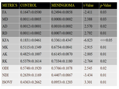 |
109 | Role of Diffusion Magnetic Resonance Imaging in Brain Meningioma
susmita shrestha1, Karuna Raya2, Nian Wang3, and Surendra Maharjan4
1Chitwan Medical College and Teaching Hospital, Chitwan, Nepal, 2Sharda University, Greater Noida, India, 3Indiana University School of medicine, Stark Neuroscience, IndianaPolis, IN, United States, 4Indiana University School of medicine, IndianaPolis, IN, United States Keywords: Tumors, Brain Meningioma as an extra-axial tumor of meninges is difficult to interpret within brain interface using normal Magnetic Resonance Imaging sequence protocol. Recently, Various advancement in diffusion Magnetic Resonance Imaging (dMRI) techniques such as DTI (Diffusion Tensor Imaging), DKI (Diffusion Kurtosis Imaging) and NODDI (Neurite Orientation Dispersion and Density Imaging) has been utilized as an emerging diagnostic tool to measure the degree of mobility of water molecules within biologic tissue and investigate micro-structural integrity of the brain tissue. Thus, we aim to differentiate meningiomas from normal brain parenchyma using DTI, DKI and NODDI metrics. |
|
1749.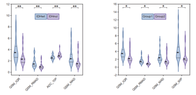 |
110 | Identification of IDH genotype and tumor subtype of adult-type diffuse gliomas based on WHO CNS5 using histogram features of QSM and ADC
Yifan Sun1, Zheting Yang1, Yang Song2, and Rifeng Jiang1
1Fujian Medical University Union Hospital, Fuzhou, China, 2MR Scientific Marketing, Siemens Healthineers Ltd., Shanghai, China Keywords: Tumors, Quantitative Susceptibility mapping, Glioma; Isocitrate dehydrogenase; WHO CNS5 Preoperative prediction of glioma histological features and biological behavior is clinically important. However, few studies reported the histogram analysis of QSM in the isocitrate dehydrogenase (IDH) genotype and subtype of gliomas. In this study, we explored the value of histogram features of QSM and ADC in predicting the IDH genotype and tumor subtype of adult-type diffuse gliomas based on the fifth edition of the World Health Organization Classification of Tumors of the Central Nervous System (WHO CNS5). We found that histogram parameters based on QSM and ADC are significantly related to the IDH genotype, tumor subtype, and proliferation of glioma. |
|
1750. |
111 | Healthy and cancerous brain ADC: diffusion- and echo time dependence with pulsed gradient spin-echo (PGSE) diffusion imaging
Jens Johansson1, Kerstin Lagerstrand2,3, Isabella M Björk-Burtscher1,4, Mats Laesser1,5, Hanna Hebelka1,4, and Stephan E Maier1,6
1Radiology, Clinical sciences,Sahlgrenska Academy, University of Gothenburg, Gothenburg, Sweden, 2Medical Radiation Sciences, Clinical sciences,Sahlgrenska Academy, University of Gothenburg, Gothenburg, Sweden, 3Medical Physics and Biomedical Engineering, Sahlgrenska University Hospital, Gothenburg, Sweden, 4Radiology, Sahlgrenska University Hospital, Gothenburg, Sweden, 5Sahlgrenska University Hospital, Gothenburg, Sweden, 6Radiology, Brigham and women's hospital, Boston, MA, United States Keywords: Tumors, Diffusion/other diffusion imaging techniques, Diffusion time Clinical systems vary widely in their gradient performance. Accordingly, effective diffusion time and echo time, which are minimized by the system software according to available gradient performance, can differ significantly among systems. A major concern is that non-Gaussian diffusion in tissues can lead to significant variation of the measured apparent diffusion coefficient (ADC), which would clearly diminish its value as biomarker. This study evaluated if diffusion and/or echo time variations in pulsed gradient spin-echo (PGSE) sequences significantly influences the ADC measured in healthy brain and brain tumors. |
|
1751. |
112 | Intravoxel Tissue Heterogeneity Revealed by Non-Gaussian Time-Dependent Diffusion MRI and Its Correlation with Histology
Guangyu Dan1,2, Weiguo Li2,3,4, Kaibao Sun1, Qingfei Luo1, Muge Karaman1,2, and Xiaohong Joe Zhou1,2,5
1Center for Magnetic Resonance Research, University of Illinois at Chicago, Chicago, IL, United States, 2Department of Biomedical Engineering, University of Illinois at Chicago, Chicago, IL, United States, 3Research Resource Center, University of Illinois at Chicago, Chicago, IL, United States, 4Department of Radiology, Northwestern University, Evanston, IL, United States, 5Departments of Radiology and Neurosurgery, University of Illinois College of Medicine, Chicago, IL, United States Keywords: Tumors, Tumor, Glioma Recently, diffusion-weighted MRI has emerged for probing intravoxel structural heterogeneity non-invasively as an alternative to gold standard histopathological analysis. A previous study demonstrated the correlation between the imaging- and histology-based tissue heterogeneities using a continuous-time random-walk (CTRW) model at a single diffusion time. In this study, we extended the study to multiple diffusion times and investigated the diffusion time dependence of the CTRW parameters and their correlation with histology-based heterogeneity on human glioma tissues. Significant time dependency was observed in the CTRW parameters, providing practical insights into the selection of diffusion time when assessing tissue heterogeneity. |
|
1752.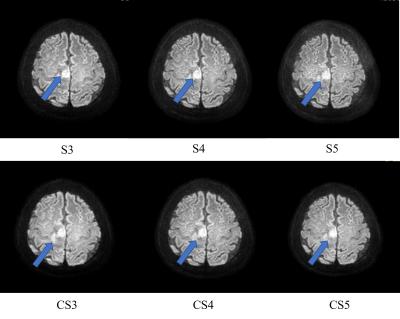 |
113 | The application of high-resolution isotropic EPI DWI by CS with different acceleration factor in brain tumor
Mengting Duan1, Yi Zhu2, Qi Wang1, and Hui Liu1
1The Fourth Hospital of Hebei Medical University, Hebei, China, 2Philips Healthcare, Beijing, China Keywords: Tumors, Cancer, EPI-DWI, Compressed SENSE It has been shown before that C-SENSE can improved the ss-EPI DWI image quality in higher spatial resolution. The aim of this paper is to achieve a high-resolution isotropic EPI DWI by CS and to investigate the impact of different acceleration factor(AF) on the diagnostic quality of brain metastases in comparison with SENSE EPI DWI. |
|
1753.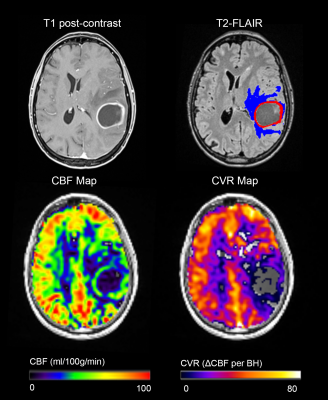 |
114 | Arterial Spin Labeling in Cerebral Gliomas During Breath-Holding
Sergio M. Solis-Barquero1,2, Marta Calvo-Imirizaldu1,2, Veronica Aramendia-Vidaurreta1,2, Rebeca Echeverria-Chasco1,2, Antonio Martinez-Simon3, Elena Cacho-Asenjo3, Marta Vidorreta4, Pablo D. Dominguez1,2, Reyes Garcia de Eulate1, and Maria A. Fernandez-Seara1,2
1Department of Radiology, Clinica Universidad de Navarra, Pamplona, Spain, 2IdiSNA, Instituto de Investigación Sanitaria de Navarra, Pamplona, Spain, 3Department of Anesthesia, Perioperative Medicine and Critical Care, Clinica Universidad de Navarra, Pamplona, Spain, 4Siemens Healthcare, Madrid, Spain Keywords: Tumors, Perfusion, Breath Holding PCASL was used to measure cerebral blood flow (CBF) in eighteen patients with high-grade gliomas (III and IV) during normal breathing followed by a breath-holding task (ten periods of 21s interleaved with periods of normal breathing) to assess baseline CBF and cerebrovascular reactivity (CVR). All patients completed the task successfully and CBF and CVR maps were generated. CBF ratio in contrast-enhanced tumor area was higher in grade IV than grade III gliomas, as expected. CVR in tumor areas was decreased compared to GM CVR values. More studies are needed to assess the CVR heterogeneity in tumoral tissue. |
|
1754.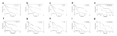 |
115 | Preoperative ASL correlates with molecular markers of IDH and HIF-1α in glioma microenvironment and is predictive of patient outcome
Ruiwen Geng1, Yang Dong1, and Lizhi Xie2
1Radiology, The Second Hospital, Dalian Medical University, Dalian, China, 2GE Healthcare, MR Research China, Beijing, China Keywords: Tumors, Arterial spin labelling This study retrospectively analyzed the association between arterial spin labeling (ASL) and the molecular markers of isocitrate-dehydrogenase (IDH) mutation and hypoxia-inducible factor (HIF) -1α in glioma. The association of patients’ overall survival (OS) and progression-free survival (PFS) with quantization cerebral blood flow (CBF) from ASL, the imaging parameters from conventional MRI, clinical and pathological parameters were investigated in glioma. |
|
1755.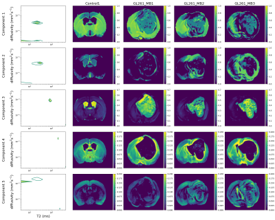 |
116 | Characterising glioma heterogeneity with diffusion-relaxation MRI and InSpect
Paddy J. Slator1, Rui V. Simoes2,3, Rafael N. Henriques2, Tânia Carvalho2, Daniel C. Alexander1, Noam Shemesh2, and Andrada Ianus2
1Centre for Medical Image Computing and Department of Computer Science, University College London, London, United Kingdom, 2Champalimaud Research, Champalimaud Foundation, Lisbon, Portugal, 3Institute for Research & Innovation in Health (i3S), University of Porto, Porto, Portugal Keywords: Tumors, Microstructure We characterise ex-vivo mouse brain tumours with combined T2-diffusion MRI and InSpect, a data-driven unsupervised learning technique that identifies distinct tissue components. We hence reveal heterogeneous tumour structures without imposing a biophysical model. |
|
1756.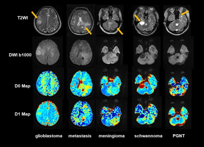 |
117 | MR Varying Diffusion Curvature Imaging on Different Brain Tumors: A Preliminary Experience
Chuanshuai Tian1, Wentao Hu2, Yongming Dai2, Sixuan Chen1, Jianan Zhou1, Xin Zhang1, and Bing Zhang1
1Department of Radiology, The Affiliated Drum Tower Hospital of Nanjing University Medical School, Nanjing, China, 2Central Research Institue, United Imaging Healthcare, Shanghai, China Keywords: Tumors, Diffusion/other diffusion imaging techniques Dozens of diffusion models have been created to describe the non-Gaussian nature of diffusion. Nevertheless, many of them imply assumptions that have not been rigorously confirmed, or includes abstract parameters. The purpose of this study was to observe the characteristics of varying diffusion curvature (VDC) indicators in several types of brain tumor. D0 and D1 distribution were obtained for each subject. This study illustrates the potential of applying a simple and pure empirical non-Gaussian diffusion model VDC on brain tumor imaging. |
|
1757.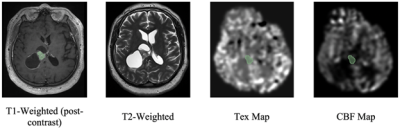 |
118 | Perfusion and Time of Exchange Measurements Using BBB-ASL in Gliomas: The Initial Experience
Gülce Turhan1, Ayşe İrem Çetin1, Amnah Mahroo2, Beatriz Padrela3, Simon Kanstandin4,5, Daniel Christopher Hoinkiss2, Nora Josefin Breutigam2, Klaus Eickel2,5, Jan Petr6, Henk Mutsaerts3, Ayca Ersen Danyeli7, Koray Ozduman8, Matthias Guenther2,5, Alp Dincer9,10, and Esin Ozturk-Isik1,10
1Institute of Biomedical Engineering, Bogazici University, Istanbul, Turkey, 2Fraunhofer Institute for Digital Medicine MEVIS, Bremen, Germany, 3Amsterdam University Medical Center, Amsterdam, Netherlands, 4Fraunhofer Institute for Digital Medicine MEVI, Bremen, Germany, 5mediri GmbH, Heidelberg, Germany, 6Helmholtz-Zentrum Dresden-Rossendorf, Dresden, Germany, 7Department of Pathology, Acibadem University, Istanbul, Turkey, 8Department of Neurosurgery, Acibadem University, Istanbul, Turkey, 9Department of Radiology, Acibadem University, Istanbul, Turkey, 10Brain Tumor Research Group, Acibadem University, Istanbul, Turkey Keywords: Tumors, Arterial spin labelling Subtle changes in BBB integrity might be missed by contrast-enhanced T1-weighted MRI. Blood-brain barrier arterial spin labeling (BBB-ASL) is a new technique to assess BBB disruptions. In this work, we measured the cerebral blood flow (CBF) and exchange time (Tex) values of the tumor, normal-appearing white matter, and normal-appearing gray matter regions in gliomas using BBB-ASL technique. Our results indicated higher CBF and leakier BBB in contrast-enhanced regions of gliomas than in the normal-appearing GM. |
|
1758. |
119 | Whole-tumor ADC Histogram Analysis in evaluating Tongue Squamous Cell Carcinoma heterogeneity and predicting Cervical Lymph Node Metastases
Xinying Li1, Ke Xue2, Xing Yang1,3, Yongming Dai2, Zhen Tian1, and Yingwei Wu1
1Shanghai Ninth People’s Hospital, affiliated to Shanghai Jiao Tong University, School of Medicine, Shanghai, China, 2MR Collaboration, Central Research Institute, Shanghai United Imaging Healthcare, Shanghai, China, 3Shanghai Pulmonary Hospital, Shanghai, China Keywords: Head & Neck/ENT, Diffusion/other diffusion imaging techniques Tumor heterogeneity occurred frequently in patients with SCC and associated with poor prognosis. Compared to clinic TNM stage or the histological grade, the histological heterogeneity has not been well addressed yet. Our results clearly demonstrated that higher incidence of CNM was observed in histological hetero-group than that in homo-group. whole-lesion ADC histogram metrics presented lower values in hetero-group and in CNM+ group. ADC75th and kurtosis were two independent prognostic factors for evaluating the CNM status. Whole-tumor ADC histogram offered an approach for detecting intra-tumoral heterogeneity and predicting cervical lymph node metastases status. |
|
1759. |
120 | Time-Dependent OGSE Diffusion MRI with MAGNUS Ultra-High-Performance Gradient Coil for Microstructure Imaging in Glioma Brain Tumor Patients
Robert Shih1, Ante Zhu2, Raymond Huang3, J Kevin DeMarco4, H Douglas Morris1, Maureen Hood1, Nastaren Abad2, Chitresh Bhushan2, Thomas Foo2, and Vincent Ho1
1Uniformed Services University, Bethesda, MD, United States, 2GE Research, Niskayuna, NY, United States, 3Brigham and Women's Hospital, Boston, MA, United States, 4Walter Reed National Military Medical Center, Bethesda, MD, United States Keywords: Tumors, Tissue Characterization The MAGNUS ultra-high-performance gradient coil delivers simultaneous 200 mT/m and 500 T/m/s performance on each axis, with higher peripheral nerve stimulation thresholds than whole-body gradient coils, which is particularly useful for diffusion MRI-based microstructure imaging. We used MAGNUS-enabled OGSE to assess shorter diffusion times and length scales <10 µm in four brain tumor patients with gliomas. We measured differences in time-dependent diffusivity between high-grade glioma and low-grade glioma, as well as between recurrent glioblastoma and treatment effects. The malignant lesions demonstrated greater time-dependence of diffusivity. Further investigation with additional subjects and histopathologic correlation may lead to future clinical applications. |
|
 Back to Meeting Home
 Back to the Program-at-a-Glance
The International Society for Magnetic Resonance in Medicine is accredited by the Accreditation Council for Continuing Medical Education to provide continuing medical education for physicians.
 Back to Meeting Home
 Back to the Program-at-a-Glance
 View Presentation Video