Digital Poster
Connectivity, Multiple Sclerosis & White Matter Degeneration
ISMRM & ISMRT Annual Meeting & Exhibition • 03-08 June 2023 • Toronto, ON, Canada

Digital Poster
Connectivity, Multiple Sclerosis & White Matter Degeneration
| Computer # | |||
|---|---|---|---|
3371.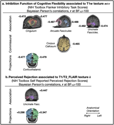 |
141 |
White matter macrostructure simultaneously predicts perceived
rejection and cognitive inflexibility in late life
Tatiana Wolfe1,
Carlos Ernesto Garrido Salmon2,
G. Andrew James1,
Laura B. Dunn1,
and Clint Kilts1
1Psychiatry, University of Arkansas for Medical Sciences, Little Rock, AR, United States, 2Physics, University of Sao Paulo, Ribeirao Preto, Brazil Keywords: White Matter, Neuroscience, Neurodegeneration, Cognitive Flexibility, Late Life, Psychiatric Disorders' Risk Factors. The interplay between brain white matter health and cognitive flexibility in late life is intimate. Understanding the age-related patterns of white matter disintegration along the cingulum network will aid in elucidating factors underlying individual susceptibility to psychiatric illness related cognitive flexibility impairment. We evaluated Bayesian Pearson correlations between a measurement of T1/T2-FLAIR kurtosis and psychiatric risk factors available in a UK Biobank sample. Our findings strongly suggest (BF10>100) that MRI apparent integrity loss in the cingulum, uncinate fascicles and corticothalamic fibers is a significant corollary of an older-age exclusive effect of increased perceived rejection that co-occurs with cognitive flexibility decline. |
|
3372. |
142 |
Associations of white matter connections with neurocognitive
development in healthy children
Rajikha Raja1,
Ruitian Song1,
John Glass1,
and Wilburn E Reddick1
1Diagnostic Imaging, St. Jude Children's Research Hospital, Memphis, TN, United States Keywords: Brain Connectivity, Normal development, Connectome We investigated the associations between structural connectivity and neurocognitive development in healthy children using MRI and behavioral data from 42 subjects belonging to Lifespan Human Connectome Project Development dataset. Diffusion MRI data were analysed using multi-shell multi-tissue constrained spherical deconvolution model to compute fiber orientation distribution functions before probabilistic fiber tracking was performed to obtain whole brain tractograms. Connectivity matrices were computed based on streamline density using nodes from HCP-MMP1 parcellation. Multiple significant correlations between connectivity and cognitive scores measuring working memory, processing speed and executive functioning were identified in this study. |
|
3373. |
143 |
Towards an enhanced characterization of white matter pathologies
with Q-FiberMapper 2.0
Tommy Boshkovski1,
Óscar Peña-Nogales1,
Paulo Rodrigues1,
Vesna Prčkovska1,
and Kire Trivodaliev1
1QMENTA Inc., Boston, MA, United States Keywords: Brain Connectivity, Software Tools FiberMapper 2.0 facilitates automatic (pre-) processing of MRI data, reconstructs major white matter tracts and derives imaging biomarkers along the tracts sensitive to a wide range of microstructural properties. Additionally, Q-FiberMapper 2.0 implements tract lesion burden analysis which together with the quantitative MR imaging biomarkers could be very useful for early diagnosis, evaluation and monitoring of different neurodegenerative and neuroinflammatory diseases that affect the white matter such as traumatic brain injury, multiple sclerosis, and others. |
|
3374.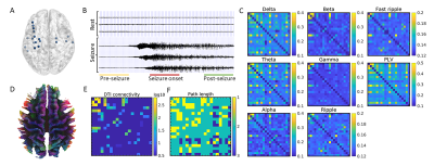 |
144 |
White Matter Structural Connectivity is Associated with Spectral
Coherence and Phase Synchronization Measured by Intracranial EEG
Bingyang Cai1,
Shize Jiang2,
Hui Huang1,
Jiwei Li1,
Siyu Yuan1,
Ya Cui1,
Lihong Tang1,
Liang Chen2,
and Jie Luo1
1School of Biomedical Engineering, Shanghai Jiao Tong University, Shanghai, China, 2Department of Neurosurgery, Huashan Hospital, Fudan University, Shanghai, China Keywords: Brain Connectivity, Diffusion Tensor Imaging Using high quality atlas of the structural connectome, and direct recording of neuronal activity by stereotactic-EEG (SEEG), this study investigated the relationship between neuronal signal connectivity at different seizure stage and the white matter scaffold. The results show electrical signal spectral coherence and phase synchronization are significantly stronger between structurally connected nodes within short distance, while at the time of pre-seizure and seizure onset, high frequency (>80Hz) signal may propagate through longer distance with structural connections. |
|
3375.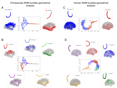 |
145 |
Morphological comparison of superficial white matter bundles
between the human and chimpanzee brain
Maëlig Chauvel1,
Marco Pascucci1,
Ivy Uszynski1,
Bastien Herlin1,
Jean-François Mangin1,
William Donald Hopkins2,
and Cyril Poupon1
1Université Paris-Saclay, CEA, CNRS, UMR 9027, BAOBAB, NeuroSpin, , Gif-sur-Yvette, France, Saclay, France, 2Michele E Keeling Center for Comparative Medicine and Research, The University of Texas MD Anderson Cancer Center, Bastrop, Texas, Texas, TX, United States Keywords: Brain Connectivity, Neuroscience, chimpanzee, white matter atlas The role of the superficial brain connections remains unclear. They appear to be important in the structural organization of the connectivity between the different brain gyri and are usually described as 'U-fibers' referring to their shape. Using an isomap algorithm, we explored in finer detail the superficial connectivity of the chimpanzee and human brains, still poorly known. This method allowed us to better capture the variability of the different shapes of the bundles that we observed and to overcome many bias due to subjective preconceptions. |
|
3376.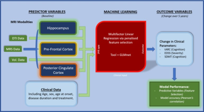 |
146 |
Multiparametric MRI neuroimaging signatures predict cognitive
decline in Multiple Sclerosis: a 5-year longitudinal study
Oun Al-iedani1,2,
Stasson Lea2,
Abdulaziz Alshehri2,3,4,
Vicki E. Maltby2,5,6,
Rodney Lea2,7,
Saadallah Ramadan2,3,
and Jeannette Lechner-Scott2,5,6
1School of Biomedical Sciences and Pharmacy, College of Health, Medicine and Wellbeing, University of Newcastle, Callaghan, Australia, 2Hunter Medical Research Institute, New Lambton Heights, Australia, 3School of Health Sciences, College of Health, Medicine and Wellbeing, University of Newcastle, Callaghan, Australia, 4Department of Radiology, King Fahad University Hospital, Dammam, Saudi Arabia, 5Department of Neurology, John Hunter Hospital, New Lambton Heights, Australia, 6School of Medicine and Public Health, College of Health, Medicine and Wellbeing, University of Newcastle, Callaghan, Australia, 7School of Biomedical Sciences, Queensland University of Technology, Brisbane, Australia Keywords: Multiple Sclerosis, Radiomics This novel longitudinal study evaluates multiparametric MRI signature for predicting cognitive decline in multiple sclerosis (MS) cohort followed for 5-years using a penalised regression machine learning approach (GLMnet). 43 MS participants were assessed at baseline and 5-years follow-up. Baseline (input) data consisted of 76 multiparametric MRI measures for different brain regions and tissues. The best performing model was for a change in tARCS (15 features; r=0.7±0.07), which was substantially higher than that for SDMT (r=0.496±0.08). These findings highlight the importance of using measures from multiple MR modalities analysed in combination with machine learning techniques when assessing cognitive decline. |
|
3377.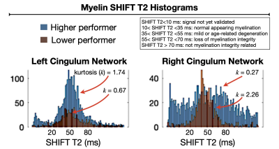 |
147 |
Cingulum network myelination structure by SHIFT MRI
characterizes higher order cognitive function in multiple
sclerosis
Tatiana Wolfe1,
Ashley Pike1,
Sienna Colonese2,
Carlos Ernesto Garrido Salmon3,
Laura B. Dunn1,
R. Lee Archer4,
Clint D. Kilts1,
and G. Andrew James1
1Psychiatry, University of Arkansas for Medical Sciences, Little Rock, AR, United States, 2Neurosciences, Colorado State University, Denver, CO, United States, 3Physics, University of Sao Paulo, Ribeirao Preto, Brazil, 4Neurology, University of Arkansas for Medical Sciences, Little Rock, AR, United States Keywords: Multiple Sclerosis, Brain Connectivity, Myelination integrity, cognitive impairment Impairment of higher-order cognition is a difficult aspect of MS pathology that poses a long-standing challenge for patient care advancement. Understanding how myelination integrity in the cingulum network – a brain pathway implicated in cognition – is related to lost or preserved cognitive functions is fundamental to enable comprehensive MS care and personalized remyelination therapeutics. We investigated if myelination integrity data has the potential to aid in characterizing individual differences in information processing in MS patients. Our findings support that myelination structure-function measured by SHIFT MRI is elucidatory variable to describe individual differences in higher-order cognitive function in MS patients. |
|
3378.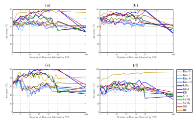 |
148 |
Identification of Subcortical White Matter Biomarkers in
Multiple Sclerosis Patients using Machine Learning
Cristian Montalba1,2,3,
Raul Caulier-Cisterna1,3,
Pamela Franco1,3,
Tomás Labbé2,
Marcelo E Andia1,2,3,
Miguel Guevara4,
Jean-François Mangin5,
Juan Pablo Cruz2,
Ethel Ciampi6,7,
Claudia Cárcamo7,8,
Pamela Guevara4,
Rodrigo Salas3,9,10,
and Sergio Uribe1,2,3
1Biomedical Imaging Center, Pontificia Universidad Catolica de Chile, Santiago, Chile, 2Radiology Department, School of Medicine, Pontificia Universidad Catolica de Chile, Santiago, Chile, 3Millennium Institute for Intelligent Healthcare Engineering - iHEALTH, Pontificia Universidad Catolica de Chile, Santiago, Chile, 4Faculty of Engineering, Universidad de Concepción, Concepción, Chile, 5UNATI, Neurospin, CEA, Université Paris-Saclay, Gif-sur-Yvette, France, 6Neurology Service, Hospital Dr. Sótero del Río, Santiago, Chile, 7Neurology Department, School of Medicine, Pontificia Universidad Catolica de Chile, Santiago, Chile, 8Interdisciplinary Center of Neurosciences, Pontificia Universidad Catolica de Chile, Santiago, Chile, 9Faculty of Engineering, Universidad de Valparaíso, Valparaíso, Chile, 10Biomedical Engineering School, Universidad de Valparaíso, Valparaíso, Chile Keywords: Multiple Sclerosis, Machine Learning/Artificial Intelligence Radiological biomarkers of cognitive impairment in Multiple Sclerosis (MS) are still scarce. This study aimed to identify subcortical white matter biomarkers of cognitive impairment related to verbal episodic memory in MS patients and healthy controls using a Machine Learning approach. |
|
3379.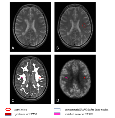 |
149 |
Hyperperfusion precedes demyelination in Multiple Sclerosis
lesion formation : a multimodal MRI and [11C]-PiB PET study
Théodore Soulier1,
Emanuele Morena2,
Arya Yazdan Panah3,
Matteo Tonietto4,
Mariem Hamzaoui5,
Vito Ricigliano6,
Giacomo Boffa5,
Michel Bottlaender7,
Benedetta Bodini8,
and Bruno Stankoff8
1Institut du Cerveau (ICM) = Paris Brain Institute - Sorbonne Université, Paris, France, 2Sapienza University of Rome, Rome, Italy, 3Paris Brain Institute - Sorbonne Université - INRIA, Paris, France, 4Centre Hospitalier Frédéric Joliot - CEA, Orsay, France, 5Paris Brain Institute - Sorbonne Université, Paris, France, 6Paris Brain Institute - Sorbonne Université - Hôpital Pitié Salpêtrière APHP, Paris, France, 7Centre Hospitalier Frédéric Joliot - CEA - Université Paris Saclay, Orsay, France, 8Paris Brain Institute - Sorbonne Université - CNRS - INSERM - Hôpital Saint Antoine APHP, Paris, France Keywords: Multiple Sclerosis, PET/MR, Prelesions, lesion formation Studying prelesions, the healthy areas preceding lesion appearance, helps to understand the Multiple Sclerosis (MS) lesion formation process. We study here prelesions in a longitudinal cohort of 19 MS patients combining MRI with Positron Emission Tomography (PET). MRI allowed to identify prelesions, whereas [11C]-PiB PET allowed to quantify myelin content as well as perfusion. We found that the 69 analyzable prelesions of our cohort were heterogeneous regarding perfusion: 24.6% were hyperperfused and 18.8% hypoperfused. Hypoperfused prelesions showed multimodal evidence of demyelination whereas hyperperfused prelesions were not demyelinated yet, suggesting that hyperperfusion precedes demyelination in the MS lesion formation process. |
|
3380.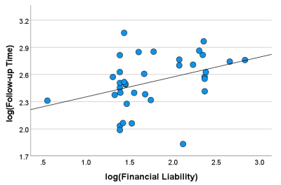 |
150 |
Influence of Financial and Socioeconomic Factors on MR Imaging
Follow-up Time in Patients with Multiple Sclerosis
Rishab Agarwal1,
Gelareh Sadigh2,3,
Jason W. Allen3,
David Sandlin4,
and Candace C. Fleischer1,3
1Department of Biomedical Engineering, Georgia Institute of Technology and Emory University, Atlanta, GA, United States, 2Department of Radiological Sciences, University of California, Irvine, Irvine, CA, United States, 3Department of Radiology and Imaging Sciences, Emory University School of Medicine, Atlanta, GA, United States, 4Department of Neurology, Emory University School of Medicine, Atlanta, GA, United States Keywords: Multiple Sclerosis, Health Care Economics, Financial Toxicity, Imaging Follow-up Time Multiple sclerosis (MS) is a chronic condition relying heavily on repeated MR imaging, and patients are particularly vulnerable to “financial toxicity”, a component of healthcare economics defined as the effects of healthcare costs on patient well-being. Our goal was to explore the effects of financial and socioeconomic factors on MRI follow-up time in patients with MS. Financial liability, or patient cost after primary insurance coverage, was associated with longer MRI follow-up time, suggesting financial toxicity may contribute to imaging frequency in MS patients. Further investigation over a longer time period is warranted. |
|
3381.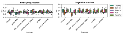 |
151 |
Machine learning based prediction of clinical progression in
multiple sclerosis
Samantha Noteboom1,
Moritz Seiler2,
Claudia Chien2,
Roshan P. Rane2,
Eva M. M. Strijbis3,
Friedemann Paul2,
Menno M. Schoonheim1,
and Kerstin Ritter2
1MS Center Amsterdam, Anatomy and Neurosciences, Vrije Universiteit Amsterdam, Amsterdam Neuroscience, Amsterdam UMC location VUmc, Amsterdam, Netherlands, 2Department of Psychiatry and Neurosciences, Charité – Universitätsmedizin Berlin, corporate member of Freie Universität Berlin and Humboldt-Universität zu Berlin, Berlin, Germany, 3MS Center Amsterdam, Neurology, Vrije Universiteit Amsterdam, Amsterdam Neuroscience, Amsterdam UMC location VUmc, Amsterdam, Netherlands Keywords: Multiple Sclerosis, Brain Machine learning may aid in individualized prediction of disease progression in multiple sclerosis (MS). In this study, we used data from 354 patients with MS to evaluate the capability of different machine learning approaches at predicting future disease worsening. Multiple clinical end points of disease worsening were tested and different combinations of clinical and structural MRI measures were used as inputs. Machine learning models were capable of discriminating between patients with low and high disability but did not perform well in predicting future disease course. |
|
3382.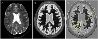 |
152 |
Clinical implementation of Multiplied, Added, Subtracted and/or
Divided (MASDIR) sequences on white matter lesions in Multiple
Sclerosis
Letizia Losa1,
Denis Peruzzo1,
Graeme Bydder2,
Andrea Salmaggi3,
and Nivedita Agarwal4
1CESNE, IRCCS E. Medea, Bosisio Parini, Italy, 2Department of Radiology, University of California, San Diego, San Diego, CA, United States, 3Neurology, Manzoni Hospital, Lecco, Lecco, Italy, 4Neuroradiology, IRCCS E. Medea, Bosisio Parini, Italy Keywords: Multiple Sclerosis, Quantitative Imaging, T1 mapping Multiple Sclerosis (MS) is a demyelinating progressive chronic disease. MR imaging is a key diagnostic tool that allows long-term monitoring of disease progression. Since T1 values can identify clinically relevant tissue properties such as increased water content, myelin loss and changes in microstructure, we assessed the whole brain using Multiplied, Added, Subtracted and/or Divided Inversion Recovery (MASDIR) in patients with MS in an attempt to improve contrast and quantify T1 of lesions. We show that MASDIR is a clinically reliable, easy to implement technique that greatly increases T1 contrast and simultaneously provides quantitative T1 maps. |
|
3383.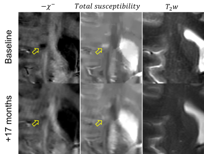 |
153 |
Mapping of Time-Dependent Myelin Changes in Multiple Sclerosis
Lesions through Quantitative Separation of Magnetic
Susceptibility Sources
Alexey Dimov1,
Thanh Nguyen1,
Shun Zhang2,
Yi Wang1,
and Susan Gauthier3
1Radiology, Weill Cornell Medicine, New York, NY, United States, 2Tongji Hospital, Wuhan, China, 3Neurology, Weill Cornell Medicine, New York, NY, United States Keywords: Multiple Sclerosis, Susceptibility, Quantitative Susceptibility Mapping, susceptibility source separation De- and remyelination are important treatment targets for multiple sclerosis therapies. In this study, we utilize susceptibility source separation to study differences in longitudinal trajectories of two important lesion types, paramagnetic rim lesions (rim+) and non-rim (rim-) lesions. We report significant difference in myelin content: while rim- lesions tend to demonstrate slow remyelination, rim+ tend to remain stable or demyelinate over time. |
|
3384.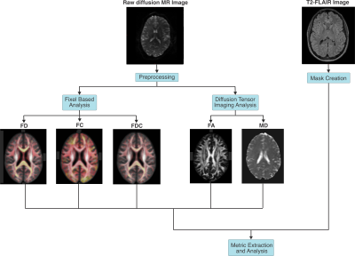 |
154 |
Fixel Based Analysis detection of peri-lesion and NAWM
abnormalities in Relapsing Remitting Multiple Sclerosis
Nikolai I Lesack1,
Sarah Levy2,
James Sumowski2,
Stephanie S. G. Brown3,
Sam Horng2,
and Rebecca Emily Feldman1
1Medical Physics, University of British Columbia, Kelowna, BC, Canada, 2Icahn School of Medicine at Mount Sinai, New York, NY, United States, 3University of Cambridge, Cambridge, United Kingdom Keywords: Multiple Sclerosis, Diffusion/other diffusion imaging techniques, fixed based analysis, DTI, brain, lesions, peri-lesion, normal appearing white matter Diffusion Tensor Imaging and Fixel Based Analysis (FBA) techniques were used to analyze microstructural properties of multiple sclerosis (MS) lesions and normal appearing white matter (NAWM) in relapsing-remitting multiple sclerosis (RRMS) subjects. FBA metric values were similar in peri-lesion white matter and MS lesions compared with bulk NAWM which may indicate FBA metric sensitivity to the infiltration of RRMS lesion pathology into surrounding NAWM. FBA fibre density in bulk NAWM was significantly different between subjects treated with Copaxone vs. Tysabri, which may indicate FBA metric sensitivity to NAWM differences between disease modifying therapies. |
|
3385.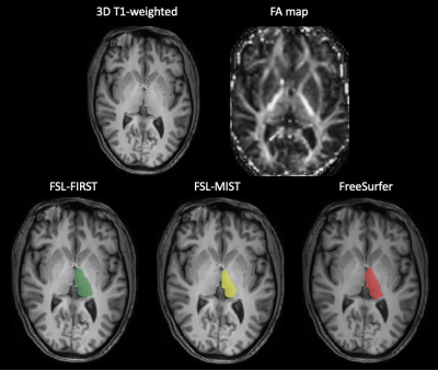 |
155 |
Quantification of Thalamic Volume in Multiple Sclerosis: From
the Multicenter INNI Dataset Towards the Clinical Application
Loredana Storelli1,
Elisabetta Pagani1,
Patrizia Pantano2,3,
Gioacchino Tedeschi4,
Nicola De Stefano5,
Maria Assunta Rocca1,6,7,
and Massimo Filippi1,6,7,8,9
1Neuroimaging Research Unit, Division of Neuroscience, IRCCS San Raffaele Scientific Institute, Milan, Italy, 2Department of Human Neurosciences, Sapienza University of Rome, Rome, Italy, 3IRCCS NEUROMED, Pozzilli, Italy, 4Department of Advanced Medical and Surgical Sciences, and 3T MRI-Center, University of Campania “Luigi Vanvitelli”, Naples, Italy, 5Department of Medicine, Surgery and Neuroscience, University of Siena, Siena, Italy, 6Neurology Unit, IRCCS San Raffaele Scientific Institute, Milan, Italy, 7Vita-Salute San Raffaele University, Milan, Italy, 8Neurorehabilitation Unit, IRCCS San Raffaele Scientific Institute, Milan, Italy, 9Neurophysiology Service, IRCCS San Raffaele Scientific Institute, Milan, Italy Keywords: Multiple Sclerosis, Neuroinflammation Thalamic atrophy has been found since the earliest phases of multiple sclerosis (MS). However, this measure is not included in clinical practice, due to the time-consuming manual segmentation and technical challenges. By comparing thalamic segmentations from three available automatic methods in a multicenter dataset, we found that the inclusion of fractional anisotropy maps facilitated the automatic identification of thalamic boundaries increasing robustness of the results. In particular, the multimodal approach (FSL-MIST) showed a better capability to detect small longitudinal variations of thalamic volumes in MS patients and a better correlation with another relevant MRI measure such as the lesion volume. |
|
3386.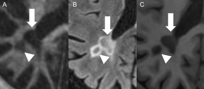 |
156 |
Signal variability of multiple sclerosis (MS) lesions on 3D
IR-UTE MRI correlates with patient’s disability
Sam Sedaghat1,
Hyungseok Jang1,
Jiyo Athertya1,
Yajun Ma1,
Jody Corey-Bloom1,
and Jiang Du1
1University of California San Diego, San Diego, CA, United States Keywords: Multiple Sclerosis, Neurodegeneration, UTE, MPRAGE, FLAIR, EDSS We found a significant correlation between the signal variability of MS lesions on IR-UTE / MPRAGE sequences and the EDSS, while the IR-UTE sequence is superior. By using direct myelin imaging, the grade of disability in MS patients could be estimated more accurately. The signal variability of MS lesions on IR-UTE could be used as a novel imaging biomarker for patient’s disability. |
|
3387.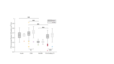 |
157 |
Improved Characterization of White Matter in Multiple Sclerosis
with Free Water-Corrected Diffusion MRI
Samantha By1,
Jean-Christophe Houde2,
Chahin Pachai1,
Loika Maltais2,
Jorge A. Torres3,
Gabrielle Grenier2,
Sujuan Huang4,
and Daniel P. Bradley3
1Bristol Myers Squibb, Lawrenceville, NJ, United States, 2Imeka Solutions, Inc., Sherbrooke, QC, Canada, 3Biogen, Cambridge, MA, United States, 4Cytel, Waltham, MA, United States Keywords: Multiple Sclerosis, Diffusion/other diffusion imaging techniques Free water (FW)-corrected metrics in diffusion MRI have shown promise in elucidating confounding pathological processes in multiple sclerosis (MS). Here, we present a retrospective analysis of FW-corrected diffusion in a cohort of 287 MS participants from a multi-center clinical trial. Our preliminary results indicate the method’s ability to distinguish different lesion types and MS subtypes. |
|
3388.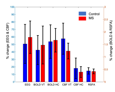 |
158 |
Investigation of cerebrovascular reactivity of multiple
sclerosis patients using multi-modal EEG-fMRI study in
hypercapnia condition
Wanyong Shin1,
Balu Krishnan2,
Ajay Nemani1,
Daniel Ontaneda3,
and Mark J Lowe1
1Radiology, Cleveland Clinic, Cleveland, OH, United States, 2Eplepsy, Cleveland Clinic, Cleveland, OH, United States, 3Mellen Center for Multiple Sclerosis, Cleveland Clinic, Cleveland, OH, United States Keywords: Multiple Sclerosis, Multimodal, EEG We investigated the change of EEG power, BOLD and CBF contrast during visual stimulation in multiple sclerosis patients and healthy controls. The result shows that cerebrovascular reactivity is disrupted in MS patients compared to controls. |
|
3389.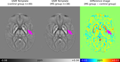 |
159 |
Automated QSM Processing in Multiple Sclerosis
Ashley Stewart1,
Po-Jui Liu2,3,
Matthias Weigel2,3,
Steffen Bollmann1,
and Cristina Granziera2,3
1School for Information Technology and Electrical Engineering, The University of Queensland, Brisbane, Australia, 2Translational Imaging in Neurology (ThINk) Basel, Department of Biomedical Engineering, Faculty of Medicing, University Hospital of Basel and University of Basel, Basel, Switzerland, Basel, Switzerland, 3Neurological Clinic and Policlinic, MS Center and Research Center for Clinical Neuroimmunology and Neuroscience Basel (RC2NB), University Hospital Basel and University of Basel, Basel, Switzerland, Basel, Switzerland Keywords: Multiple Sclerosis, Susceptibility In this work, an automated and scalable processing tool for Quantitative Susceptibility Mapping (QSM), QSMxT, is applied to a large dataset of 3D echo-planar imaging (3D-EPI) acquisitions of Multiple Sclerosis (MS) patients and volunteer controls. This is used to reconstruct QSMs for all subjects and generate a group space for the study. MS lesions are depicted and compared across multiple contrasts, including FLAIR, MP2RAGE, 3D-EPI magnitude and QSM. The group space is used to generate average QSM images for the MS and control groups along with a difference image to investigate iron accumulation and demyelination patterns. |
|
3390.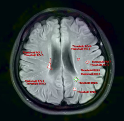 |
160 |
Altered amide proton transfer weighted signal and diffusion in
multiple sclerosis: correlation with neuroflament light and
disease duration
Jing Huang1,
Zhiwei Shen2,
and Jie Lu1
1Xuanwu Hospital, Capital Medical University, Beijing, China, 2Philips Healthcare, Beijing, China Keywords: Multiple Sclerosis, CEST & MT APTw imaging can help us understand the pathological changes in MS more sensitively and accurately. |
|
 Back to Meeting Home
 Back to the Program-at-a-Glance
The International Society for Magnetic Resonance in Medicine is accredited by the Accreditation Council for Continuing Medical Education to provide continuing medical education for physicians.
 Back to Meeting Home
 Back to the Program-at-a-Glance
 View Presentation Video