Digital Poster
Exploring Tumors Based on Digits: Omics, Machine Learning & Quantification I
ISMRM & ISMRT Annual Meeting & Exhibition • 03-08 June 2023 • Toronto, ON, Canada

Digital Poster
Exploring Tumors Based on Digits: Omics, Machine Learning & Quantification I
| Computer # | |||
|---|---|---|---|
1958.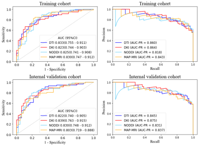 |
141 | Predicting Adult-type Diffuse Gliomas Grade: A Comparison of Four Radiomics-based Diffusion Prediction Models
Peng Wang1,2, Jinlong He1, Qiong Wu1, Shenghui Xie1, Lixin Weng2, Shaoyu Wang3, Huapeng Zhang3, Yang Song3, and Yang Gao1
1Affiliated Hospital of Inner Mongolia Medical University, Hohhot, China, 2Inner Mongolia Medical University, Hohhot, China, 3Siemens Healthineers, Shanghai, China Keywords: Tumors, Diffusion/other diffusion imaging techniques Application of MRI diffusion models can aid in the formulation of logical treatment strategies by non-invasively predicting gliomas pathogenic grade. However, it appears that the thorough comparison of diffusion models has not been addressed or has only been partially addressed. The objective of this study was to compare the potential clinical uses of four radiomics-based diffusion prediction models for predicting adult-type diffuse gliomas grade. According to the findings, the relatively simple model (i.e. diffusion tensor imaging) had the same potential for clinical use than the more sophisticated diffusion models. This suggests that additional point-to-point research is required. |
|
1959.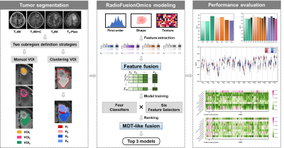 |
142 | A Subregion-based RadioFusionOmics Model Discriminates between Grade 4 Astrocytoma and Glioblastoma on Multisequence MRI
Ruili Wei1, Xinrui Pang1, Ye Wang1, Fangrong Liang1, Yongzhou Xu2, and Ruimeng Yang1
1Department of Radiology, Guangzhou First People's Hospital, Guangzhou, China, 2Philips Healthcare, Guangzhou, China Keywords: Tumors, Radiomics
The 2021 update of WHO CNS5 underlines the importance of IDH genotype prediction in the setting of adult-type grade 4 glioma. We developed a RFO model to discriminate between grade 4 astrocytoma and glioblastoma using subregional radiomics signatures from conventional MRI sequences. The fusion models from multiparametric MR images outperformed that from single sequence. The comparison between two different subregion manners revealed that voxel-wise habitats defined by clustering procedure yielded a higher discriminative capability. Our results also implied that tumor edema may contain underlying heterogeneous metrics between grade 4 astrocytoma and GBM.
|
|
1960.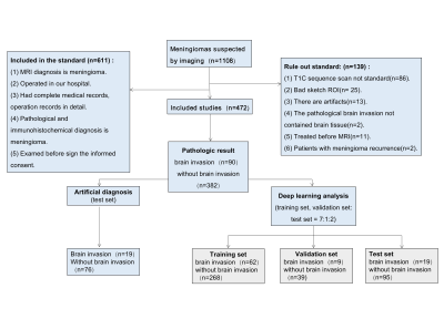 |
143 | Prediction of brain invasion in meningioma by preoperative MRI based on U-NET and DenseNet convolutional neural network
Juan Yu1, Yujian Liu2, kan Deng3, Fan Lin4, Guanghui Yue2, Jie Du2, and Liangping Luo5
1Radiology Department, Shenzhen Second People's Hospital, Shenzhen, China, 2Shenzhen University, Shenzhen, China, 3Philips Healthcare, Guangzhou, China, 4Shenzhen Second People's Hospital, Shenzhen, China, 5The First Affiliated Hospital of Jinan University, Guangzhou, China Keywords: Tumors, Tumor, meningioma The new guidelines suggest that brain invasion is not a unique feature of malignant meningiomas, but may be a pathologic condition of benign meningiomas with a potential risk of recurrence. However, maximum preservation of normal brain tissue is essential for surgery. Accurate preoperative MRI evaluation of meningioma brain invasion is expected to solve the dilemma faced by neurosurgeons. |
|
1961.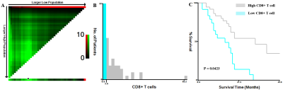 |
144 | MRI histogram analysis of tumor-infiltrating CD8+ T cell levels in patients with glioblastoma
Caiqiang Xue1, Qing Zhou1, and Junlin Zhou1
1Lanzhou University Second Hospital, Lanzhou, China Keywords: Tumors, Tumor CD8+ T cell infiltration in tumors is a powerful predictor of the clinical and postoperative prognosis of GBM patients. Immunohistochemical staining was used to assess tumor-infiltrating CD8+ T cell expression in patient-derived tumor tissue samples. Histogram analysis of GBM was performed using Firevoxel software. Among the T1C histogram features, the CV, mean, 5th, 10th, 25th, and 50th percentiles were correlated with the levels of CD8+ T cells. The ROC curve analysis revealed that the CV had the highest AUC value (0.783). Histogram analysis is an efficient non-invasive imaging modality for the prediction of tumor-infiltrating CD8+ T cells in glioblastoma. |
|
1962.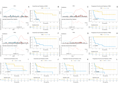 |
145 | Radiomics Based on Diffusion and Perfusion MRI Image: New Prognostic Biomarkers in PCNSL patients
Liu Guo LI1, Jun Zhang1, and Lin Ma1
1Department of Radiology, The First Medical Center, Chinese PLA General Hospital, Beijing, China Keywords: Tumors, Nervous system, Lymphoma Prognostic prediction based on clinical data and laboratory tests are facing reliability challenges for immunocompetent primary central nervous system lymphoma (PCNSL) . In this study, we attempted to explore new prognostic biomarkers based on MRI-radiomics features to predict the outcomes in PCNSL. Our results showed that radio-score based on the ADC map and ASL was independent risk factors in OS and PFS predictions. Meanwhile adding radiomics features improved the model performance compared with traditional ones among which model based on ASL had better performance than ADC. So radiomics features can effectively classify different prognosis and improve the model performance in PCNSL. |
|
1963.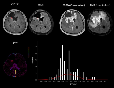 |
146 | Histogram model of DCE-MRI in predicting progression of enhancing non-measurable disease after chemoradiotherapy in high-grade glioma
Haimei Cao1, Zhousan Huang1, Ruowei Qiu1, Zhiyong Li2, Kan Deng3, Jay J Pillai4, Guanglong Huang2, Yikai Xu1, Jun Hua5,6, and Yuankui Wu1
1Department of Medical Imaging, Nanfang Hospital, Southern Medical University, Guangzhou, China, 2Department of Neurosurgery, Nanfang Hospital, Southern Medical University, Guangzhou, China, 3Philips Healthcare, Guangzhou, China, 4Division of Neuroradiology, Russell H. Morgan Department of Radiology and Radiological Science, Johns Hopkins University School of Medicine, Baltimore, MD, United States, 5Neurosection, Division of MRI Research, Department of Radiology, Johns Hopkins University School of Medicine, Baltimore, MD, United States, 6F.M. Kirby Research Center for Functional Brain Imaging, Kennedy Krieger Institute, Baltimore, MD, United States Keywords: Tumors, Perfusion, enhancing non-measurable disease (NMD) Early prediction of disease progression is of potential clinical significance for the management of high-grade glioma (HGG) patients. We investigated the value of histogram models based on volume transfer constant (Ktrans) between the plasma and extravascular extracellular space and extravascular volume (Ve) in predicting the progression of enhancing non-measurable diseases (NMD) of HGG after chemoradiotherapy. Our results showed that histogram models based on Ktrans and Ve can accurately predict the progression of enhancing NMD of HGG following chemoradiotherapy, and combining Ktrans and Ve helps improve the prediction performance. |
|
1964.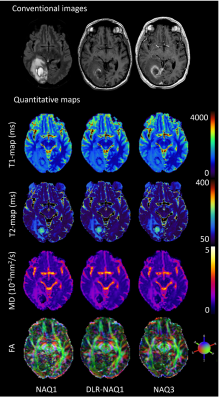 |
147 | Deep Learning-based noise reduction for advanced brain MR imaging: Application to quantitative biomarkers in brain tumors
Clement Debacker1,2, Geoffroy Pouliquen1,2, Sylvain Charron2, Anna Fayolle1,2, Valentin H. Prevost3, Wolter de Graaf4, Alexandre Roux2,5, Johan Pallud2,5, and Catherine Oppenheim1,2
1Radiology department, GHU Paris Psychiatrie et Neurosciences, Site Sainte-Anne, Paris, France, 2IMA-BRAIN, Université Paris Cité, Institute of Psychiatry and Neuroscience of Paris (IPNP), INSERM U1266, Paris, France, 3Canon Medical Systems Corporation, Tochigi, Japan, 4Canon Medical Systems Europe, Zoetermeer, Netherlands, 5Neuro-surgery department, GHU Paris Psychiatrie et Neurosciences, Site Sainte-Anne, Paris, France Keywords: Tumors, Tumor Our work is the clinical validation of a deep-learning algorithm (DLR) used to denoise MR images on quantitative MR biomarkers. Since it has been trained on including T1- and T2-weighted conventional images, in healthy volunteers, its effects on multiparametric quantitative MRI in patients, are uncertain. It could potentially improve brain tumors characterization by providing quantitative biomarkers under clinical time-constraints. |
|
1965. |
148 | Histogram analysis based on T2 mapping can predict the molecular markers in meningiomas
Zongye Li1, Yijie Yang2, Yue Zhang1, Yanhong Lin2, Xiao Wang1, Yuchuan Zhuang3, Qinqin Yang2, Eryuan Gao1, Yanan Ren1, Yong Zhang1, Shuhui Cai2, Zhong Chen2, Congbo Cai2, Jingliang Cheng1, and Jianfeng Bao1
1Department of Magnetic Resonance Imaging, the First Affiliated Hospital of Zhengzhou University, Zhengzhou, China, 2Department of Electronic Science, Xiamen University, Xiamen, China, 3Department of Imaging Sciences, University of Rochester, Rochester, NY, United States Keywords: Tumors, Relaxometry There is an immediate need for neurosurgeons to be able to identify certain molecular markers’ status of meningioma preoperatively, such as progesterone receptors and S100. In this study, we explored the use of histogram analysis of T2 maps and ADC maps in predicting such molecular markers. T2 maps and ADC maps could serve as useful tools for predicting PR and S100 status of meningiomas. Similar studies may be extended to investigate other pathological markers. |
|
1966.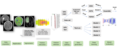 |
149 | MRI-Based Radiomics Approach with Deep Learning for Distinguishing IDH-Mutant from IDH Wild-type Grade-4 Astrocytomas
Seyyed Ali Hosseini1,2, Elahe Hosseini3, Isaac Shiri4, Ghasem Hajianfar5, Stijn Servaes1,2, Pedro Rosa-Neto1,2, Laiz Godoy6, Stephen Bagley7, MacLean Nasrallah8, Donald M O’Rourke9, Suyash Mohan6, and SANJEEV CHAWLA6
1Department of Neurology and Neurosurgery, Faculty of Medicine, McGill University, Montréal, QC, Canada, 2Translational Neuroimaging Laboratory, The McGill University Research Centre for Studies in Aging, Douglas Hospital,McGill University, Montreal, QC, Canada, 3Electrical and Computer Engineering, Kharazmi University, Tehran, Iran (Islamic Republic of), 4Division of Nuclear Medicine and Molecular Imaging, Geneva University Hospital, Geneva, Switzerland, 5Rajaie Cardiovascular Medical and Research Center, Iran University of Medical Science, Tehran, Iran (Islamic Republic of), 6Radiology, Perelman School of Medicine at the University of Pennsylvania, Philadelphia, PA, United States, 7Hematology-Oncology, Perelman School of Medicine at the University of Pennsylvania, Philadelphia, PA, United States, 8Clinical Pathology and Laboratory Medicine, Perelman School of Medicine at the University of Pennsylvania, Philadelphia, PA, United States, 9Perelman School of Medicine at the University of Pennsylvania, Philadelphia, PA, United States Keywords: Tumors, Radiomics Patients (n=57) with isocitrate dehydrogenase (IDH) mutant grade-4 astrocytomas and IDH wild-type glioblastomas underwent anatomical imaging (post-contrast T1 and T2-FLAIR) on 3T magnet. Neoplasms were segmented into 5 ROIs and 105 radiomics features were extracted from each ROI. Features were subsequently selected using various algorithms. Patients were divided into two groups (50%-training and 50%-testing). A GAN-based deep learning algorithm was used to generate 1000 synthesized data-sets and four distinct deep-learning modules were implemented. The best model for differentiating two genotypes of neoplasms was obtained from core tumor regions by using K-best feature selection and Ensembled algorithm with high diagnostic performance. |
|
1967.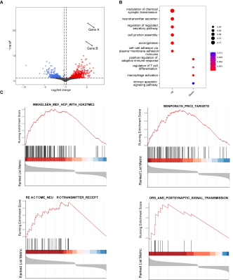 |
150 | Radiogenomic-based model identifies prognostic-related biological pathways in diffuse midline gliomas
Xiaorui Su1, Xibiao Yang1, Shuang Li1, Hanbing Shao1, Yukun Liu2, and Qiang Yue1
1Huaxi MR Research Center (HMRRC), Department of Radiology, West China Hospital of Sichuan University, Chengdu, China, 2West China School of Medicine &West China Hospital, Chengdu, China Keywords: Tumors, Radiomics To explore biological interpretation of prognosis-related radiomic signatures in diffuse midline gliomas, forty-one cases with paired MRI and RNA sequencing were screened in the public database TCGA-TCIA. We extracted radiomics features from preoperative MRI and divided these patients into high- and low-risk groups. Differential and enrichment analysis showed that high-expression genes of the high-risk group were mainly activated and involved in biological processes such as axon extension, neurotransmitter signaling, and cell connection. Moreover, typical driver molecular changes known in DMG were observed. It suggested that biological interpretation of prognosis-related radiomic signatures could be achieved by integrating MRI and RNA data |
|
1968.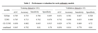 |
151 | The Value of Multi-parametric MRI-Based Radiomics Model in Predicting the IDH1 Mutation in Glioma
Shao ru zhang1, Zhi qiang Chen2, Xiao hua Chen1, Zhuo Wang1, Yun shu Zhou1, Shi li Liu1, Ruo di Zhang1, Yu hui Xiong3, and Bing Chen4
1Clinical medicine school of Ningxia Medical University, Yinchuan, China, 2Department of Radiology ,the First Hospital Affiliated to Hainan Medical College, Haikou, China, 3GE Healthcare, Beijing, China, 4Department of Radiology, General Hospital of Ningxia Medical University, Yinchuan, China Keywords: Tumors, Radiomics This study aims to explore the value of multi-parametric MRI-based radiomics model for non-invasively predicting IDH1 mutation in glioma. It was shown that among various single-sequence radiomics models, the contrast-enhanced T1-weighted image radiomics model should be considered as an optimal model in predicting IDH1 mutation, while the combined model based on three sequences could further improve the predicting performance. |
|
1969.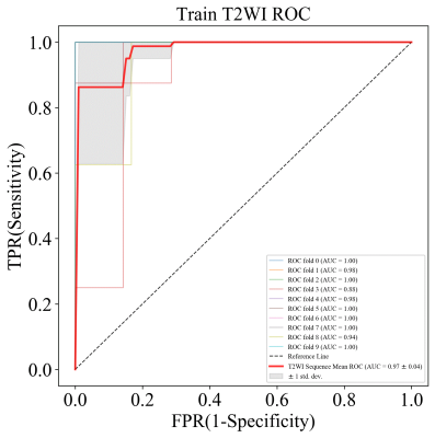 |
152 | Multi-Parametric MRI-based radiomics for noninvasively predicting Tert genotype in Oligodendroglomas
Jun Zhao1, Tiejun Gan2, Xiaoai Ke1, Wanjun Hu2, Qing Zhou1, Jing Zhang2, and Junlin Zhou1
1Department of Radiology, Lanzhou University Second Hospital, Lanzhou, China, 2Department of Magnetic Resonance, Lanzhou University Second Hospital, Lanzhou, China Keywords: Tumors, Radiomics, Multimodal,Quantitative Imaging In this study, radiomic based predictive models using MRI can non-invasively assess Tert genotype of Oligodendrogliomas(ODs).Compared With histopathological assessment,radiomic based predictive models has its unique advantages in assessing Tert genotype of ODs. such as noninvasive, more comprehensive information about tumor heterogeneity and repeatable.Furthermore,radiomic based predictive models can clearly and directly show Tert genotype of ODs.Therefore, it is a potential alternative to invasive biopsy. |
|
1970.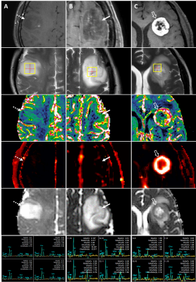 |
153 | Glioma grading using multiparametric MRI: head-to-head comparison among four different advanced techniques
Minkook Seo1, Yangsean Choi1, Youn Soo Lee2, Kook-Jin Ahn1, Bum-soo Kim1, Jae-Sung Park3, and Sin-soo Jeon3
1Radiology, Seoul St. Mary's Hospital, Seoul, Korea, Republic of, 2Hospital Pathology, Seoul St. Mary's Hospital, Seoul, Korea, Republic of, 3Neurosurgery, Seoul St. Mary's Hospital, Seoul, Korea, Republic of Keywords: Tumors, Tumor, Glioma Diagnostic performances differentiating low-grade (LGGs) from high-grade gliomas (HGGs) were compared between the four advanced techniques of multiparametric MRI—DSC, DCE, MRS and DWI. Sixty-four patients with pathologically confirmed glioma underwent preoperative multiparametric MRI with the four techniques, followed by histogram analysis of the tumors. HGGs showed significantly higher Ktrans, rCBV, and Cho/Cr than LGGs. The AUROC of the 95th percentile Ktrans was 0.83, being the most helpful parameter, followed by 95th percentile rCBV (0.72). Significant correlations were observed between the 95th percentile rCBV and Ktrans. Our findings demonstrate higher diagnostic utility of DCE and DSC MRI in glioma grading. |
|
1971.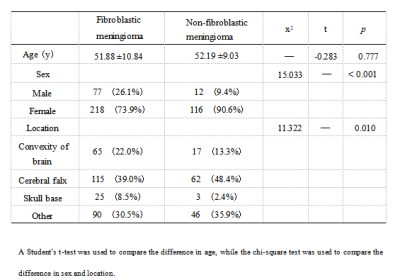 |
154 | A clini-radiomics model based magnetic resonance imaging for differentiating fibroblastic from non-fibroblastic meningioma
Tao Han1, Xianwang Liu1, and Junlin Zhou1
1Lanzhou University Second Hospital, LanZhou, China Keywords: Tumors, Machine Learning/Artificial Intelligence This study evaluated the feasibility of noninvasive preoperative differentiation fibroblastic meningioma (FM) from non-fibroblastic meningiomas (nFM) based a clini-radiomics model. A total of thirteen radiomics features were included after Selectpercentile and Lasso feature screening. Our results showed that Random forest is the most efficient among the six radiomics models in differentiating FM from nFM, and the diagnostic efficacy of clini-radiomics models in training and validation group is further improved. Therefore, we believe that the clini-radiomics model is of great value in noninvasive preoperative differentiation of FM and nFM and contributes to the selection of individualized treatment options for meningioma patients. |
|
1972.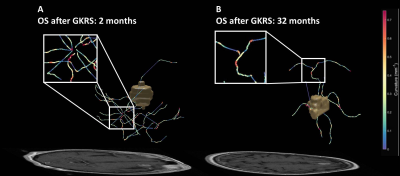 |
155 | The Association of Peritumoral Vasculature Radiomics of Lung Cancer Brain Metastasis with Patient Outcome after Radiosurgery
Chien-Yi Liao1, Cheng-Chia Lee2, Huai-Che Yang2, Wen-Yuh Chung2, Hsiu-Mei Wu3, Wan-Yuo Guo3, Ren-Shyan Liu4, and Chia-Feng Lu1
1National Yang Ming Chiao Tung University, Taipei, Taiwan, Taipei, Taiwan, 2Department of Neurosurgery, Neurological Institute, Taipei Veteran General Hospital, Taipei, Taiwan, Taipei, Taiwan, 3Department of Radiology, Taipei Veteran General Hospital, Taipei, Taiwan, Taipei, Taiwan, 4Department of Medical Imaging, Cheng-Hsin General Hospital, Taipei, Taiwan, Taipei, Taiwan Keywords: Tumors, Radiotherapy Gamma Knife radiosurgery (GKRS) is a first-line treatment for brain metastases (BMs) from non-small cell lung cancer (NSCLC). The convolutedness, leakiness and disorganized structure of the peritumoral vasculature have been suggested to be related to the treatment resistance of the tumor. In this study, radiomic features that measure the morphology and spatial organization of peritumoral vasculature were extracted from pre-GKRS MRI. These features were applied to predict overall survival after GKRS in NSCLC-BM patients. We suggested that peritumoral vasculature radiomics could facilitate patient management by identifying potential benefits of GKRS. |
|
1973.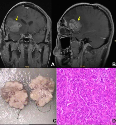 |
156 | The efficacy of preoperative MRI features in the diagnosis of meningioma WHO grade and brain invasion
Jun Jiang1, Juan Yu1, Xiajing Liu1, Kan Deng2, Fan Lin1, and Liangping Luo3
1Shenzhen Second People's Hospital, Shenzhen, China, 2Philips Healthcare, Guangzhou, China, 3The First Affiliated Hospital of Jinan University, Guangzhou, China Keywords: Tumors, MR Value The purpose of this study is to explore the efficacy of clinical and MRI-specific features for tumor grading and the brain invasion assessment in patient with meningioma. Our results showed that the tumor-brain interface is considered as a key factor, preoperative MRI has excellent performance in diagnosing meningioma WHO grade and brain invasion. |
|
1974.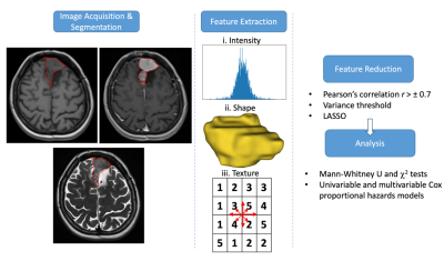 |
157 | Correlation of MRI Radiomics Features and Recurrence After Gamma Knife Treatment in Parasagittal and Falx Meningiomas
Esra Sümer1, O Artunç Türe2, Meriç Şengöz3,4, M Necmettin Pamir3,4, Alp Dinçer4,5, Koray Özduman3,4, and Esin Ozturk-Isik1,4
1Institute of Biomedical Engineering, Boğaziçi University, İstanbul, Turkey, 2Department of Radiation Oncology, Acıbadem University, İstanbul, Turkey, 3Department of Neurosurgery, Acıbadem University, İstanbul, Turkey, 4Brain Tumor Research Group, Acıbadem University, İstanbul, Turkey, 5Department of Radiology, Acıbadem University, İstanbul, Turkey Keywords: Tumors, Radiomics, Gamma Knife, meningioma Meningiomas located at and around the falx and superior sagittal sinus are known to have a more aggressive clinical behavior. Additionally, their localization and invasion characteristics make complete resection difficult, creating a need for radiosurgery. This study assessed the relationship between radiomics/clinical features of parasagittal and falx meningiomas and their recurrence-free survival after Gamma Knife. The radiomics and clinical features (age, pre-op tumor volume, coverage, and sensitivity) were analyzed together. Univariable Cox analysis associated maximum and skewness of T1 lesion, age, TV, and coverage, whereas multivariable Cox analysis associated skewness of T2 lesion, coverage, and selectivity with RFS. |
|
1975. |
158 | Prediction of Astrocytoma Pathological Grade Using Radiomics Extracted from Pre-operative Multiparametric MRI
Esra Sümer1, Ayça Ersen Danyeli2,3, M. Necmettin Pamir3,4, Koray Özduman3,4, Alp Dinçer3,5, and Esin Ozturk-Isik1,3
1Institution of Biomedical Engineering, Boğaziçi University, İstanbul, Turkey, 2Department of Medical Pathology, Acıbadem University, İstanbul, Turkey, 3Brain Tumor Research Group, Acıbadem University, İstanbul, Turkey, 4Department of Neurosurgery, Acıbadem University, İstanbul, Turkey, 5Department of Radiology, Acıbadem University, İstanbul, Turkey Keywords: Tumors, Radiomics, astrocytoma, pathological grade, machine learning The astrocytomas are currently graded from 2 to 4 according to World Health Organization 2021 central nervous system tumor classification. Increasing grade defines increasing malignancy. This study aims to explore the potential of radiomics features extracted from multiparametric pre-operative MRI to predict the grade of astrocytomas. For differentiation of grade 2 from grades 3&4, the FLAIR radiomics were predominantly determined by the feature selection procedure. The highest accuracy for differentiating grade 2 from grades 3&4 was 88.04±0.03% (92.37±0.04% precision and 86.76±0.04% recall). |
|
1976. |
159 | Histogram-based analysis of recurrence pattern correlations with MGMT methylation status in glioblastoma using synthetic MRI and 3D-ASL
Pei Dang1, Xueying Huang2, Xin Ge3, Lidong Wang4, Aijun Wang2, Minglei Wang2, Xuhong Yang5, Yuhui Xiong6, and Xiaodong Wang7
1Radiology, General Hospital of Ningxia Medical University, yinchuan, China, 2General Hospital of Ningxia Medical University, Yinchuan, China, 3Lanzhou University, Lanzhou, China, 4Radiology, Yinchuan Hospital of Traditional Chinese Medicine, Yinchuan, China, 5Ningxia Medical University, Yinchuan, China, 6GE Healthcare MR Research, Beijing, China, 7Radiology, General Hospital of Ningxia Medical University, Yinchuan, China Keywords: Tumors, Cancer, Glioblastoma; Synthetic MRI; 3D-ASL; Histogram; MGMT; Recurrence patterns At least 10% of glioblastoma relapses occur at distant and even contralateral locations. This disseminated growth limits surgical intervention and contributes to neurologicalmorbidity.The aim of the present study was to evaluate factors predicting the recurrencepattern determined.The results revealed that the pattern of recurrence in glioblastoma patients after combined radio-chemotherapy treated are strictly correlated with age at diagnosis and MGMT methylation status.Whole-tumor histogram analysis of quantitative parameters from synthetic MRI and 3D-ASL was to evaluate the value of predicting MGMT methylation status .Combining T110th, T210th ,and CBFentropy may explore as an effective strategy to predict of MGMT methylation status. |
|
 Back to Meeting Home
 Back to the Program-at-a-Glance
The International Society for Magnetic Resonance in Medicine is accredited by the Accreditation Council for Continuing Medical Education to provide continuing medical education for physicians.
 Back to Meeting Home
 Back to the Program-at-a-Glance
 View Presentation Video