Digital Poster
Exploring Tumors Based on Digits: Omics, Machine Learning & Quantification II
ISMRM & ISMRT Annual Meeting & Exhibition • 03-08 June 2023 • Toronto, ON, Canada

Digital Poster
Exploring Tumors Based on Digits: Omics, Machine Learning & Quantification II
| Computer # | |||
|---|---|---|---|
2115.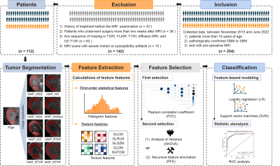 |
121 | Texture analysis of mean apparent propagator-magnetic resonance imaging in distinguishing glioblastoma and solitary brain metastasis
Guohua Zhao1, Yizhou Su2, Yusong Lin2, Jie Dong3, Eryuan Gao1, Xiaoyue Ma1, Jie Bai1, Huiting Zhang4, Xu Yan4, Guang Yang5, and Jingliang Cheng1
1The Department of Magnetic Resonance Imaging, The First Affiliated Hospital of Zhengzhou University, Zhengzhou, China, 2Collaborative Innovation Center for Internet Healthcare, Zhengzhou, China, 3School of Information Engineering, North China University of Water Resources and Electric Power, Zhengzhou, China, 4MR Scientific Marketing, Siemens Healthineers, Shanghai, China, 5Shanghai Key Laboratory of Magnetic Resonance, East China Normal University, Shanghai, China Keywords: Tumors, Diffusion/other diffusion imaging techniques Preoperative differentiation between glioblastomas (GBM) and solitary brain metastases (SBM) would aid in appropriate treatment planning and follow-up. Mean apparent propagator (MAP) MRI is effective in evaluating the inhomogeneity of brain microstructure. Texture analysis can be used to extract and quantify these inhomogeneities. In this study, we extracted the texture features from MAP-MRI, and validated the candidate features in discriminating between GBM and SBM. The RTAP model achieved the best discriminative power for the single metric model, and performed similarly to the MAP-MRI combined model. Texture analysis based on MAP-MRI has great potential to distinguish between the two entities. |
|
2116.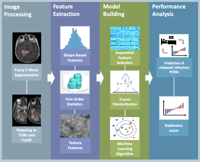 |
122 | Radiomics and Machine Learning for Prediction of Relapsed and Refractory Primary Central Nervous System Lymphoma
Yan-Lin Liu1, Ching-Chung Ko2,3, Yang Zhang1, Lee-Ren Yeh4, Jeon-Hor Chen1, and Min-Ying Su1
1Department of Radiological Sciences, University of California, Irvine, CA, United States, 2Department of Medical Imaging, Chi Mei Medical Center, Tainan, Taiwan, 3Department of Health and Nutrition, Chia Nan University of Pharmacy and Science, Tainan, Taiwan, 4Department of Radiology, E-DA Hospital, I-Shou University, Kaohsiung, Taiwan Keywords: Tumors, Machine Learning/Artificial Intelligence A subset of primary central nervous system lymphoma (PCNSL) may show early relapsed/refractory (R/R) disease after treatments. This study investigated the role of radiomics and machine learning for the prediction of R/R in PCNSL after treatments. Total 46 patients with pathologically confirmed PCNSL were included. Total 321 radiomic features were extracted from various pre-treatment MR sequences in each patient to build prediction models. Among various machine learning algorithms, the best predictive performance with accuracy of 82.6%, precision of 80%, and AUC of 0.85 were obtained in support vector machine. |
|
2117.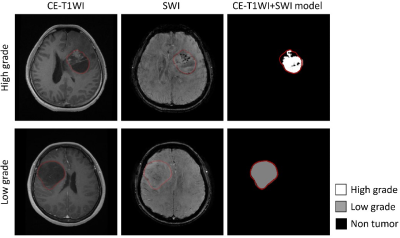 |
123 | Grading brain astrocytoma using convolutional neural network: contrast-enhanced T1 and susceptibility-weighted imaging
Zong-Ze Chen1, Yong-Han Lai1, Ching-An Liao1, Teng-Yi Huang2, Ping-Hong Lai1,3,4, and Tzu-Chao Chuang1
1National Sun Yat-Sen University, Kaohsiung, Taiwan, 2National Taiwan University of Science and Technology, Taipei, Taiwan, 3Kaohsiung Veterans General Hospital, Kaohsiung, Taiwan, 4National Yang-Ming University, Taipei, Taiwan Keywords: Tumors, Susceptibility Susceptibility-weighted imaging (SWI) has shown its potential to discriminate between high-grade and low-grade astrocytoma. In this study, we developed a fully automatic diagnosis system for astrocytoma grading by using convolutional neural network with contrast-enhanced T1-weighted images and SWI, separately or jointly, as input data. The results show that the model with both imaging modalities as input data provides high accuracy in astrocytoma grading and is potentially helpful for clinical diagnosis. |
|
2118.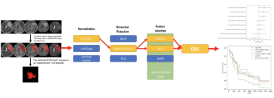 |
124 | The application value of Radiomics combined with clinical features and genomics in predicting glioma survival
Jinlong He1, Yang Gao1, Shaoyu Wang2, and Huapeng Zhang2
1Department of Imaging Diagnosis, The Affiliated Hospital of Inner Mongolia Medical University, Hohhot, China, 2MR Scientific Marketing, Siemens Healthineers, Shanghai, China Keywords: Tumors, Radiomics This study aimed to explore the value of MR multi-sequence radiomics combined with clinical features and genomics in predicting the survival of patients with glioma. Results showed that the clinical and imaging characteristics, radiomics features, and genotype status were important risk factors for glioma survival. The combination of multiple factors can better predict and evaluate the prognosis of glioma. Multi sequence based radiomics combined with clinical and imaging features and genotype status can better reflect the heterogeneity and prognosis of glioma. |
|
2119.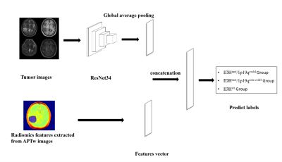 |
125 | Prediction of glioma genotypes by APTw-derived radiomic features combined with deep learning networks
Xinying Ren1,2, Diaohan Xiong1,2, Yujing Li1,2, Kai Ai3, and Jing Zhang1
1Lanzhou University Second Hospital, Lanzhou, China, 2Second Clinical School, Lanzhou University, Lanzhou, China, 3Philips Healthcare, Xi'an, China Keywords: Tumors, Machine Learning/Artificial Intelligence, Radiomics The study aimed to predict glioma genotypes combined with radiomic features and deep learning networks by using amide proton transfer (APT) imaging. The genetic subtypes of gliomas can be predicted by radiomics and deep learning networks using conventional MRI, however there are still problems with low accuracy and insufficient generalization. This study puts the screened APT radiomics features into a neural network and compares it with traditional radiomic. The results demonstrated that the proposed model had better performance. Therefore, APTw-derived radiomic features have good ability to predict 3-class molecular typing, providing novel classification tool for non-invasive evaluation for glioma genotypes. |
|
2120.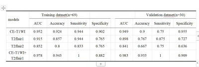 |
126 | The Value of Multiparametric MRI-based Radiomics Features in Distinguishing Primary Central Nervous System Lymphoma from High-grade Glioma
Shao ru zhang1, Zhi qiang Chen2, Xiao hua Chen1, Zhuo Wang1, Shi li liu1, Yun shu Zhou1, Ruo di Zhang1, Yu hui Xiong3, and Bing Chen4
1Clinical medicine school of Ningxia Medical University, Yinchuan, China, 2Department of Radiology ,the First Hospital Affiliated to Hainan Medical College, Haikou, China, 3GE Healthcare, Beijing, China, 4Department of Radiology, General Hospital of Ningxia Medical University, Yinchuan, China Keywords: Tumors, Brain In this study, we aim to explore the radiomics model constructed by a single sequence or a combined sequences whether has the same or better performance, and to explore the effect of different choices of region of interest to the performance of radionmics model. |
|
2121.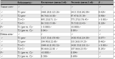 |
127 | Evaluation of relaxometry in differentiating recurrence and necrosis of high-grade glioma after radiotherapy using synthetic MR
LIU YANLING1, CUI YUELONG1, NIU ZHEN1, GUO JINXIA2, ZHANG SHASHA1, ZHANG JUNLI1, and WANG YONG1
1Anyang District Hospital, Anyang City, China, 2GE Healthcare, Beijing, China Keywords: Tumors, Cancer This study is to utilize the relaxometry generated by unenhanced and Gd-enhanced synthetic MRI to identify the recurrence and necrosis in patient with high grade glioma after radiotherapy. The results indicated enhanced T1 was significantly shortened in tumor and peripheral edema region for recurrent tissue in compare with necrosis and showed well ability of differentiation |
|
2122.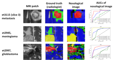 |
128 | Nosological images of brain tumor MV-MRS 3T data based on classifiers trained with SV-MRS 1.5T data, a proof-of-concept
Gulnur Semahat Ungan1,2, Albert Pons-Escoda3, Daniel Ulinic2, Carles Arus1,2, Alfredo Vellido1,4, and Margarida Julia-Sape1,2
1Centro de Investigacion Biomedica en Red (CIBER), Cerdanyola del Valles, Spain, 2Universitat Autonoma de Barcelona, Cerdanyola del Valles, Spain, 3Hospital de Bellvitge, L'Hospitalet del Llobregat, Spain, 4Universitat Politecnica de Catalunya, Barcelona, Spain Keywords: Tumors, Spectroscopy, visualization, data analysis, MRS, brain tumours We used SV-MRS 1.5T data of patients with brain tumors to create colored-classification images of MV-MRS 3T grids of an independent cohort of patients. In 10 out of 15 MV cases the solid tumor region corresponded to the correct class. In the remaining 5 cases, the reasons for (partial) misclassification included heterogeneity and bad spectral quality. |
|
2123.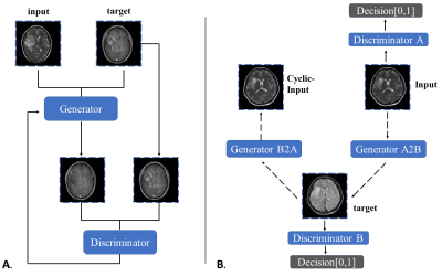 |
129 | Synthesize conventional MRI Sequences by Generative Adversarial Networks with only T2 for Use in a Multisequence gliomas classification Model
Diaohan Xiong1, Xinying Ren1, Yujing Li1, Rui Wang1, Kai Ai2, and Jing Zhang1
1Lanzhou University Second Hospital, Lanzhou, China, 2Philips Healthcare, Xi'an, China Keywords: Tumors, Machine Learning/Artificial Intelligence The aim of this study was to test deep learning classification models of glioma subtypes using the generated images. GANs were created based on the two frameworks, pix2pix and cycleGAN. The source domain was T2 and the target domain was T1c, T2-FLAIR or ADC. The results demonstrated that the T2 to T1c pix2pix model has the highest PSNR and SSI. When only the T2-flair or T1c sequence is replaced with the generated image, the classification accuracy is same as the original image. Therefore, depending solely on T2 sequences, GANs networks could generate other sequences for Use in gliomas classification Model. |
|
2124.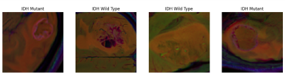 |
130 | Boosting The Deep Learning Performance in Predicting IDH Mutation in Gliomas Using Multiparametric MRI Including SWI, FLAIR and CE-T1WI
Sena Azamat1,2, Buse Buz-Yaluğ1, Alpay Ozcan3, Ayça Ersen Danyeli4,5,6, Necmettin Pamir5,7, Alp Dinçer4,8, Koray Ozduman4,7, and Esin Ozturk-Isik1,4
1Institute of Biomedical Engineering, Bogazici University, Istanbul, Turkey, 2Basaksehir Cam and Sakura City Hospital, Istanbul, Turkey, 3Electric and Electronic Engineering Department, Bogazici University, Istanbul, Turkey, 4Brain Tumor Research Group, Acibadem University, Istanbul, Turkey, 5Center for Neuroradiological Applications and Reseach, Acibadem University, Istanbul, Turkey, 6Department of Medical Pathology, Acibadem University, Istanbul, Turkey, 7Department of Neurosurgery, Acibadem University, Istanbul, Turkey, 8Department of Radiology, Acibadem University, Istanbul, Turkey Keywords: Tumors, Machine Learning/Artificial Intelligence Gliomas with IDH mutations tend to have a better prognosis regardless of the histopathological grade. The main aim of this study was to identify isocitrate dehydrogenase (IDH) mutations in glioma patients using deep learning models based on SWI, FLAIR and CE-T1W images separately and together. As a result, a 2D CNN model based on multiparametric MRI resulted in an accuracy of 92.8%, while CNN based on just SWI had 72.2% and CNN based on just FLAIR had 61.1% accuracies for predicting IDH mutation in gliomas. |
|
2125.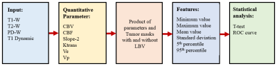 |
131 | Automatic Segmentation of Vasculature in DCE-MRI of Brain Tumors and its Influence in Grading
Anshika Kesari1, Virendra Kumar Yadav1, Raufiya Jafari1, Rakesh Kumar Gupta2, Rana Patir3, Sandeep Vaishya3, Sunita Ahlawat4, and Anup Singh1,5,6
1Centre for Biomedical Engineering, Indian Institute of Technology Delhi, New Delhi, India, 2Department of Radiology, Fortis Memorial Research Institute, Gurugram, India, 3Department of Neurosurgery, Fortis Memorial Research Institute, Gurugram, India, 4SRL Diagnostics, Fortis Memorial Research Institute, Gurugram, India, 5Yardi School for Artificial Intelligence, Indian Institute of Technology Delhi, New Delhi, India, 6Department of Biomedical Engineering, All India Institute of Medical Sciences, New Delhi, India Keywords: Tumors, Brain Quantitative DCE-MRI parameters are sensitive to tumor microvasculature and have shown potential in tumor grading. Generally, value of some of these parameters increases with tumor grades. During analysis of tumor region, large-blood-vessels (LBV) can influence the actual changes due to tumor microvasculature and hence provide erroneous results. Objectives of this study were to develop an automatic framework for segmentation of the LBV present within and outside of the tumor region and to reduce errors in tumor grading. The statistical analysis of parameters in tumor region showed significant differences between with and without LBV. Moreover, LBV removal improved tumor classification accuracy. |
|
2126.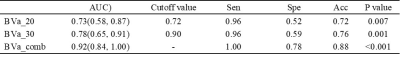 |
132 | Histogram model of MRI arteriolar blood volume in detecting subclinical recurrence of high-grade glioma after chemoradiotherapy
Yuankui Wu1, Zhousan Huang1, Yingxia Huang1, Mingxi Li1, Haimei Cao1, Guanglong Huang2, Hao Zhang1, and Yikai Xu1
1Department of Medical Imaging, Nanfang Hospital, Southern Medical University, Guangzhou, China, 2Department of Neurosurgery, Nanfang Hospital, Southern Medical University, Guangzhou, China Keywords: Tumors, Perfusion, Inflow-based vascular space occupancy (iVASO) Early detection of disease progression is of important relevance for the management of high-grade glioma (HGG) patients. Related studies showed that perfusion-weighted MR imaging (PWI) has power to detect early recurrence of glioblastoma. Inflow-based vascular space occupancy (iVASO) is a noninvasive perfusion technology that can provide absolute blood volume of precapillary arterioles (arteriolar blood volume, BVa). In this preliminary study, the potential value of BVa in detecting subclinical recurrence of HGG was investigated. The results showed that the histogram features of BVa might have the potential to detect subclinical recurrence. |
|
2127.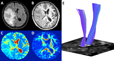 |
133 | Automated Fiber Quantification Predicts Motor Weakness in Patients Following Resection of Primary and Metastatic Brain Lesions
George Russell Glenn1, Sneha Sai Venkata Ka Mannam2, Chibueze Nwagwu3, Subir Goyal4, Gustavo Pradilla2, Edjah Kweku-Ebura Nduom2, Jeffery James Olson 2, David Painton Bray 2, and Hoang Bojanowski Kimberly2
1Diagnostic Radiology, Emory University, Lilburn, GA, United States, 2Neurosurgery, Emory University, Atlanta, GA, United States, 3College of Medicine, Emory University, Atlanta, GA, United States, 4Biostatistics and Bioinformatics, Emory University, Alanta, GA, United States Keywords: Tumors, Surgery Preoperative diffusion tensor imaging (DTI) data was analyzed using Automated Fiber Quantification (AFQ) along the corticospinal tract (CST) for 58 patients undergoing surgical resection of primary or metastatic brain lesions. For each patient, DTI parameters were analyzed along the ipsilateral CST, the side of the pathological lesion, and the contralateral CST, the side opposite the pathological lesion. Patients were then separated based on the presence or absence of postoperative motor weakness. Patients with post operative motor weakness were found to have significantly different diffusion parameters along their ipsilateral CST compared to the ipsilateral CST of patients without postoperative motor weakness. |
|
2128.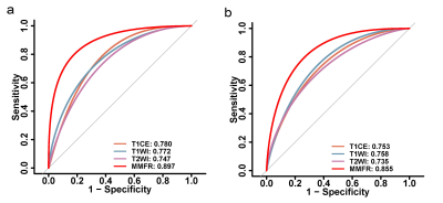 |
134 | Multiparametric MRI-based fusion radiomics for preoperatively predicting TERT promoter mutation status and survival in glioblastoma patients
Hongbo Zhang1, Hanwen Zhang2, Beibei Zhou3, Yuze Zhang1, Lei Wu1, Yi Lei2, and Biao Huang1
1Guangdong Provincial Key Laboratory of Artificial Intelligence in Medical Image Analysis and Application, Guangdong Provincial People's Hospital, Guangdong Academy of Medical Sciences, Guangzhou, China, 2Department of Radiology, The First Affiliated Hospital of Shenzhen University, Health Science Center, Shenzhen Second People's Hospital, Shenzhen, China, 3Department of Radiology, Department of Radiology,The Seventh Affiliated Hospital, Sun Yat-sen University, Shenzhen, China Keywords: Tumors, Radiomics Radiomics uses computer software to mine massive quantitative image features from medical imaging images and then screens the most valuable radiomics features using statistical and/or machine learning methods. Furthermore, it is used to parse clinical information for disease characterisation, tumour grading and staging, efficacy evaluation, and prognosis prediction. In our study, we demonstrated that multiparametric MRI-based fusion radiomics model is an effective preoperative non-invasive method to predict telomerase reverse transcriptase promoter mutations and progression-free survival in glioblastoma patients. |
|
2129.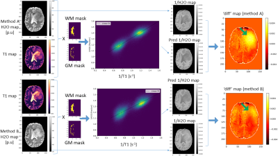 |
135 | An investigation into the derangement of the linear relationship between 1/T1 and 1/H2O in brain tumours
Dennis C. Thomas1,2,3,4, Ralf Deichmann5, Elke Hattingen1,2,3,4, Seyma Alcicek1,2,3,4, Ulrich Pilatus1,2,3,4, and Katharina J. Wenger1,2,3,4
1Institute of Neuroradiology, University Hospital Frankfurt, Frankfurt, Germany, 2University Cancer Center Frankfurt (UCT), Frankfurt, Frankfurt, Germany, 3Frankfurt Cancer Institute (FCI), Frankfurt, Germany, 4German Cancer Research Center (DKFZ) and German Cancer Consortium (DKTK), Heidelberg, Germany, 55 Brain Imaging Center, Goethe University, Frankfurt, Germany Keywords: Tumors, Brain, Water content mapping, T1 mapping Quantitative MRI was applied to assess T1 and water content (H2O) in brain tumors. 1/T1 and 1/H2O are linearly related in healthy WM and GM. Here, we explore deviations from this linear relationship in the tumor regions in 3 glioblastoma patients. Linear regression between 1/T1 and 1/H2O was performed in the healthy brain in the contralateral hemisphere and the obtained values were used to calculate predicted 1/H2O maps for the whole brain. Difference maps between predicted and true 1/H2O were calculated . Further, the importance of robust bias field correction techniques is demonstrated here by comparing two bias field correction approaches. |
|
2130.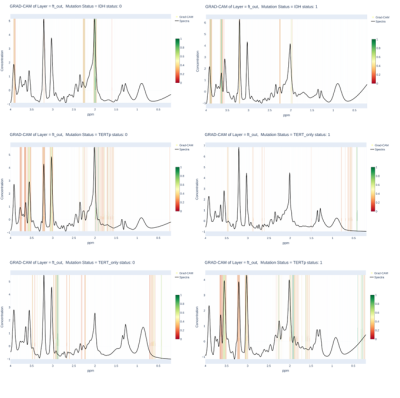 |
136 | Attention Deep-Shallow Network (ADSN): A Deep Learning Model for IDH and TERTp Mutation Detection in Gliomas using 1H-MRS
Abdullah Bas1, Banu Sacli-Bilmez1, Ayca Ersen Danyeli2,3, Ozge Can2,4, Koray Ozduman2,5, Alp Dincer2,6, and Esin Ozturk-Isik1,2
1Institute of Biomedical Engineering, Bogazici University, Istanbul, Turkey, 2Brain Tumor Research Group, Acibadem University, Istanbul, Turkey, 3Department of Medical Pathology, Acibadem University, Istanbul, Turkey, 4Department of Biomedical Engineering, Acibadem University, Istanbul, Turkey, 5Department of Neurosurgery, Acibadem University, Istanbul, Turkey, 6Department of Radiology, Acıbadem University, Istanbul, Turkey Keywords: Tumors, Machine Learning/Artificial Intelligence, Deep Learning Isocitrate dehydrogenase (IDH) and telomerase reverse transcriptase promoter (TERTp) mutations affect the clinical behavior and survival rate of diffuse gliomas. According to the latest WHO 2021 brain tumor classification, IDH mutation is an important factor for grouping adult-type diffuse gliomas. The preoperative detection of these mutations is very critical for treatment planning. In this study, we propose enhanced 1D-CNN models by adding an attention mechanism as a prior network to focus on relevant spectral frequencies of 1H-MRS to identify IDH-mutant (IDH-mut), TERTp-mutant (TERTp-mut), and IDH-wt, TERTp-mut (TERTp-only) gliomas using three binary models |
|
2131.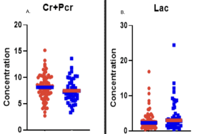 |
137 | Comparative Evaluation of Metabolite Composition in Brain Tumor Epilepsy Patients
Natasha Najam1, Steven Tobochnik2, Huijin Liao1, Sanghoon Kim1, Jong Woo Lee2, and Alexander Lin1
1Center for Clinical Spectroscopy, Department of Radiology, Brigham and Women's Hospital , Harvard Medical School, Boston, MA, United States, 2Department of Neurology, Brigham and Women's Hospital , Harvard Medical School,, Boston, MA, United States Keywords: Tumors, Spectroscopy, Brain Tumor-Related Epilepsy, Isocitrate Dehydrogenase Mutation Brain Tumor Related Epilepsy (BTRE) is a multi-factorial condition with unclear pathophysiology. It has a robust prevalence in tumor patients with 20-40% experiencing onset seizures and a further 20-45% experiencing refractory seizures to treatment.1 This work examines the tumor microenvironment using magnetic resonance spectroscopy (MRS) to learn more about the epileptogenic mechanisms through a comparison of metabolite composition in BTRE patients and non-seizure tumor patients. Increasing knowledge about the pathophysiology of BTRE might aid the management of anti-tumor and BTRE treatment in the future. |
|
2132.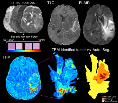 |
138 | Autopsy-based radio-pathomic maps of tumor probability delineate tumor presence within radiological segmentations
Samuel A Bobholz1, Allison K Lowman1, Savannah R Duenweg2, Aleksandra Winiarz2, Margaret Stebbins2, Fitzgerald Kyereme1, Jennifer Connelly3, Dylan Coss4, Wade M Mueller5, Mohit Agarwal1, Anjishnu Banerjee6, and Peter S LaViolette1,7
1Radiology, Medical College of Wisconsin, Milwaukee, WI, United States, 2Biophysics, Medical College of Wisconsin, Milwaukee, WI, United States, 3Neurology, Medical College of Wisconsin, Milwaukee, WI, United States, 4Pathology, Medical College of Wisconsin, Milwaukee, WI, United States, 5Neurosurgery, Medical College of Wisconsin, Milwaukee, WI, United States, 6Biostatistics, Medical College of Wisconsin, Milwaukee, WI, United States, 7Biomedical Engineering, Medical College of Wisconsin, Milwaukee, WI, United States Keywords: Tumors, Cancer, Machine learning, glioblastoma, radio-pathomics This study applied autopsy-based radio-pathomic maps to the pre-surgical PENN-GBM dataset to test the hypothesis that the predicted tumor composition of the contrast-enhancing and FLAIR-hyperintense regions identify distinct pathological features of glioblastoma. We find that greater predicted tumor within the contrast-enhancing region is indicative of IDH1-wildtype mutation status, and show that larger tumors tend to have less predicted tumor within contrast-enhancement and more tumor within non-enhancing FLAIR hyperintensity. This technique could be used to non-invasively identify more aggressive tumors. |
|
2133.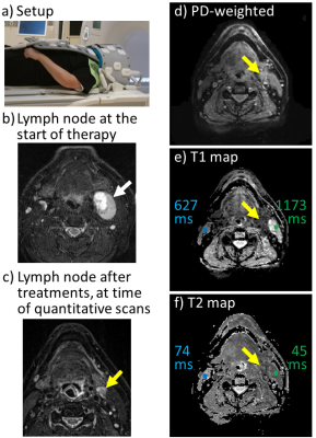 |
139 | Quantitative MRI for radiation oncology patients with head and neck squamous cell carcinoma
Bruno Madore1, Jeffrey P. Guenette1, Jonathan D. Schoenfeld1, and Evangelia Kaza1
1BWH, Harvard Medical School, Boston, MA, United States Keywords: Head & Neck/ENT, Relaxometry, Radiotherapy, treatment, tumor Radiation oncology patients often undergo longitudinal scans to evaluate the effect of treatments on lesions, and to monitor surrounding tissues. We are considering the inclusion of quantitative MRI in current imaging protocols. To this end, we implemented a simple quantitative imaging technique and tested it in a small cohort of five patients. Results captured differences in T1 and T2 between tissues on the treated side vs. the contralateral tissues. Little work has been done so far with MRI relaxometry for monitoring treatment in head and neck squamous cell carcinoma, and this work represents an early step in this direction. |
|
2134.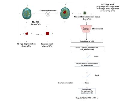 |
140 | Identification of NF2 loss in meningiomas using T2-weighted MRI and Deep Learning
Abdullah Bas1, Ayca Ersen Danyeli2,3, Ozge Can2,4, Koray Ozduman2,5, Alp Dincer2,6, and Esin Ozturk-Isik1,2
1Institute of Biomedical Engineering, Bogazici University, Istanbul, Turkey, 2Brain Tumor Research Group, Acibadem University, Istanbul, Turkey, 3Department of Medical Pathology, Acibadem University, Istanbul, Turkey, 4Department of Biomedical Engineering, Acibadem University, Istanbul, Turkey, 5Department of Neurosurgery, Acibadem University, Istanbul, Turkey, 6Department of Radiology, Acıbadem University, Istanbul, Turkey Keywords: Tumors, Machine Learning/Artificial Intelligence, Deep Learning NF2-L in meningiomas is a relevant indicator of prognosis. NF2-L is one of the most common genetic mutations in meningiomas. As a result of that situation, developing a non-invasive approach may assist the current clinical procedures. To our knowledge, some studies try to predict NF2-L using MRI modalities but either they use registration or performed using the modalities of MRI that are not in default MRI scanning procedures. Hence, the solutions that they provide are not clinically feasible. In this study, we aim to develop new approaches that are capable to implement directly into clinical procedures.[1] |
|
 Back to Meeting Home
 Back to the Program-at-a-Glance
The International Society for Magnetic Resonance in Medicine is accredited by the Accreditation Council for Continuing Medical Education to provide continuing medical education for physicians.
 Back to Meeting Home
 Back to the Program-at-a-Glance
 View Presentation Video