Digital Poster
Multiple Sclerosis
ISMRM & ISMRT Annual Meeting & Exhibition • 03-08 June 2023 • Toronto, ON, Canada

| Computer # | |||
|---|---|---|---|
2824.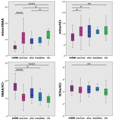 |
121 |
SWI and MRSI Unite! Metabolic Insights into Iron Deposition in
Relapsing-Remitting MS via 7T Magnetic Resonance Spectroscopic
Imaging
Alexandra Lipka1,
Wolfgang Bogner1,
Assunta Dal-Bianco2,
Gilbert Hangel1,3,
Paulus Rommer2,
Bernhard Strasser1,
Stanislav Motyka1,
Lukas Hingerl1,
Thomas Berger2,
Fritz Leutmezer2,
Stephan Gruber1,
Siegfried Trattnig1,4,
and Eva Niess1
1Department of Biomedical Imaging and Image-guided Therapy, Medical University of Vienna, High Field MR Centre, Vienna, Austria, 2Medical University of Vienna, Department of Neurology, Vienna, Austria, 3Medical University of Vienna, Department of Neurosurgery, Vienna, Austria, 4Karl Landsteiner Institute for Clinical Molecular MRI in Musculoskeletal System, Vienna, Austria Keywords: Multiple Sclerosis, Multiple Sclerosis Besides clinical examination, conventional T1/T2-weighted magnetic resonance imaging (cMRI) is the method of choice for diagnosis and treatment monitoring of Multiple Sclerosis (MS). In contrast to cMRI, which - in MS - can only depict the severity of irreversible tissue damage and is not able to explain underlying pathological processes, MR Spectroscopic Imaging (MRSI) can detect pathologies on a biochemical level, while Susceptibility Weighted Imaging (SWI) provides information about iron deposition. In 31 relapsing-remitting (RRMS) patients, we investigate - via ultra-high resolution FID-MRSI at 7T - metabolic characteristics of different types of iron accumulation and the metabolic distribution within lesions and their vicinity. |
|
2825.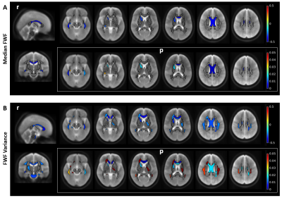 |
122 |
Tissue damage and impaired processing speed are related to brain
parenchyma free water fraction in multiple sclerosis
Alessandra S. Caporale1,2,
Antonio M. Chiarelli1,2,
Emma Biondetti1,2,
Alessandro Villani1,2,
Ilona Lipp3,
Valentina Tomassini1,2,4,5,
and Richard G. Wise1,2,5
1Department of Neurosciences, Imaging and Clinical Sciences, 'G. d'Annunzio University' of Chieti-Pescara, Chieti, Italy, 2Institute for Advanced Biomedical Technologies (ITAB), 'G. d'Annunzio University' of Chieti-Pescara, Chieti, Italy, 3Department of Neurophysics, Max Planck Institute for Human Cognitive and Brain Sciences, Leipzig, Germany, 4MS Centre, Department of Clinical Neurology, ‘SS. Annunziata’ University Hospital, Chieti, Italy, 5Cardiff University Brain Research Imaging Centre (CUBRIC), School of Psychology, Cardiff University, Cardiff, United Kingdom Keywords: Multiple Sclerosis, White Matter, normal appearing white matter, neurodegeneration, cognitive impairment In 99 relapsing-remitting multiple sclerosis (MS) patients and 25 healthy controls we quantified cerebral free water fraction (FWF) and investigated its relationship with lesion burden and information processing speed, measured with Symbol Digit Modalities Test (SDMT). FWF was obtained from the mcDESPOT method, fitting a three-compartment relaxation model to spoiled-gradient-echo and balanced-SSFP signals with varying flip angles. MS patients showed higher FWF than controls, in the lesioned tissue and in normal appearing white matter (NAWM). In NAWM and perilesional tissue, FWF correlated with lesion load. FWF spatial heterogeneity increased with worsening SDMT performance in regions involved in MS-related cognitive impairment. |
|
2826.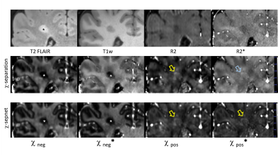 |
123 |
Comparison of chi-separation and chi-separation*: A clinical
feasibility study
Jinhee Jang1,
Hyeong-Geol Shin2,3,
Minjun Kim4,
Hyebin Lee1,
and Woojun Kim5
1Radiology, Seoul St. Mary's Hospital, Seoul, Korea, Republic of, 2Radiology, Johns Hopkins University, Baltimore, MD, United States, 3F.M. Kirby Research Center for Functional Brain Imaging, Kennedy Krieger Institute, Baltimore, MD, United States, 4Electrical and Computer Engineering, Seoul National University, Seoul, Korea, Republic of, 5Neurology, Seoul St. Mary's Hospital, Seoul, Korea, Republic of Keywords: Multiple Sclerosis, Quantitative Susceptibility mapping While χ-separation gives unique and valuable information, requirement of R2 limited its clinical application. This study assessed the clinical feasibility of two R2*-based magnetic source separation. From 24 MR scans of MS subjects, four sets of magnetic source separation maps were generated; (1) χ-separation (originally proposed method), (2) χ-separation* (exploiting R2*), (3) χ-sepnet (DL-based), and (4) χ-sepnet* (DL-based, utilizing R2*). χ-separation* and χ-sepnet* showed good agreement to χ-separation and χ-sepnet in MS lesions and the brain. However, there are small but consistent biases in each method, which are important caveats for R2*-based χ-separation. These biases were relatively smaller in χ-sepnet*. |
|
2827.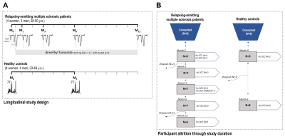 |
124 |
7-Tesla in vivo 1H-MRS-measured prefrontal glutathione increases
during oral fumarate therapy in relapsing-remitting multiple
sclerosis
Kelley M. Swanberg1,
Hetty Prinsen2,
Daniel Pelletier2,3,
and Christoph Juchem1,2,4,5
1Biomedical Engineering, Columbia University, New York, NY, United States, 2Radiology and Biomedical Imaging, Yale University School of Medicine, New Haven, CT, United States, 3Neurology, University of Southern California Keck School of Medicine, Los Angeles, CA, United States, 4Neurology, Yale University School of Medicine, New Haven, CT, United States, 5Radiology, Columbia University Irving Medical Center, New York, NY, United States Keywords: Multiple Sclerosis, Spectroscopy, dimethyl fumarate, glutathione, oxidative stress Multiple sclerosis (MS) is an autoimmune disease that damages the central nervous system. Oxidative stress, thought to play a role in MS-related pathophysiology, can be modulated in the cell by endogenous antioxidants such as glutathione (GSH), hypothesized to participate in the therapeutic effect of MS disease-modifying therapy dimethyl fumarate. We used proton magnetic resonance spectroscopy (1H MRS) to measure in vivo cortical glutathione concentrations in individuals with relapsing-remitting multiple sclerosis (RR-MS) before and during 12 months of dimethyl fumarate therapy and observed a significant positive effect of time on prefrontal cortex glutathione. No such change was shown in healthy controls. |
|
2828. |
125 |
Time-dependent Diffusion in Acute and Chronic Plaques of
Multiple Sclerosis Investigated with Oscillating-gradient
Spin-echo.
Tomoko Maekawa1,
Masaaki Hori1,2,
Katsutoshi Murata3,
Kouhei Kamiya1,2,
Christina Andica1,4,
Akifumi Hagiwara1,
Shohei Fujita1,5,
Koji Kamagata1,
Akihiko Wada1,
and Shigeki Aoki1
1Juntendo University School of Medicine, Tokyo, Japan, 2Toho University Omori Medical Center, Tokyo, Japan, 3Siemens Healthcare K.K., Tokyo, Japan, 4Faculty of Health Data Science, Juntendo University, Chiba, Japan, 5The University of Tokyo, Tokyo, Japan Keywords: Multiple Sclerosis, Diffusion/other diffusion imaging techniques, Microstructure The purpose of our study was to investigate the utility of changes in diffusivity between short and long diffusion time in evaluating acute and chronic plaques of multiple sclerosis. Chronic plaques showed weak diffusion time-dependence with diffusion times between 6.5 ms and 35.2 ms. The differences in diffusion time-dependence were attributed to the internal structure of plaques. DWI with a short diffusion time may provide additional information about the microstructure of MS plaques. |
|
2829. |
126 |
Myelin Volume Fraction Estimation in Multiple Sclerosis Lesions
Mert Şişman1,2,
Thanh D. Nguyen2,
Alexey V. Dimov2,
Melanie Marcille 3,
Pascal Spincemaille2,
Susan A. Gauthier3,
and Yi Wang2,4
1Electrical and Computer Engineering, Cornell University, Ithaca, NY, United States, 2Department of Radiology, Weill Cornell Medicine, New York, NY, United States, 3Department of Neurology, Weill Cornell Medicine, New York, NY, United States, 4Biomedical Engineering, Cornell University, Ithaca, NY, United States Keywords: Multiple Sclerosis, White Matter Monitoring myelin content quantitatively is important in the study of Multiple Sclerosis (MS). Here, a novel approach is proposed to estimate myelin volume fraction (MVF) of MS lesions from routine multi-echo gradient echo (mGRE) using biophysical modeling of the myelin sheaths. The obtained MVF values correlated significantly with the myelin water fraction (MWF) values obtained in the same lesions using conventional multicomponent T2 relaxometry. Moreover, the change in MVF values in newly enhancing lesions over the first year also correlated significantly with the change in MWF values over the same period. |
|
2830. |
127 |
Z-score maps to characterize spinal cord lesions in MS patients:
Toward a semi-automated lesion identification/segmentation
method
Samira Mchinda1,2,3,
Arnaud Le Troter1,2,
Sarah Demortière4,
Nilser Laines Medina1,2,3,
Lauriane PINI1,2,
Jean Pelletier1,2,4,
Bertrand Audoin1,2,4,
and Virginie Callot1,2,3
1Aix Marseille Univ, CNRS, CRMBM, Marseille, France, 2APHM, Hôpital Universitaire Timone, CEMEREM, Marseille, France, 3iLab-Spine, International Associated Laboratory, Montreal, Canada, Marseille, France, 4APHM, Hôpital Universitaire Timone, Département de Neurologie, Marseille, France Keywords: Multiple Sclerosis, Spinal Cord, z-score maps Manual segmentation, considered as ground truth, remains time consuming and operator dependent. In this work, a strategy to identify and potentially segment individual cervical spinal cord lesions based on z-score maps(ZM) from 3T quantitative T1-MP2RAGE(T1q) imaging is proposed. All manually segmented lesions could be visually identified in ZM with a lower threshold(LT) of 2. Conversely, Sixty-nine% of the lesions could be isolated automatically using LT=3 and analyzing the characteristics of the ZM clusters in combination with ZM gradient maps. Four patients (/4) without lesions, and 21/23 controls showed no significant cluster. |
|
2831. |
128 |
Quantitative T2 measurements of diffusely-abnormal white matter
in relapsing-remitting MS patients at baseline
Benjamin Charles Musall1,
Yanyu Yang2,
Arash Kamali1,
John A Lincoln3,
Vi Ly2,
Xi Luo2,
Ponnada A Narayana1,
Refaat E Gabr1,
and Khader M Hasan1
1Diagnostic and Interventional Imaging, UTHealth McGovern Medical School, Houston, TX, United States, 2Department of Biostatistics and Data Science, University of Texas School of Public Health, Houston, TX, United States, 3Department of Neurology, UTHealth McGovern Medical School, Houston, TX, United States Keywords: Multiple Sclerosis, Quantitative Imaging Quantitative T2 of diffusely-abnormal white matter (DAWM), assessed at baseline in a population of 800 patients with relapsing-remitting MS with U-Net segmentation and T2 mapping, is distinct and intermediate to T2-hyperintense focal lesions and normal-appearing white matter (NAWM). |
|
2832.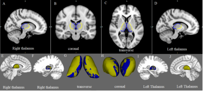 |
129 |
Association of subcortical structural shapes with fatigue in
Multiple Sclerosis.
Yujing Li1,2,
Tingli Yang3,
Min Li3,
Rui Wang3,
Xinying Ren3,
Diaohan Xiong3,
Kai Ai4,
and Jing Zhang3
1Department of Magnetic Resonance, Lanzhou University Second Hospital, Lanzhou, China, 2Second Clinical School, Lanzhou University, Lanzhou, China, 3Lanzhou University Second Hospital, Lanzhou, China, 4Philips Healthcare, Xi’an, China Keywords: Multiple Sclerosis, Gray Matter This study investigated the shape changes in the subcortical structure of patients with multiple sclerosis (PwMS) and how they are associated with MS-related fatigue. Surface-based vertex analysis was performed on 25 fatigued RRMS (F-MS), 25 non-fatigued RRMS (NF-MS), and 40 healthy controls (HCs). Compared to HCs, PwMS showed regional shape contractions in the areas of bilateral thalamus. The morphological changes in the bilateral thalamus occur only on the side closest to the ventricles. Compared to the NF-MS subgroup, F-MS patients showed regional surface atrophy in the areas of left caudate nucleus, which was negatively correlated with the severity of fatigue. |
|
2833.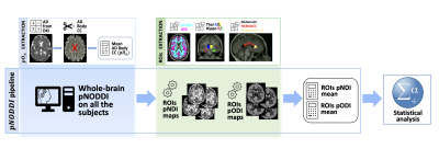 |
130 |
Personalized-NODDI (pNODDI) discriminates between healthy and
multiple sclerosis subjects
Elena Grosso1,
Carmen Tur2,3,
Alberto Calvi2,
Sara Collorone2,
Francesco Grussu2,4,
Marco Battiston2,
Ferran Prados Carrasco2,5,6,
Baris Kanber2,5,
Rebecca Samson2,
Egidio D'Angelo1,7,
Claudia AM Gandini Wheeler-Kingshott1,2,7,
and Fulvia Palesi1
1Department of Brain and Behavioral Sciences, University of Pavia, Pavia, Italy, 2NMR Research Unit, Department of Neuroinflammation, Queen Square Multiple Sclerosis Centre, UCL Queen Square Institute of Neurology, University College London (UCL), London, United Kingdom, 3Neurology-Neuroimmunology Department Multiple Sclerosis Centre of Catalonia (Cemcat), Vall d’Hebron Barcelona Hospital Campus, Barcelona, Spain, 4Radiomics Group, Vall d’Hebron Institute of Oncology, Vall d’Hebron Barcelona Hospital Campus, Barcelona, Spain, 5Department of Medical Physics and Biomedical Engineering, Centre for Medical Image Computing (CMIC), University College London, London, United Kingdom, 6E-Health Center, Universitat Oberta de Catalunya, Barcelona, Spain, 7Brain Connectivity Centre Research Unit, IRCCS Mondino Foundation, Pavia, Italy Keywords: Multiple Sclerosis, Diffusion/other diffusion imaging techniques, personalised-NODDI Neurite Orientation Dispersion and Density Imaging (NODDI) is a multi-compartmental model for microstructure characterization utilising diffusion-weighted MRI. Here, a recently proposed personalized-NODDI (pNODDI) modelling technique was used to assess microstructural alterations in a cohort of Multiple Sclerosis (MS) patients. Our findings showed that pNODDI metrics were sensitive to MS pathology and could discriminate between healthy controls and MS. Furthermore, pNODDI metrics of key brain regions increased the discriminative power between the two groups. Overall, pNODDI provides a personalized and clinically feasible model for microstructure, increasing sensitivity to pathological alterations. |
|
2834.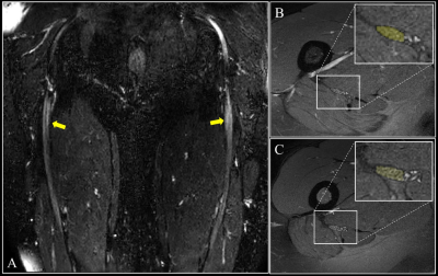 |
131 |
A pilot in vivo study of the sciatic nerve in multiple sclerosis
using quantitative magnetic resonance imaging
Ratthaporn Boonsuth1,2,
Marco Battiston1,
Rebecca S. Samson1,
Alberto Calvi1,3,
Claudia A. M. Gandini Wheeler-Kingshott1,4,5,
and Marios C. Yiannakas1
1NMR Research Unit, Queen Square MS Centre, Department of Neuroinflammation, UCL Queen Square Institute of Neurology, University College London, London, United Kingdom, 2Department of Radiologic Technology, Faculty of Associated Medical Sciences, Chiang Mai University, Chiang Mai, Thailand, 3Center of Neuroimmunology, Laboratory of Advanced Imaging in Neuroimmunological Diseases; Institut d’Investigacions Biomèdiques August Pi i Sunyer (IDIBAPS), Barcelona, Spain, 4Brain Connectivity Research Centre, IRCCS Mondino Foundation, Pavia, Italy, 5Department of Brain and Behavioural Sciences, University of Pavia, Pavia, Italy Keywords: Multiple Sclerosis, Quantitative Imaging, Nerves, Multimodal, Neurography The peripheral nervous system is not routinely examined objectively in multiple sclerosis (MS), despite evidence from neuropathology that demonstrates its implication. In this pilot in vivo study, the sciatic nerve was examined using multi-shell diffusion-weighted imaging, quantitative magnetisation transfer and T1 relaxometry to investigate whether pathological neural tissue damage could be detected in people with relapsing-remitting MS as compared to healthy controls. |
|
2835.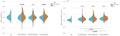 |
132 |
Lesional and non-lesional cortical integrity in cognitive
networks are linked to cognitive impairment in multiple
sclerosis.
Eva A. Krijnen1,2,
Albulena Bajrami1,3,
Tommy A.A. Broeders1,
Samantha Noteboom1,
Piet M. Bouman1,
Frederik Barkhof4,5,
Bernard M.J. Uitdehaag6,
Eric C. Klawiter2,
Ismail Koubiyr1,
and Menno M. Schoonheim1
1MS Center Amsterdam, Anatomy and Neurosciences, Amsterdam Neuroscience, Amsterdam UMC location VUmc, Amsterdam, Netherlands, 2Department of Neurology, Massachusetts General Hospital, Harvard Medical School, Boston, MA, United States, 3Multiple Sclerosis Specialist Center, Neurology B, Department of Neurosciences, Biomedicine and Movement Sciences, University of Verona, Verona, Italy, 4MS Center Amsterdam, Radiology and Nuclear Medicine, Amsterdam Neuroscience, Amsterdam UMC location VUmc, Amsterdam, Netherlands, 5Institutes of Neurology and Healthcare Engineering, University College London, London, United Kingdom, 6MS Center Amsterdam, Neurology, Amsterdam Neuroscience, Amsterdam UMC location VUmc, Amsterdam, Netherlands Keywords: Multiple Sclerosis, Gray Matter, Diffusion/Other Diffusion Imaging Techniques Cognitive functioning in people with multiple sclerosis (MS) is strongly related to cortical lesions (CLs), although the severity of demyelination remains difficult to assess in-vivo. In addition, how the pathological microstructural changes in normal-appearing gray matter relate to cognition also remains unclear. This study assessed how microstructural integrity (based on diffusion MRI) in CLs and normal-appearing cortex relates to cognition in MS. Microstructural integrity changes were most evident in cognitively-impaired MS, especially in the normal-appearing cortex. Regionally, damage was especially related to cognition within cognitive functional networks such as the ventral attention network. |
|
2836.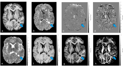 |
133 |
Characterization of Paramagnetic Rim Lesions using quantitative
and semi-quantitative MRI modalities
Agnese Tamanti1,
Annalisa Colombi1,
Angela Peloso1,
Nicola Serafin1,
Valentina Camera1,
Francesca Benedetta Pizzini2,
Marco Castellaro3,
and Massimiliano Calabrese2
1Department of Neurosciences, Biomedicine and Movement sciences, University of Verona, Verona, Italy, 2Neuroradiology Unit, Department of Diagnostics and Pathology, University of Verona, Verona, Italy, 3Department of Information Engineering, University of Padova, Padova, Italy Keywords: Multiple Sclerosis, Multi-Contrast, paramagnetic rim lesions Chronic active lesions have been recently investigated in multiple sclerosis (MS) and associated with the detection of paramagnetic rims on susceptibility-based MRI images (filtered phase and quantitative susceptibility mapping). Quantitative MRI approaches have the potential to reveal the degree of tissue damage in paramagnetic rim lesions. In this study a significant increase in T2* relaxation time and Quantitative Susceptibility Mapping (QSM) as well as a decrease in Magnetization Transfer Ratio (MTR) and Magnetization Transfer Saturation (MTSat) was revealed in PRL+ lesions compared to PRL-, thus supporting the use of these MRI metrics to detect tissue damage associated with chronic activity. |
|
2837. |
134 |
Hidden lesions may confound 1H-MRS metabolite quantification by
internal water referencing in individuals with multiple
sclerosis
Catherine M. Medeiros1,
Kay C. Igwe1,2,
Hetty Prinsen3,
Abhinav V. Kurada1,
Kelley M. Swanberg1,3,
and Christoph Juchem1,3,4,5
1Biomedical Engineering, Columbia University School of Engineering and Applied Science, New York, NY, United States, 2Neurology, Columbia University Irving Medical Center, New York, NY, United States, 3Radiology and Biomedical Imaging, Yale University School of Medicine, New Haven, CT, United States, 4Neurology, Yale University School of Medicine, New Haven, CT, United States, 5Radiology, Columbia University Irving Medical Center, New York, NY, United States Keywords: Multiple Sclerosis, Segmentation, Quantification, Water Referencing A current standard practice in proton magnetic resonance spectroscopy (1H-MRS) uses water as an internal reference for absolute metabolite quantification. We investigate how apparent metabolite concentrations might be affected by tissue-level changes, specifically multiple sclerosis lesions, via water versus creatine referencing in voxels considered normal-appearing at time of scan. Our results suggest that multiple possible routes of confound by hidden lesions on metabolite quantification in even normal-appearing tissue should be considered when applying internal water referencing to 1H-MRS studies in cohorts with multiple sclerosis. |
|
2838.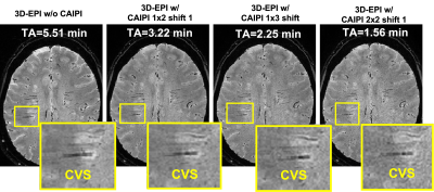 |
135 |
Feasibility of ultra-fast, high-resolution, T2*-weighted brain
imaging using 3D echo-planar imaging and CAIPIRINHA
Sreekanth Madhusoodhanan Nair1,
Jin Jin2,
Fei Han3,
Brian Renner1,
Elaina Gombos1,
Ke Cheng Liu4,
Sunil Patil5,
John A Derbyshire6,
Ken Sakaie7,
Emmanuel Obusez7,
Jonathan Lee7,
Mark Elliott8,
Russell T Shinohara9,
Matthew K Schindler10,
Jae W Song8,
Michel Bilello8,
Marwa Kaisey1,
Omar Al-Louzi1,
Nader Binesh11,
Marcel Maya11,
Javier Galvan11,
Hui Han12,
Debiao Li12,
Andrew Solomon13,
Daniel S Reich14,
Nancy L Sicotte1,
Mark Lowe7,
Daniel Ontaneda15,
and Pascal Sati1,12
1Department of Neurology, Cedars Sinai Medical Center, Los Angeles, CA, United States, 2Siemens Healthcare Pty Ltd, Brisbane, Australia, 3Siemens Medical Solutions, Los Angeles, CA, United States, 4Siemens Medical Solutions, Malvern, PA, United States, 5Siemens Medical Solutions, Baltimore, MD, United States, 6Functional MRI Facility, National Institute of Mental Health, Bethesda, MD, United States, 7Imaging Institute, Cleveland Clinic, Cleveland, OH, United States, 8Department of Radiology, University of Pennsylvania, Philadelphia, PA, United States, 9Department of Biostatistics, Epidemiology, and Informatics, University of Pennsylvania Perelman School of Medicine, Philadelphia, PA, United States, 10Department of Neurology, University of Pennsylvania, Philadelphia, PA, United States, 11Department of Imaging, Cedars-Sinai Medical Center, Los Angeles, CO, United States, 12Biomedical Imaging Research Institute, Cedars-Sinai Medical Center, Los Angeles, CA, United States, 13Larner College of Medicine, The University of Vermont, Burlington, VT, United States, 14Translational Neuroradiology Section, National Institute of Neurological Disorders and Stroke, National Institutes of Health, Bethesda, CA, United States, 15Mellen Center, Department of Neurology, Neurological Institute, Cleveland Clinic, Cleveland, OH, United States Keywords: Multiple Sclerosis, Data Analysis, Parallel Imaging High-resolution T2*-weighted brain imaging using 3D echo planar imaging (3D-EPI) at 3T allows detection of new biomarkers of neurological disorders, such as the central vein sign in multiple sclerosis. However, current 3D-EPI scan times are suboptimal for a widespread implementation in hospitals and private imaging centers. In this study, we evaluated the feasibility of combining 3D-EPI acquisition with different CAIPIRINHA undersampling patterns. A significant reduction in scan time (up to 70% reduction) was achieved without any obvious aliasing artifacts, confirming the feasibility of ultra-fast, high-resolution T2* brain imaging for future clinical applications at 3T. |
|
2839.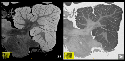 |
136 |
Time- and Signal-Efficient 3T Ultra-High-Resolution Imaging of
the Ex Vivo Cerebellum and Entire Human Brain
Matthias Weigel1,2,3,
Peter Dechent4,
Riccardo Galbusera1,2,
Erik Bahn5,
Ludwig Kappos1,2,
Wolfgang Brück5,
Christine Stadelmann5,
and Cristina Granziera1,2
1Translational Imaging in Neurology (ThINk) Basel, Department of Biomedical Engineering, Faculty of Medicine, University Hospital Basel and University of Basel, Basel, Switzerland, 2Neurological Clinic and Policlinic, MS Center and Research Center for Clinical Neuroimmunology and Neuroscience Basel (RC2NB), University Hospital Basel and University of Basel, Basel, Switzerland, 3Division of Radiological Physics, Dept. of Radiology, University Hospital Basel, Basel, Switzerland, 4MR-Research in Neurosciences, Department of Cognitive Neurology, University Medical Center Göttingen, Göttingen, Germany, 5Institute of Neuropathology, University Medical Center Göttingen, Göttingen, Germany Keywords: Multiple Sclerosis, Ex-Vivo Applications Ex Vivo MRI of the entire human brain facilitates new and fascinating insights into cerebral and cerebellar morphology and pathology. One key factor for achieving ultra-high spatial resolutions is a signal- and time-efficient MRI sequence, particularly, at 3T field strength. Though counterintuitive at first notion, we suggest a “slow” balanced steady-state free precession (bSSFP) approach with phase-cycling and very low receiver bandwidth (“LoBa-bSSFP”) as a highly signal- and time-efficient scheme for ex vivo acquisitions. LoBa-bSSFP can support spatial resolutions as high as 98-microns isotropic for covering the entire cerebellum on a common 3T MR system. |
|
2840.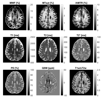 |
137 |
Assessment of multiple sclerosis pathology with quantitative
magnetic resonance imaging
Sarah Schlaeger1,
Mark Mühlau2,
Guillaume Gilbert3,
Irene Vavasour4,
Thomas Amthor5,
Mariya Doneva5,
Aurore Menegaux1,
Markus Lauerer2,
Viola Pongratz2,
Claus Zimmer1,
Benedikt Wiestler1,
Christine Preibisch1,2,
Jan Kirschke1,
and Ronja C. Berg1,2
1Department of Diagnostic and Interventional Neuroradiology, Klinikum rechts der Isar, Technical University of Munich, Munich, Germany, 2Department of Neurology, Klinikum rechts der Isar, Technical University of Munich, Munich, Germany, 3MR Clinical Science, Philips Healthcare, Mississauga, ON, Canada, 4Department of Radiology, University of British Columbia, Vancouver, BC, Canada, 5Philips Research Europe, Hamburg, Germany Keywords: Multiple Sclerosis, Brain Quantitative MRI in patients with multiple sclerosis (MS) allows for the objective identification of even subtle pathological changes in lesional, perilesional, NAGM, and NAWM tissue. Routine clinical scan protocols need to be tailored to the most diagnostically relevant MR biomarkers, however, to date only a few studies have investigated the particular diagnostic sensitivity of different MR biomarkers to different structural alterations of brain tissue affected by MS. Therefore, we studied 13 MS patients and 14 healthy controls using multiparametric, quantitative MR: we evaluated and compared the ability of nine different MR biomarkers to detect and characterize MS pathology. |
|
2841.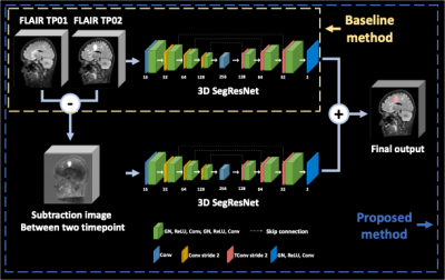 |
138 |
New Multiple Sclerosis Lesion Segmentation in Longitudinal FLAIR
MR Images using Subtraction Image
Junghwa Kang1,
Jeongmin Yim2,
Jinhee Jang3,
and Yoonho Nam1
1Department of Biomedical engineering, Hankuk University of Foreign Studies, Yongin, Korea, Republic of, 2College of Medicine, The Catholic University of Korea, Seoul, Korea, Republic of, 3Department of Radiology, Seoul St. Mary's Hospital, Seoul, Korea, Republic of Keywords: Multiple Sclerosis, Multiple Sclerosis, longitudinal MR, new lesion Measurement of new lesions in longitudinal images is crucial in the evaluation of MS but is laborious and time-consuming. In this study, wWe investigate the effect of subtraction images between two time point FLAIR images on new MS lesion segmentation in longitudinal FLAIR images. The results showed that our method with combination of longitudinal FLAIR images and their subtraction images together helps to improve the accuracy of segmentation of new MS lesion. |
|
2842.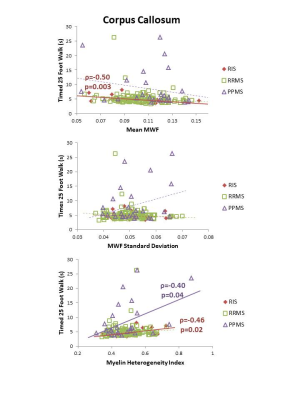 |
139 |
White Matter Myelin Content and Motor Function in Radiologically
Isolated Syndrome and Multiple Sclerosis
Irene Margaret Vavasour1,
Connor Keane2,
Poljanka Johnson3,
Joshua Lee2,
Adelia Adelia2,
Jiwon Oh4,
Anthony Traboulsee2,
and Shannon Kolind1,2,5
1Radiology, University of British Columbia, Vancouver, BC, Canada, 2Medicine, University of British Columbia, Vancouver, BC, Canada, 3Neuroscience, University of British Columbia, Vancouver, BC, Canada, 4Medicine, University of Toronto, Toronto, ON, Canada, 5Physics and Astronomy, University of British Columbia, Vancouver, BC, Canada Keywords: Multiple Sclerosis, Relaxometry, radiologically isolated syndrome, normal-appearing, myelin water fraction, myelin heterogeneity index white matter, motor function Regional myelin water fraction (MWF) in radiologically isolated syndrome (RIS) white matter were compared to relapsing-remitting (RR) and primary-progressive (PP) multiple sclerosis (MS). Results showed myelin damage in some white matter regions of RIS. Early myelin damage in RIS may be associated with subclinical ambulatory dysfunction. Manual dexterity correlated with myelin content in the cortical spinal tract and corpus callosum in PPMS whereas it correlated with myelin heterogeneity in RRMS. These findings suggest that myelin changes in critical white matter tracts have differing impact in gross and fine motor function in RIS and MS subtypes. |
|
 Back to Meeting Home
 Back to the Program-at-a-Glance
The International Society for Magnetic Resonance in Medicine is accredited by the Accreditation Council for Continuing Medical Education to provide continuing medical education for physicians.
 Back to Meeting Home
 Back to the Program-at-a-Glance
 View Presentation Video