Digital Poster
Seeking Further Mechanisms in Tumors I
ISMRM & ISMRT Annual Meeting & Exhibition • 03-08 June 2023 • Toronto, ON, Canada

Digital Poster
Seeking Further Mechanisms in Tumors I
| Computer # | |||
|---|---|---|---|
2314.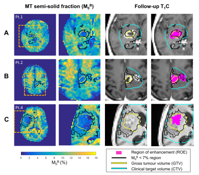 |
141 | Characterizing Quantitative Magnetization Transfer Maps from an MR-Linac in Regions of Progression in Biopsy-Only Glioblastoma
Rachel W Chan1, Liam SP Lawrence2, Hany Soliman3, Mark Ruschin4, James Stewart4, Aimee Theriault4, Chia-Lin Tseng5, Sten Myrehaug3, Jay Detsky4, Pejman J Maralani6, Hanbo Chen4, Brandon Tran2, Mary Jane Lim-Fat7, Sunit Das8, Greg J Stanisz1,2,9, Arjun Sahgal4, and Angus Z Lau1
1Physical Sciences Platform, Sunnybrook Research Institute, Toronto, ON, Canada, 2Medical Biophysics, University of Toronto, Toronto, ON, Canada, 3Sunnybrook Health Sciences Centre, Toronto, ON, Canada, 4Department of Radiation Oncology, Sunnybrook Health Sciences Centre, Toronto, ON, Canada, 5Department of Radiation Oncolog, Sunnybrook Health Sciences Centre, Toronto, ON, Canada, 6Department of Medical Imaging, University of Toronto, Sunnybrook Health Sciences Centre, Toronto, ON, Canada, 7Division of Neurology, Department of Medicine, Sunnybrook Health Sciences Centre, University of Toronto, Toronto, ON, Canada, 8Department of Surgery, St. Michael’s Hospital, Toronto, ON, Canada, 9Department of Neurosurgery and Paediatric Neurosurgery, Medical University, Lublin, Poland Keywords: Tumors, CEST & MT, Radiotherapy In biopsy-only glioblastoma patients scanned and treated on a 1.5T MR-Linac, this study quantifies the relationship between the quantitative magnetization transfer (qMT) semi-solid fraction and enhancing regions seen on post-contrast T1-weighted imaging on follow-up MRI scans. Metrics were computed to assess the spatial overlap between low semi-solid fraction regions and enhancing regions. In certain patients, the low semi-solid fraction region at the time of treatment correlated with enhancing region at follow up imaging. Our results suggest reduced semi-solid fraction values precede tumour progression, which could be used for guiding dose adaptation in radiation therapy. |
|
2315.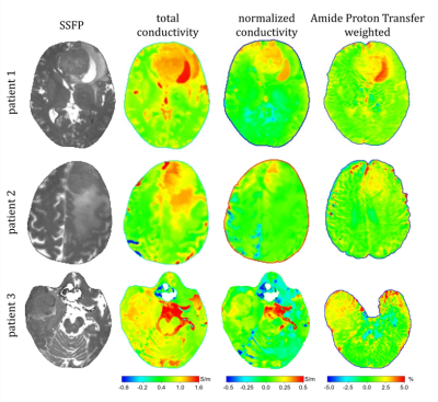 |
142 | Relationship between electric conductivity, water content, and protein content in brain tumors
Ulrich Katscher1 and Khin Khin Tha2
1Philips Research Europe, Hamburg, Germany, 2Hokkaido University, Sapporo, Japan Keywords: Tumors, Brain Strong mutual relation is observed between tissue electrical conductivity, water content, and sodium concentration. However, it was recently reported that the relation between conductivity and water content might hold only for healthy tissue but be violated in tumors. It was furthermore reported that water content relates only to free sodium, while conductivity additionally relates also to sodium bound to proteins. A discrepancy between water content and conductivity might thus indicate protein concentration. The current study confirms this hypothesis by comparing the discrepancy between water content and conductivity with Amide Proton Transfer imaging as a marker of protein concentration. |
|
2316.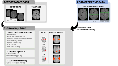 |
143 | Functional mapping in gliomas: a comparison between presurgical resting-state fMRI and intraoperative Direct Electrical Stimulation
Manuela Moretto1, Beatrice Federica Luciani1, Francesca Saviola1, Stefano Tambalo1, Luca Zigiotto2,3, Donna Gift Cabalo1,4, Luciano Annicchiarico2,3, Martina Venturini2,3, Silvio Sarubbo2,3, and Jorge Jovicich1
1Center for Mind/Brain Sciences (CIMeC), University of Trento, Trento, Italy, 2Department of Neuroscience, Division of Neurosurgery, S.Chiara Hospital, APSS Trento, Trento, Italy, 3Structural and Functional Connectivity Lab, S.Chiara Hospital, APSS Trento, Trento, Italy, 4Multimodal Imaging and Connectome Analysis Laboratory, McConnell Brain Imaging Centre, Montreal Neurological Institute and Hospital, McGill University, Montreal, QC, Canada Keywords: Tumors, fMRI (resting state), Brain Connectivity Resting-state functional MRI (rs-fMRI) has been proposed as a non-invasive technique for the presurgical mapping of functional networks, in contrast to intraoperative Direct Electrical Stimulation (DES) mapping. While previous studies have investigated the agreement between a predefined DES atlas and resting-state networks in low-grade glioma patients, here we exploit patient-specific DES points and the ReStNeuMap tool to investigate the agreement between sensorimotor, language and speech-articulation networks in a cohort of high- and low-grade glioma patients. We found a good overlap between ReStNeuMap-derived networks and DES points, while low spatial similarity between networks and functional connectivity maps derived from DES. |
|
2317.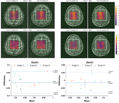 |
144 | A robust MRSI protocol to investigate glutamatergic mechanisms in glioma
Seyma Alcicek1,2,3,4, Michael W. Ronellenfitsch2,3,4,5, Dennis C. Thomas1,2,3,4, Joachim P. Steinbach2,3,4,5, Vincent Prinz6, Marie-Thérèse Forster6, Elke Hattingen1,2,3,4, Ulrich Pilatus1,2,3,4, and Katharina J. Wenger1,2,3,4
1Institute of Neuroradiology, University Hospital Frankfurt, Goethe University, Frankfurt am Main, Germany, 2University Cancer Center Frankfurt (UCT), Frankfurt am Main, Germany, 3Frankfurt Cancer Institute (FCI), Frankfurt am Main, Germany, 4German Cancer Research Center (DKFZ) Heidelberg, Germany and German Cancer Consortium (DKTK), Partner Site Frankfurt/Mainz, Frankfurt am Main, Germany, 5Dr. Senckenberg Institute of Neurooncology, University Hospital Frankfurt, Goethe University, Frankfurt am Main, Germany, 6Department of Neurosurgery, University Hospital Frankfurt, Goethe University, Frankfurt am Main, Germany Keywords: Tumors, Spectroscopy Glutamate metabolism plays a significant role in glioma invasion and growth and may lead to glioma-associated epileptic discharges and excitotoxicity. In in vivo MR spectroscopy, the molecular similarity of glutamate and glutamine results in overlapped, hence indistinguishable, spectral peaks and hinders their quantification. Here, we propose the use of J-modulation at optimum echo time for accurate and separate quantification of glutamate and glutamine. To study the reproducibility of this approach, in vivo MRSI sLASER measurement at 120 ms echo time was evaluated in three healthy subjects in two separate sessions, and the findings are reported. |
|
2318.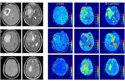 |
145 | Differentiation of tumor-infiltrated edema and pure vasogenic edema in patients with brain tumors by using a fractional order calculus model
Qinyuan Zhang1, Jianing Li1, Yun Su1, Zhuoheng Yan1, Zhixuan Hu1, Weike Zeng1, Mengzhu Wang2, Xu Yan2, Jiaji Mao1, and Jun Shen1
1Department of Radiology, Sun Yat-Sen Memorial Hospital, Sun Yat-Sen University, Guangzhou, China, 2MR Scientific Marketing, Siemens Healthineers Ltd, Guangzhou, China Keywords: Tumors, Diffusion/other diffusion imaging techniques A fractional order calculus model (FROC) holds promise in differentiating tumor-infiltrated edema from pure vasogenic edema of brain tumors. This study used a q-space Cartesian grid sampling scheme, diffusion spectrum imaging, in patients with high-grade gliomas, meningiomas and brain metastases to generate FROC-based parameters, including D, β and μ. The μ showed the best individual parameter performance in differentiating tumor-infiltrated edema from pure vasogenic edema. |
|
2319.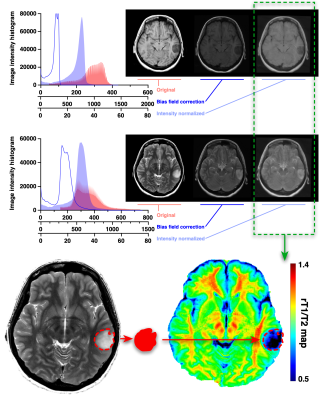 |
146 | Correlation of T1- to T2-weighted signal intensity ratio with T1- and T2-relaxation time and IDH mutation status in glioma
Takahiro Sanada1, Shota Yamamoto1,2, Mio Sakai3, Toru Umehara2,4, Hirotaka Sato1,5, Masato Saito1, Nobuyuki Mitsui1, Satoru Hiroshima1, Ryogo Anei6, Yonehiro Kanemura7, Mishie Tanino8, Katsuyuki Nakanishi3, Haruhiko Kishima2, and Manabu Kinoshita1,9
1Neurosurgery, Asahikawa Medical University, Asahikawa, Japan, 2Neurosurgery, Osaka University Graduate School of Medicine, Suita, Japan, 3Diagnostic Radiology, Osaka International Cancer Institute, Osaka, Japan, 4Neurosurgery, Hanwa Memorial Hospital, Osaka, Japan, 5Neurosurgery, Japanese Red Cross Kitami Hospital, Kitami, Japan, 6Neurosugery, Moriyama Hospital, Asahikawa, Japan, 7Biomedical Research and Innovation, Institute for Clinical Research, National Hospital Organization Osaka National Hospital, Osaka, Japan, 8Diagnostic Pathology, Asahikawa Medical University, Asahikawa, Japan, 9Neurosugery, Osaka International Cancer Institute, Osaka, Japan Keywords: Tumors, Radiomics The current study aimed to test whether the ratio of T1-weighted to T2-weighted signal intensity (rT1/T2) derived from conventional MRI could act as a surrogate relaxation time predictive of IDH mutation status in histologically lower-grade gliomas. The findings showed that rT1/T2 strongly correlates with T1- and T2-relaxation times in histologically LrGGs. The mean value of rT1/T2 was able to discriminate IDHwt and IDHmt tumors in two domestic cohorts, with statistical significance. However, this result was not validated in the original TCIA/TCGA cohort due to the wide variety of imaging characteristics in the cohort. |
|
2320.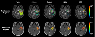 |
147 | DCE-MRI in Distinguishing Solitary Brain Metastasis from Glioblastoma: Evaluation of Multiple Pharmacokinetic Models
Bao Wang1, Yuan Yao1, Jingzhen He1, and Dmytro Pylypenko2
1Radiology, Qilu Hospital of Shandong University, Jinan, China, 2GE Healthcare, Beijing, China Keywords: Tumors, DSC & DCE Perfusion This study aimed to explore performances of parameters derived from DCE-MRI with multiple pharmacokinetic models regarding this differential diagnosis, as well as overall performances of models. Eighty-eight patients with suspected GBM and SBM underwent DCE-MRI in this study. Peritumoral vp from shutter-speed model achieved the best single-parameter’s performance. Additionally, shutter-speed model achieved the highest overall separating performance. Shutter-speed is the recommended DCE-MRI pharmacokinetic model for differential diagnosis between GBM and SBM. |
|
2321.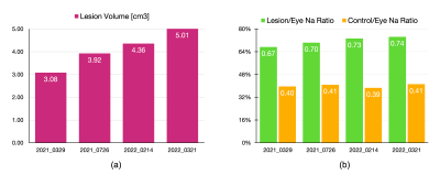 |
148 | Quantitative Sodium Imaging Report for Monitoring Brain Tumor Lesion Progress
Sanghoon Kim1 and Alexander Lin1
1BWH, Boston, MA, United States Keywords: Tumors, Quantitative Imaging, Sodium Imaging ▶︎ MR has been commonly used for the brain tumor patient monitoring. However, most of the MR images does not provide quantitative physiologic information, which can be ambiguous with longitudinal monitoring. ▶︎ Sodium MR imaging can quantitatively provide tumor cell viability information. ▶︎ This presentation introduces a brain tumor patient’s longitudinal quantitative sodium imaging report. ▶︎ Potentially, this quantitative report can be attributed to the tumor patient’s treatment plan. |
|
2322.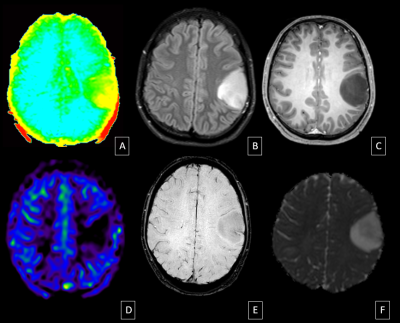 |
149 | Comprehensive Analysis Of APTw Imaging In Diagnosis And Grading Of Gliomas – Bridging The Gap Between Imaging And Histopathological Outcomes
Lavanya Yegnaraman1, Natesan Chidambaranathan1, Adhityan Rajendran2, Rashmi Rao3, Manikandan Mariyapillai3, and Narayana Krishna Rolla3
1Radiodiagnosis, Apollo Hospitals Chennai, Chennai, India, 2Radiology, Apollo Hospitals, Chennai, India, 3Philips India Ltd, Gurgaon, India Keywords: Tumors, CEST & MT, amide proton transfer The utility of Amide Proton Transfer weighted imaging in preoperative grading of gliomas was studied. 64 patients with various primary intraaxial tumours were included in the study. Mean APTw was able to differentiate between IDH mutant grade 2 and grade 3 gliomas (astrocytomas and oligodendrogliomas) with statistical significance (p<0.05). APTw imaging can well delineate between the tumour core and the tumour periphery and the difference in the APT means between the two regions is statistically significant. APTw imaging also identifies grade 3 tumours misclassified as grade 2 on other sequences. |
|
2323.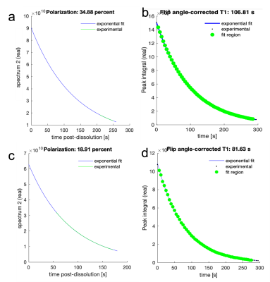 |
150 | Deuterated Vitamin C as a new probe for the assessment of in vivo oxidative stress using deuterium metabolic imaging and 13C HP MRS
Saket Patel1, Nathaniel Kim1, Thasin Peyear1, and Kayvan R Keshari1
1Radiology, Memorial Sloan Kettering Cancer Center, Manhattan, NY, United States Keywords: Tumors, Deuterium, DMI, HP, Metabolism, Probes, Imaging agent A chemical synthesis of C-4 deuterated ascorbic acid as a potential new deuterium metabolic imaging marker for in vivo VitC metabolism. Additionally, we have developed new HP methods for Vit C as well as its oxidized analogue, DHA, to be used as a 13C HP MRS substrates. Furthermore, deuteration increases the 13C T1 of HP DHA and improved polarization from optimized HP formulations along with enhanced 13C T1 will result in better quantification of oxidative stress in vivo. The aim is to implement both DMI and HP MRS in brain tumor models to understand the anticancer activity of VitC. |
|
2324.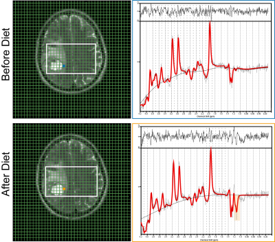 |
151 | Analysis of fasting-induced metabolic changes in glioma tissue detected by non-invasive 1H/31P MR-Spectroscopy
Seyma Alcicek1,2,3,4, Iris Divé2,3,4,5, Ulrich Pilatus1,2,3,4, Vincent Prinz6, Joachim P. Steinbach2,3,4,5, Marie-Thérèse Forster6, Elke Hattingen1,2,3,4, Michael W. Ronellenfitsch2,3,4,5, and Katharina J. Wenger1,2,3,4
1Institute of Neuroradiology, University Hospital Frankfurt, Goethe University, Frankfurt am Main, Germany, 2University Cancer Center Frankfurt (UCT), Frankfurt am Main, Germany, 3Frankfurt Cancer Institute (FCI), Frankfurt am Main, Germany, 4German Cancer Research Center (DKFZ) Heidelberg, Germany and German Cancer Consortium (DKTK), Partner Site Frankfurt/Mainz, Frankfurt am Main, Germany, 5Dr. Senckenberg Institute of Neurooncology, University Hospital Frankfurt, Goethe University, Frankfurt am Main, Germany, 6Department of Neurosurgery, University Hospital Frankfurt, Goethe University, Frankfurt am Main, Germany Keywords: Tumors, Spectroscopy Nutritional interventions such as fasting are currently under evaluation as anti-cancer treatment. In glioma patient cohorts, the feasibility and safety of fasting in addition to antitumor treatment has been shown. However, it is still unclear whether fasting exerts effects on the glioma tumor tissue at all, and whether fasting causes metabolic or immunological changes in the glioma microenvironment that could be exploited therapeutically. This is a report on the metabolic flexibility of tumor cells induced by a fasting cycle of 72 hours prior to biopsy or resection analyzed by MR-Spectroscopy (MRS) as part of the ERGO3-trial. |
|
2325.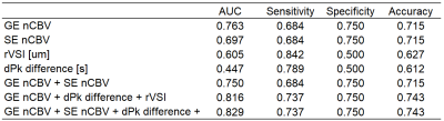 |
152 | Benefit of Vessel Size/Architecture Imaging with DSC-MRI for Differentiation of Tumor Recurrence versus Pseudoprogression in Glioma Patients
Jian Ming Teo1,2, Jason M Johnson3, Halyna Pokhylevych3, Kinsey Lano3, Ping Hou1, Xinzeng Wang4, and Ho-Ling Liu1
1Department of Imaging Physics, The University of Texas MD Anderson Cancer Center, Houston, TX, United States, 2The University of Texas MD Anderson Cancer Center UTHealth Graduate School of Biomedical Sciences, Houston, TX, United States, 3Department of Neuroradiology, The University of Texas MD Anderson Cancer Center, Houston, TX, United States, 4GE Healthcare, Houston, TX, United States Keywords: Tumors, DSC & DCE Perfusion, Gliomas Vessel Size Imaging was performed on twenty-five glioma patients to obtain voxel-wise relative vessel size index and peak shift maps. This study aimed to investigate if relative vessel size index and difference in peak shift between lesion vs normal appearing white matter voxels will assist in differentiating pseudoprogression vs tumor recurrence for post-treatment gliomas. It was shown that AUC and accuracy increases when incorporating rVSI and difference in peak shift. |
|
2326.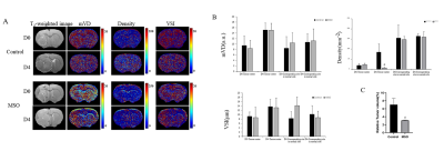 |
153 | Effects of glutamine synthetase on neovascularization in glioma studied by In vivo MR vessel size imaging
tianwei song1, yanan zhang1, chen zhou1, and Junchao Qian1
1Institute of Health and Medical Technology, Hefei Institutes of Physical Science, Hefei Cancer Hospital, Chinese Academy of Sciences, Hefei 230031, P. R. China, Hefei, China Keywords: Tumors, Blood vessels Glioblastoma (GBM) is a highly invasive and vascularized primary malignant brain tumor. Glutamine synthetase (GS) could induce vessel sprouting through the improvement of endothelial cell migration in inflammatory diseases. MR vessel size imaging was supposed to visualize angiogenesis potentially in the brain. In the study we research the role of GS on neovascularization in glioma by in vivo vessel size imaging. Our results indicate that the vascular changes mainly occur in the tumor center and significant decreases of microvascular density values in the MSO group compared to the control. GS inhibitor could be a potential drug to treat glioma. |
|
2327.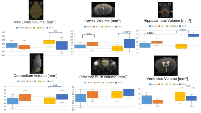 |
154 | Differences in Brain Structural and Metabolic Phenotype in Mouse Models of Down Syndrome and Brain Tumors
Natalie Julie Serkova1, Paula Araya2, Sharon Cain3, Jenna Steiner1, Kelly Sullivan4, and Joaquin Espinoza2
1Radiology, University of Colorado Anschutz, Aurora, CO, United States, 2Pharmacology, University of Colorado Anschutz, Aurora, CO, United States, 3Biochemistry, Chaminade University of Honolulu, Honolulu, HI, United States, 4Pediatrics, University of Colorado Anschutz, Aurora, CO, United States Keywords: Tumors, Animals, Down Syndrome Gliomas are exceedingly rare in Down Syndrome (DS). This study characterizes metabolic biomarkers and structural differences in the brains of mouse DS models and compare them to glioma models. Analysis of the age-dependent differences in the metabolic profile was carried out by 1H-NMR metabolomics and compartmental brain volumes of mouse brains were assessed from T2-weighted MRI. A significant age-dependent neuronal loss in the cortex and hippocampus, with a hyperventricular profile, was observed in DS mice. Highly decreased lactate and amino acid concentrations were observed in DS, in contrast to glioma extracts, indicative of decreased metabolic dependency on glycolysis and proteolysis. |
|
2328. |
155 | Evaluations of tumor acidosis that monitors early response to radiotherapy in a glioblastoma model using acidoCEST MRI
Michelle Zalles1, F William Schuler1, Jorge de la Cerda1, Alia Khaled1, Sanhita Sinharay2, and Mark D Pagel1
1Cancer Systems Imaging, UT MD Anderson Cancer Center, Houston, TX, United States, 2Center for Biosystems Science and Engineering, Indian Institute of Science, Bangalore, India Keywords: Cancer, CEST & MT, pH imaging The treatment of glioblastoma potentially causes tumor necrosis, which has reduced glycolytic metabolism. AcidoCEST MRI measures tumor acidosis caused by tumor glycolysis. Our study demonstrated that acidoCEST MRI, a molecular imaging method that measures tumor extracellular pH, can detect a decrease in pHe < 7.0 for tumors that resisted radiotherapy or were untreated, and can detect an increase in pHe > 7.0 for tumors that were successfully treated or showed evidence for necrosis. |
|
2329.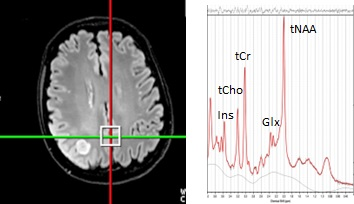 |
156 | Metabolic Alternations in Normal Appearing Brain in Brain Tumor Patients: Potential Mechanism of Chemo-Brain
Natasha Najam1, Sam Jiang1, Huijun Liao1, and Alexander Peter Lin1
1Center for Clinical Spectroscopy, Department of Radiology, Brigham and Women's Hospital / Harvard Medical School, Boston, MA, United States Keywords: Tumors, Spectroscopy, chemotherapy, normal-appearing brain Patients that undergo chemotherapy have been shown to develop cognitive dysfunction after treatment. Previous studies have shown changes in white matter metabolism; however, few have examined grey matter metabolism. Our results show that glutamate, glutathione, creatine, N-acetylaspartate, and myoinositol were reduced in the posterior cingulate in tumor patients that underwent chemotherapy when compared to healthy controls. The effect of radiotherapy was also examined but did not show metabolic differences. |
|
2330.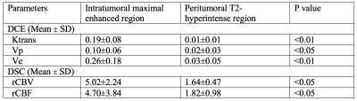 |
157 | MR multitasking-based dynamic imaging forcerebrovascular evaluation (MT-DICE) in brain tumors
Jiayu Xiao1, Yang Chen1, Jason Ye2, Zhehao Hu1, Anthony Christodoulou3, Mark Shiroshi1, Debiao Li3, and Zhaoyang Fan1,2,4
1Radiology, Keck School of Medicine of USC, Los Angeles, CA, United States, 2Radiation Oncology, Keck School of Medicine of USC, Los Angeles, CA, United States, 3Cedars-Sinai Medical Center, Los Angeles, CA, United States, 4Viterbi School of Engineering, University of Southern California, Los Angeles, CA, United States Keywords: Tumors, DSC & DCE Perfusion Brain tumors represent a great therapeutic challenge and tend to impact blood brain barrier (BBB) integrity, angiogenesis, and vascularity. Dynamic contrast-enhanced (DCE) MRI is widely used to quantify BBB permeability and dynamic susceptibility contrast MRI (DSC) is used to quantify cerebral perfusion. We incorporated the newly developed MR multitasking-based dynamic imaging for cerebrovascular evaluation (MT-DICE) technique to achieve simultaneous DCE and DSC acquisition with a single-dose injection. MT-DICE could quantify DCE and DSC parameters with good inter-reader agreement in patients with brain tumors. The intratumoral enhanced region showed more severe BBB disruption and higher perfusion than the peritumoral T2-hyperintense regions. |
|
2331. |
158 | Prediction of IDH status of Glioma using Diffusion Tensor Imaging and Clinical features
Yuxia Liang1, Yuhan Ren2, Yu Shang3, Xiang Liu3, Maode Wang3, Ming Zhang3, and Chen Niu4
1The first affiliated hospital of Xi'an Jiaotong University, Xi'An, China, 2Hospital of Stomatology Xi'an Jiaotong University, Xi'an, China, 3The first affiliated hospital of Xi'an Jiaotong University, Xi'an, China, 4The first affiliated hospital of Xi'an Jiaotong University, XI'an, China Keywords: Tumors, Diffusion Tensor Imaging, glioma Isocitrate dehydrogenase (IDH) is critical to prognosis of glioma. While, reliable techniques for preoperative assessment of IDH status remain scarce. In this study, we investigated mean diffusivity (MD) and anisotropy fraction (FA) using Diffusion Tensor Imaging (DTI) combined with the clinical features to predict IDH status. Our results found significant differences in FAmean/FAnawm, MDmin, NLR, and age between IDH mutant and IDH wild groups. The model incorporating FAmean/FAnawm, MDmin, NLR, and age predicted IDH status with area under ROC curve of 0.85, 95% CI: 74.3%~95.7%. Our findings suggested that DTI combined with clinical features can non-invasively prediction of IDH status. |
|
 Back to Meeting Home
 Back to the Program-at-a-Glance
The International Society for Magnetic Resonance in Medicine is accredited by the Accreditation Council for Continuing Medical Education to provide continuing medical education for physicians.
 Back to Meeting Home
 Back to the Program-at-a-Glance
 View Presentation Video