Digital Poster
Seeking Further Mechanisms in Tumors II
ISMRM & ISMRT Annual Meeting & Exhibition • 03-08 June 2023 • Toronto, ON, Canada

Digital Poster
Seeking Further Mechanisms in Tumors II
| Computer # | |||
|---|---|---|---|
2490.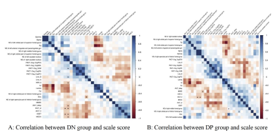 |
141 | The effect of different expressions of Delta-catenin on the properties of small world brain network in breast cancer patients before chemotherapy
Mingtuan Xue1, Jiajun Cao1, Wei Du1, Wenjia Wang2, and Yanwei Miao1
1The department of radiology, the first Affiliated Hospital of Dalian Medical University, Dalian, China, 2GE Healthcare, Beijing, China Keywords: Tumors, Cancer, BRCA patients, Delta-catenin, Resting-state fMRI This work aimed to explore the effects of different expressions of Delta-catenin in breast cancer(BRCA) patients based on small-world brain network. The results showed that the expression status of Delta-catenin proteins had significant differences on the small-world brain network properties of patients. |
|
2491.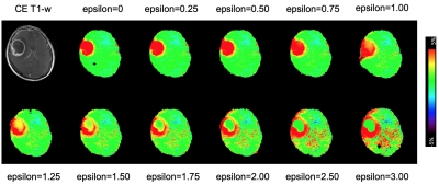 |
142 | Amide proton transfer-weighted MRI of brain tumors with fluid & solid compartment corrections using background magnetization transfer effects
Osamu Togao1, Jochen Keupp2, Koji Yamashita3, Kazufumi Kikuchi4, Tatsuhiro Wada5, and Kousei Ishigami4
1Molecular Imaging & Diagnosis, Graduate School of Medical Sciences, Kyushu University, Fukuoka, Japan, 2Philips Research, Hamburg, Germany, 3Department of Radiology Informatics and Network, Graduate School of Medical Sciences, Kyushu University, Fukuoka, Japan, 4Department of Clinical Radiology, Graduate School of Medical Sciences, Kyushu University, Fukuoka, Japan, 5Division of Radiology, Department of Medical Technology, Graduate School of Medical Sciences, Kyushu University, Fukuoka, Japan Keywords: Tumors, CEST & MT We propose a simple and efficient metric for APT-weighted MRI, which suppresses fluid signals and enhances signals form solid components based on the spectral shape of the background MT ratio. No extra acquisition or mathematical fitting is needed because the metric can be computed from the minimum Z-spectral data required for standard APT-weighted MRI. This post-processing normalizing for background MT effects may facilitate the quantitative evaluation of the APT-weighted signal of the active solid component in brain tumors. |
|
2492.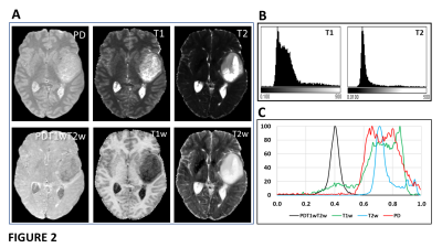 |
143 | Optimisation of 1H MRS quantitation with tissue water referencing in the presence of brain tumour heterogeneity
Franklyn A Howe1, Timothy L Jones2, Philip Rich3, Felix Raschke4, Jamshid Dehmeshki5, and Thomas R Barrick1
1Molecular and Clinical Sciences Research Institute, St George's, University of London, London, United Kingdom, 2Neurosurgery, St George's University Hospital Foundation Trust, London, United Kingdom, 3Neuroradiology, St George's University Hospital Foundation Trust, London, United Kingdom, 4Faculty of Medicine and University Hospital Carl Gustav Carus, OncoRay - National Center for Radiation Research in Oncology, Dresden, Germany, 5Image Analysis Group, London, United Kingdom Keywords: Tumors, Spectroscopy, quantitation A tissue water signal is frequently used as a reference for metabolite quantitation by 1H MRS. We have determined the variability of T1, T2 and PD in glial brain tumours and normal brain to assess how tumour heterogeneity affects the reference signal at 3T. The typical short echo time MRS parameters TR 2000ms and TE 32ms reside within a “sweet spot” of minimum variation in the product PD*exp(-TE/T2)*[1-exp(-TR/T1)] that determines the water reference signal across a wide range of tissues (NWM, tumour core, necrosis and oedema). This choice also provides good SNR per unit time for the key metabolite singlets. |
|
2493.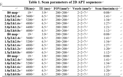 |
144 | Optimization of 2D-APT imaging of brain tumor based on saturated RF pulse parameters
na liu1, ailian liu2, peng sun1, qingwei song1, and yanwei miao3
1The First Affiliated Hospital of Dalian Medical University, DaLian, China, 2The First Affiliated Hospital of Dalian Medical University, Dalian, China, 3The First Affiliated Hospital of Dalian Medical University, dalian, China Keywords: Tumors, Molecular Imaging 2D APTw imaging is a single-slice imaging method focusing on the selected plane, with low spatial coverage and a much shorter scan time than its 3D counterpart, which makes it an optional scan sequence for patients who cannot tolerate long scan times. However, there is still a lack of an optimal setting for 2D APTw imaging in clinics for human brain tumors. Therefore, this study aims to explore the different combinations of pulse power and duration of saturation RF pulse in 2D APTw imaging on brain tumors and to find an optimal setting suitable for clinical use. |
|
2494.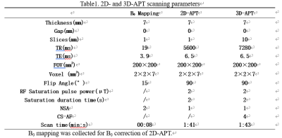 |
145 | Consistency between 2D and 3D amide proton weighted imaging on brain tumors
na liu1, liangjie lin2, yukun zhang1, ailian liu3, qingwei song1, and yanwei miao1
1The First Affiliated Hospital of Dalian Medical University, DaLian, China, 2Philips Healthcare, BeiJing, China, 3The First Affiliated Hospital of Dalian Medical University, Dalian, China Keywords: Tumors, Molecular Imaging Amide Proton Transfer (APT) weighted imaging is a new type of magnetic resonance molecular imaging technology derived from Chemical Exchange Saturation Transfer (CEST) technology. APT weighted imaging can be carried out with 2D and 3D acquisition mode, so called as 2D- and 3D-APT respectively. There is no relevant report on the comparison of 2D and 3D-APT imaging on brain tumors. This study intends to compare and analyze the performance of 2D- and 3D-APT imaging on brain tumors through imaging and pathological data, and recommend a better APT imaging scheme to improve the diagnosis of brain tumors. |
|
2495.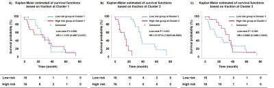 |
146 | Spatial Habitats Analysis based on Multiparametric Physiologic MR Imaging Predicts Survival of Patients with Glioblastoma
Mengqiu Cao1, Liu Fang1, Xiaoqing Wang1, Xinyue Liang2, Yongming Dai2, and Zhou Yan1
1Department of Radiology, Renji Hospital, School of Medicine, Shanghai Jiao Tong University, Shanghai, China, 2MR Collaboration, Central Research Institute, United Imaging Healthcare, Shanghai, China Keywords: Tumors, Tumor, glioblastoma Glioblastoma (GBM) is characterized remarkably high tumors heterogeneity which contribute to its poor survival and effective therapies. We build the spatial habitats based on multiparametric physiologic MRI (diffusion-weighted and perfusion-weighted MRI) to describe the intratumoral heterogeneity of gliomas and explore the possibility of the link between theses habitats and progression-free survival (PFS). This shows the spatial correspondence with molecular histological features and MRI images. This also provide valuable information to identify tumor progression areas that may require biopsy and potential treatment resistance subregions which tend to be the target areas of treatment. |
|
2496.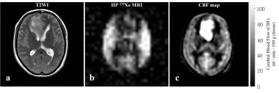 |
147 | Hyperpolarized 129Xe Magnetic Resonance Imaging in Patients with Brain Tumors
Kun Cheng1,2, Chenxi Li1, Caohui Duan1, Xiangbing Bian1, Haidong Li3, Xin Zhou3, and Xin Lou1
1Department of Radiology, Chinese PLA General Hospital, Beijing, China, 2School of Medical Imaging, Guizhou Medical University, Guiyang, China, 3Key Laboratory of Magnetic Resonance in Biological Systems, State Key Laboratory of Magnetic Resonance and Atomic and Molecular Physics, National Center for Magnetic Resonance in Wuhan, Wuhan Institute of Physics and Mathematics, Innovation Academy for Precision Measurement Science and Technology, Chinese Academy of Sciences-Wuhan National Laboratory for Optoelectronics, Wuhan, China Keywords: Tumors, Hyperpolarized MR (Gas), Xenon-129 This study explored the application of hyperpolarized 129Xe (HP 129Xe) MR imaging in patients with brain tumors. HP129Xe MR and 1H MR imaging was performed on three subjects. The results showed that HP 129Xe MRI has the ability to detect brain tumors, but the HP 129Xe brain MR imaging showed the lesion range was mismatched with T2 weighted image (T2WI) and arterial spin labeling (ASL)image, these are needed to be addressed in the future. |
|
2497.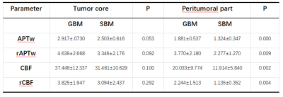 |
148 | Using 3D APTw and ASL imaging to differentiate solitary brain metastases from glioblastomas
Ling Chen1, Tao Li1, Zisan Zeng2, Yao Li1, Kan Deng3, Shuanghong Li1, Jinhuan Zhang1, and Lifang Tang1
1The Fourth Affiliated Hospital, Guangxi Medical University, Liuzhou, China, 2The First Affiliated Hospital, Guangxi Medical University, Nanning, China, 3Philips Healthcare, Guangzhou, China Keywords: Tumors, Brain This study is to determine whether APTw and ASL imaging are useful for distinguishing GBMs and SBMs. The differences in APT and CBF values between GBMs and SBMs were compared.The result showed that 3D APTw is an important MRI technique for distinguishing SBMs and GBMs. Particularly, APTw combined with ASL demonstrated a more satisfactory discrimination and a higher diagnostic performance. |
|
2498.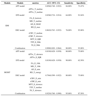 |
149 | Histogram analysis of APT Combined with DTI and DSC MRI for Predicting IDH Mutation and MGMT promotor methylation Status in Glioma
Lan Zhang1, Xinli Zhang1, Xiaona Fu1, Yuxi Jia1, Xiaoming Liu1, Lan Cheng1, Zhengwu Tan1, Xudong Li2, Zhengyin Cheng3, Xiaochuan Dong4, Peng Sun5, Xiaoxiao Zhang5, Xiaobin Jiang2, Chuansheng Zheng1, Xuan Wang2, and Jing Wang1
1Department of Radiology, Union hospital, Tongji Medical College, Huazhong University of Science and Technology, Wuhan, 430022, China., Wuhan, China, 2Department of Neurosurgery, Union hospital, Tongji Medical College, Huazhong University of Science and Technology, Wuhan, 430022, China., Wuhan, China, 3People's Hospital of Dongxihu District, Wuhan, Hubei, China., Wuhan, China, 4Department of Pathology, Union hospital, Tongji Medical College, Huazhong University of Science and Technology, Wuhan, 430022, China., Wuhan, China, 5Clinical & Technical Solutions, Philips Healthcare, Beijing 100600, China., Wuhan, China Keywords: Tumors, Multi-Contrast In this study, the diagnostic performance of histogram features of APT, DTI and DSC in predicting IDH mutation and MGMT promoter methylation status of gliomas was compared. Secondly, the histogram parameters from the signal-time intensity curve of DSC significantly improved the predictive performance of DSC model. Most importantly, the combined logistic regression model combined with APT, DTI and DSC can evaluate the tumor nature of glioma more comprehensively and obtain better diagnostic performance, which is expected to become an imaging molecular marker for the prediction of glioma genotyping in the future. |
|
2499.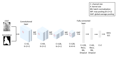 |
150 | Convolutional neural network to predict IDH mutation status in glioma from 7T chemical exchange saturation transfer imaging
Yifan Yuan1, Yang Yu2, Jun Chang1, Ying-Hua Chu3, Yi-Cheng Hsu3, Mianxin Liu4, and Qi Yue1
1Department of neurosurgery, Huashan Hospital Fudan University, Shanghai, China, 2Department of radiology, Huashan Hospital Fudan University, Shanghai, China, 3MR Collaboration, Siemens Healthineers Ltd, Shanghai, China, 4School of Biomedical Engineering, ShanghaiTech University, shanghai, China Keywords: Tumors, CEST & MT Noninvasive prediction of isocitrate dehydrogenase (IDH) mutation status in glioma guides surgical strategies and individualized management. We explored the capability of preoperatively identifying IDH status by combining a 2D convolutional neural network (CNN) and amide proton transfer chemical exchange saturation transfer (APT-CEST) imaging. Five-fold cross-validation suggested the APT-CEST with the tumor shape information predicts IDH status optimally. The novel CNN model designed for 7T APT-CEST offers improved discriminatory accuracy in predicting the IDH status of glioma, holding great potential for facilitating decision-making in clinical practice. |
|
2500.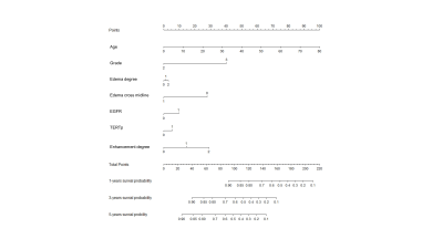 |
151 | Combined new molecular signature and radiological signature for risk stratification in IDH wild-type lower-grade glioma
Zhiyi Zhang1, Hui Zhang1, and Yan Tan1
1Shanxi Medical University, Taiyuan, China Keywords: Tumors, Cancer, Iower-grade glioma When radiological signature was combined with new molecular signature, higher performance was achieved in predicting the prognosis of IDH wild-type lower-grade glioma patients. The combined model showed great risk stratification ability, which improving survival prediction in patients with IDH wild-type lower-grade glioma. |
|
2501.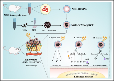 |
152 | A Novel Biomimetic Nanodrug with High Reactive Oxygen Species Production for Precision Combination Therapy of Glioblastoma
Haiyan Gao1 and Meiyun Wang1,2
1Henan provincial People’s Hospital, Zhengzhou, China, 2Laboratory of Brain Science and Brain-Like Intelligence Technology, Institute for Integrated Medical Science and Engineering, Henan Academy of Sciences, Zhengzhou, China Keywords: Tumors, Brain This project intends to innovatively design and synthesize novel radiotherapy sensitization and enhanced CDT dual-effect sensitization nanodrugs, further wrap them with engineered red blood cell membrane vesicles, and construct a high-performance biomimetic nanomedicine synergistic treatment system with high targeting, high specificity and high yield of reactive oxygen species, in order to achieve the specificity and high efficiency accumulation of nanomedicines in the GBM, while reducing toxicity to normal tissues. Magnetic resonance and other imaging methods are used to guide the efficient treatment of drugs and monitor the effect of tumor ablation. |
|
2502.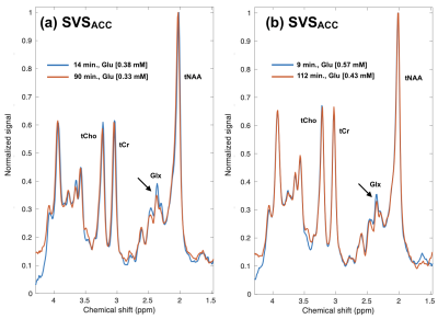 |
153 | Indirect detection of deuterated glucose brain metabolism at 3 Tesla: preliminary findings in volunteers and patients with gliomas
Adam W Autry1, Marisa LaFontaine1, Javier Villanueva-Meyer1, Susan M Chang2, Henk M de Feyter3, and Yan Li1
1Department of Radiology and Biomedical Imaging, University of California San Francisco, San Francisco, CA, United States, 2Department of Neurological Surgery, University of California San Francisco, San Francisco, CA, United States, 3Department of Radiology and Biomedical Imaging, Magnetic Resonance Research Center, Yale University, New Haven, CT, United States Keywords: Tumors, Brain, deuterated glucose This study aimed to perform indirect detection of deuterated glucose metabolism at 3 Tesla in healthy subjects and patients with brain tumors using time-series data acquired from atlas-based single-voxel spectroscopy and whole-brain 3D magnetic resonance spectroscopic imaging (MRSI). Following 2H-glucose consumption, subjects generally displayed reduced levels of glutamate in the anterior cingulate cortex (ACC) and midline gray matter, while patient T2 lesions did not appear to show the same trend. These preliminary data demonstrate the ability to detect deuterated glucose metabolism at 3 Tesla. |
|
2503.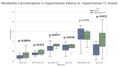 |
154 | Mapping MRI and MR Spectroscopy to Quantify Tumor and Edema Metabolism in Glioma Patients
Sribindu Sreepada1, Wufan Zhao1, Eduardo Coello1, Vicky Liao1, and Alexander Lin1
1Center for Clinical Spectroscopy, Department of Radiology, Brigham and Women's Hospital / Harvard Medical School, Boston, MA, United States Keywords: Tumors, Spectroscopy, chemical shift imaging, glioma This work examines the differences in metabolite concentrations in brain cancer lesions between the glioma and edema hyperintensities. Metabolites that showed statistically significant differences in this work are 2HG, total choline, glutamate plus glutamine, myoinositol, and lactate. Correlations between the metabolites and the partial volume fractions of hyperintense regions from MRIs revealed that the metabolite concentrations are higher in glioma than in edema. These results provide a simple method for fusing metabolic data with imaging findings which can provide added value for clinicians. |
|
2504.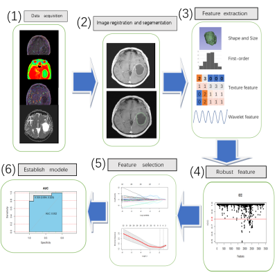 |
155 | Application of imaging omics based on DCE-MRI,DWI, and APT in glioma IDH-1 and Ki-67 prediction
zhenguo Yuan1, hexin Liang1, and Yuhan Wang2
1Shandong Provincial Hospital, Shandong, China, 2Philips Healthcare, Shanghai, China Keywords: Tumors, Brain We developed and validated radiomic models based on DWI, DCE, and APTw sequences to evaluate Ki-67 proliferation and IDH-1 mutation in glioma |
|
2505.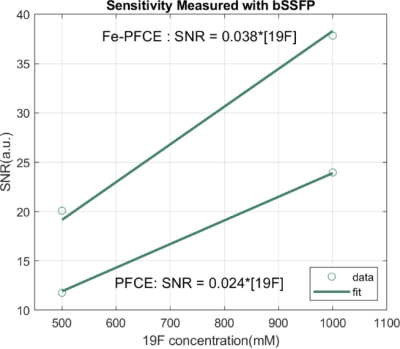 |
156 | Initial sensitivity evaluation of a new 19F nanoformulation at 3 Tesla using a novel human head coil
Gary Martinez1, Zhan Xu1, Dmitry Nevozhay1, Collin J. Harlan1, Christopher M. Walker1, Keith A. Michel1, Tyler King2, Emily Que2, Konstantin Sokolov1, and James A Bankson1
1Imaging Physics, The University of Texas MD Anderson Cancer Center, Houston, TX, United States, 2Chemistry, The University of Texas at Austin, Austin, TX, United States Keywords: Tumors, Cell Tracking & Reporter Genes, immune therapy In order to track immune cells loaded with 19F compound, a nanoformulation based on iron-doped perfluoro-15-crown-5-ether, called Fe-PFCE was developed for Fluorine-19 MRI. It’s relaxation time was characterized with a custom designed 8 channel Fluorine transmit/receive head coil on clinical 3Tesla MR scanner. Its relaxation time (T1/T2) was 39ms/7ms as comparing to 894ms/196ms nanoformulation based on original (undoped) perfluoro-15-crown-5-ether, called “PFCE”. The SNR/19F-concentration ratio of 1ml Fe-PFCE nanoformulation measured by bSSFP was found to be 53% stronger than that of PFCE. A good signal profile was yielded within the coil receiving field. |
|
| 2506. | WITHDRAWN | ||
2507.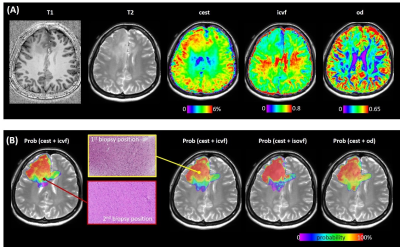 |
157 | Combing APT-CEST imaging and NODDI for an early distinction between pure edema and infiltrative tumor in glioma at 7T
Yifan Yuan1, Yang Yu2, Jun Chang1, Ying-Hua Chu3, Yi-Cheng Hsu3, He Wang4, Patrick Alexander Liebig5, Qi Yue1, Liang Chen1, and Ying Mao1
1Department of neurosurgery, Huashan Hospital Fudan University, Shanghai, China, 2Department of radiology, Huashan Hospital Fudan University, Shanghai, China, 3MR Collaboration, Siemens Healthineers Ltd, Shanghai, China, 4Key Laboratory of Computational Neuroscience and Brain-Inspired Intelligence, Fudan University, Shanghai, China, 5MR Collaboration, Siemens Healthineers Ltd, Erlangen, Germany Keywords: Tumors, CEST & MT, NODDI Glioma grows infiltratively along fiber tracts, making it difficult to determine the tumor boundary. Extended resection may impair eloquent brain areas and cause functional disorders, while conservative resection often leaves tumor residues at the cutting edge, leading to early recurrence. This study uses APT-CEST imaging and NODDI to explore glioma's microstructural and metabolic characteristics. We trained a model from 100 biopsies to predict tumor presence in non-enhancing areas. This model shows potential for guiding precise glioma resection and radiotherapy. |
|
2508. |
158 | Clinical significance of leakage corrected hemodynamic parameters computed from DCE-MRI on quantitative glioma grading
Dinil Sasi Sankaralayam1,2, Rakesh Kumar Gupta3, Anandh K Ramaniharan4,5, Rana Patir3, Suneeta Ahlawat3, and Anup Singh1,6
1Indian Institute of Technology Delhi, New Delhi, India, 2Johns Hopkins University, Baltimore, MD, United States, 3Fortis Memorial Research Institute, Gurugram, India, 4Philips Innovation Campus, Bengaluru, India, 5Cincinnati Children's Hospital Medical Center, Cincinnati, OH, United States, 6All India Institute of Medical Sciences, New Delhi, India Keywords: Tumors, DSC & DCE Perfusion Absolute quantification of cerebral-blood-volume (CBV) and cerebral-blood-flow (CBF) from dynamic-susceptibility-contrast (DSC)-MRI has shown wide clinical applications. Recent developments in dynamic-contrast-enhanced (DCE)-MRI have shown computation of CBV and CBF along with permeability information with and without model dependent analyses. The model free approach computed CBV and CBF with inherent leakage information and it is still unclear about the clinical significance of leakage correction. So this study is designed to evaluate the impact of leakage correction of CBV and CBF into glioma grading. Results of the study shows that leakage correction underperformed in terms of noise sensitivity and quantitatively differentiating glioma. |
|
 Back to Meeting Home
 Back to the Program-at-a-Glance
The International Society for Magnetic Resonance in Medicine is accredited by the Accreditation Council for Continuing Medical Education to provide continuing medical education for physicians.
 Back to Meeting Home
 Back to the Program-at-a-Glance
 View Presentation Video