Digital Poster
Applications in Tumors
ISMRM & ISMRT Annual Meeting & Exhibition • 03-08 June 2023 • Toronto, ON, Canada

| Computer # | |||
|---|---|---|---|
3213.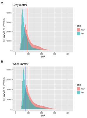 |
161 |
Advanced intraoperative image quality using multiple surface
coils.
Pien E.J. Jellema1,2,
Fredy Visser2,
Niels Blanken3,
Alberto de Luca2,4,
Eelco W. Hoving1,5,
Kirsten M. van Baarsen1,5,
Maarten H. Lequin1,3,
and Jannie Wijnen2
1Pediatric Neuro-Oncology, Princess Máxima Centre, Utrecht, Netherlands, 2Centre for Image Sciences, University Medical Centre Utrecht, Utrecht, Netherlands, 3Department of Radiology, University Medical Centre Utrecht, Utrecht, Netherlands, 4Department of Neurology, University Medical Centre Utrecht, Utrecht, Netherlands, 5Department of Neurosurgery, University Medical Centre Utrecht, Utrecht, Netherlands Keywords: Tumors, Surgery Advanced intraoperative MRI (io-MRI) could provide additional guidance during pediatric neurosurgery. Currently, io-MRI sequences are acquired with two surface coils when using the DORO head-frame. We investigated if the addition of two surface coils and higher SENSE-factors would increase signal- and contrast-to-noise ratios (SNR & CNR), image quality, and decrease artefacts. The four coils set-up resulted in higher T1w SNR and CNR that was more homogeneously distributed. Image quality of T1w, T2w, and fiber-tracks did improve with four coils. Higher SENSE factors with four coils of most ASL and diffusion MRI images did not reduce EPI artefacts or improve image quality. |
|
3214.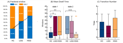 |
162 |
Dynamic characteristics of functional network activity in brains
with left-frontal glioma
Siqi Cai1,2,
Yuchao Liang3,
Yinyan Wang3,
Yufei Liu4,
Fanfan Chen4,
Chunxiang Jiang1,2,
Lei Wang3,
and Lijuan Zhang1,2
1Shenzhen Institute of Advanced Technology, Chinese Academy of Sciences, Shenzhen, China, 2University of Chinese Academy of Science, Beijing, China, 3Beijing Tiantan Hospital of Capital Medical University, Beijing, China, 4Shenzhen Second People's Hospital, Shenzhen, China Keywords: Tumors, fMRI (resting state) This study investigated the temporal features of the brain network activity with dynamic fractional amplitude of low-frequency fluctuation (dfALFF) and sliding window approach. The time-varying brain activity was clustered into two distinct states. Patients’ brains featured high occurrence of the weak state, low occurrence of the strong state, and network dALFF that decreased but spatially extended with the tumor malignancy. The dfALFF variance of networks was attenuated in HGGs but not LGGs. These malignancy-specific alterations indicate a diverse neuropathological profile of gliomas, and highlight the importance of temporal features of brain activity in the disease characterization of glioma. |
|
3215.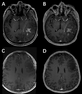 |
163 |
Evaluation of postcontrast images of glioma using 7T and 3T
magnetic resonance imaging: an intraindividual comparison study
Chenxi Li1,
Kun Cheng1,
Jianxun Qu2,
Xiaoxiao Ma1,
and Xin Lou1
1Department of Radiology, Chinese PLA General Hospital, Beijing, China, 2MR Collaboration, Siemens Healthineers Ltd., Beijing, China Keywords: Tumors, Brain With the advent of the 7T device meeting the need for ultra-high resolution, we planned to explore the advantages of 7T over 3T in terms of the internal detail display of gliomas. Seventeen patients with glioma were individually rated for internal tumor structure and feeding artery by 6 radiologists at 7T and 3T MR, and the DCS score was calculated. 7T revealed better details of internal tumor structure and higher diagnostic confidence than 3T. |
|
3216.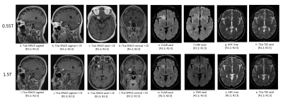 |
164 |
Retrospective Comparison of Brain Imaging in Patients at 0.55T
and 1.5/3T
Anna Lavrova1,
John Kim1,
Remy Lobo1,
Maria Masotti2,
Jacob Richardson1,
Pedro Itriago-Leon3,
Vikas Gulani1,
Katherine Wright1,
Ashok Srinivasan1,
and Nicole Seiberlich1
1Radiology, University of Michigan, Ann Arbor, MI, United States, 2Biostatistics, University of Michigan, Ann Arbor, MI, United States, 3Siemens Medical Solutions USA Inc., Houston, TX, United States Keywords: Tumors, Low-Field MRI This study aims to compare the image quality of clinical brain imaging on an FDA-approved commercial low-field 0.55T MRI scanner and conventional higher field 1.5/3T MRI systems. The image quality of 205 compatible image series in 30 patients acquired at 0.55T and 1.5T/3T MRI systems was rated by two neuroradiologists. Despite significant differences in image quality ratings between low- and higher-field scanners, all brain sequences performed at 0.55T received ratings indicating that they were acceptable for diagnostic use. This work indicates that the commercial 0.55T system can be used for routine brain imaging in clinical practice. |
|
3217.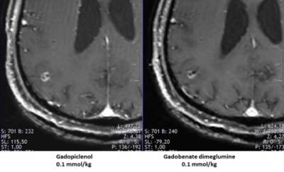 |
165 |
Impact of gadopiclenol on decision making in patients with brain
metastases
Marco Essig1,
Frank A Giordano2,
and Gustavo Sarria3
1Radiology, University of Manitoba, Winnipeg, MB, Canada, 2Radiation Oncology, University of Mannheim, Mannheim, Germany, 3Radiation Oncology, University of Bonn, Bonn, Germany Keywords: Tumors, Contrast Agent, Brain Tumor, Relativity New gadolinium-based contrast agents (GBCAs) with high relaxivity could have an impact on decision making and treatment planning of brain metastases (BM). In this post-hoc analysis , patients with BM who underwent two separate magnetic resonance imaging (MRI) examinations, one with gadopiclenol and one with gadobenate dimeglumine, both at 0.1 mmol/kg, were included. This study showed that gadopiclenol at the dose of 0.1 mmol/kg improved detection and characterization of BM, and led to a change in treatment decisions in 2 of 13 patients. |
|
3218.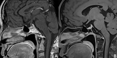 |
166 |
Detection of hormone-producing pituitary microadenoma by
Contrast-Enhanced Submillimeter level 3D T1w SPACE: Comparison
with 2D T1w Images
Cheng Cheng1,
Shengzhang Ji2,
Rui Zhao1,
Chen Zhang3,
and Guang hui Huo1
1Radiology Department, Mesclor Medical Imaging Diagnostic Center, Tianjin, China, 2Medical Imaging Department, Tianjin Fourth Central Hospital, Tianjin, China, 3MR Scientific Marketing, Siemens Healthineers, Beijing, China Keywords: Tumors, Tumor, pituitary, T1w SPACE By compared the images quality and diagnostic efficacy of enhanced 3D T1w SPACE and 2D T1w TSE, this study aims to investigate the value of Contrast-Enhanced submillimeter slice 3D T1w SPACE sequence in hormone-producing pituitary microadenoma. |
|
3219. |
167 |
Clinical application of direct signal control and other B1
shimming methods for infratentorial region T2 weighted imaging
at 7T
Xiaoyu Wang1,
Jianxun Qu2,
Tomi-Tricot Raphael3,
Shaihan J Malik4,
Song Wang1,
Xiangbing Bian1,
and Xin Lou1
1Department of Radiology, Chinese PLA General Hospital, Beijing, China, 2Siemens Healthineers Ltd, Beijing, China, 3Siemens Healthineers Ltd, United Kingdom, United Kingdom, 4King's College London, United Kingdom, United Kingdom Keywords: Tumors, Data Acquisition, direct signal control This was a clinical application study of direct signal control (DSC) technology on 7T MRI. The result suggests that DSC-T2w provides good-quality for whole-brain imaging. When imaging region is restricted, we can also use static B1 shimming with a dedicated shimming region. This study demonstrated that DSC technology can improve the image quality in 7T brain scan. |
|
3220.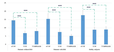 |
168 |
Reliability Verification and Diagnostic Value Based on Brain
Metastases of STAGE at 1.5T
Huijing Xiang1,
Yi Li2,
Yuqi Duan1,
Xiaoyun Liang2,
Feng Huang2,
Peian Hu3,
Dongbing Zhang1,
Zhengrong Zhou1,
and Lei Chen1
1Department of Radiology, Minhang Branch, Fudan University Shanghai Cancer Center, Shanghai, China, 2Neusoft Medical Systems Co.Ltd., Shanghai, China, 3Department of Radiology, Children's Hospital of Fudan University, National Children's Medical Center, Shanghai, China Keywords: Tumors, Tumor, Brain metastases, Reliability, Quantitative maps As the first choice to identify brain metastases (BM), the anatomical, functional and metabolic information extracted by various MR sequences is the basis. However, synthetic MR technique mostly relies on expensive equipment and high field strength, which is challenging for primary institutions to implement. In this study, we assessment the reliability and diagnostic value based on BM of Strategically Acquired Gradient Echo (STAGE) at 1.5T. Enhanced T1W map of STAGE showed better GM/WM CNR compare with T1MPRAGE for BM identification. Reliable values and inconspicuous physiological variability were observed in the quantitative maps for various brain tissues. |
|
3221.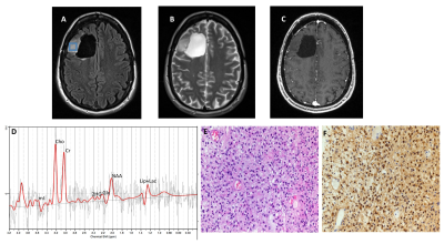 |
169 |
Non-invasive Detection of IDH-mutant Gliomas using Single and
Multi-voxel Point-resolved Spectroscopy
Laiz Laura de Godoy1,
Gaurav Verma2,
Donald M O’Rourke3,
MacLean P Nasrallah4,
Steven Brem3,
Arati Desai5,
Laurie A Loevner1,
Suyash Mohan1,
and Sanjeev Chawla1
1Radiology, Perelman School of Medicine at the University of Pennsylvania, Philadelphia, PA, United States, 2Radiology, Icahn School of Medicine at Mount Sinai, New York, NY, United States, 3Neurosurgery, Perelman School of Medicine at the University of Pennsylvania, Philadelphia, PA, United States, 4Clinical Pathology and Laboratory Medicine, Perelman School of Medicine at the University of Pennsylvania, Philadelphia, PA, United States, 5Medicine, University of pennsylvania, Philadelphia, PA, United States Keywords: Tumors, Spectroscopy, IDH mutant; gliomas; 2-hydroxyglutarate; SVS; 1H-MRSI IDH mutation has become one of the most important prognostic biomarkers in glioma management, regardless of histopathological features. The oncometabolite 2HG has been proposed as a biomarker for IDH-specific genetic profiles for gliomas. We report clinical utility of SVS and 1H-MRSI using a long TE (97 ms) in assessing IDH-mutant gliomas by detecting the characteristic resonances of 2HG. Our results from 25 patients showed sensitivity and specificity of 77% and 83%, respectively. In conclusion, 1H-MRS with optimized TE can accurately detect 2HG levels, which has significant clinical implications for determining prognosis and evaluating therapeutic efficacy for targeted and/or alternative therapies. |
|
3222.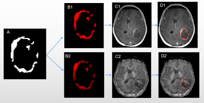 |
170 |
Spatial habitats features derived from multiparametric MR
imaging predict long versus short-term survival in gliomas:
Preliminary findings
Hui Ma1,
Zuliwei Ma1,
Shanmei Zeng1,
Mengzhu Wang2,
Yang Song2,
Chengxiu Zhang3,
Zhiyun Yang1,
and Jing Zhao1
1Department of Radiology, The First Affiliated Hospital, Sun Yat-sen University, Guangzhou, China, 2MR Scientific Marketing, Siemens Healthineers Ltd., Guangzhou, China, 3Shanghai Key Laboratory of Magneitc Resonance, East China Normal University, Shanghai, China Keywords: Tumors, Brain, high-grade gliomas, tumor habitats, habitat imaging We developed spatial habitats based on multiparametric MRI and evaluated associations between features in these habitats and survival time in patients with high-grade gliomas. The voxels in MR images were grouped into 2 clusters using the K-means clustering algorithm of in-house software nnFAE (V.0.0.10). Structural MRI habitats were defined on CE-T1WI and T2-Flair images, and physiologic MRI habitats were defined on MK derived from DKI and Ktrans derived from DCE imaging. Results showed physiologic habitats weighed more than structural ones, and suggested low vascular-permeability-and-tissue-complexity habitats may play an important role in distinguishing long- and short-term survival of high-grade gliomas patients. |
|
3223.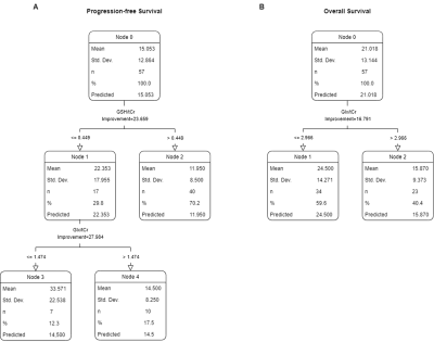 |
171 |
Magnetic Resonance Spectroscopic Correlates of Survival in
IDH-wt, TERTp-mut Gliomas
Banu Sacli-Bilmez1,
Ayça Erşen Danyeli2,3,
Cengiz Yakicier4,
M.Necmettin Pamir3,5,
Koray Özduman3,5,
Alp Dinçer3,6,
and Esin Ozturk-Isik1,3
1Institute of Biomedical Engineering, Boğaziçi University, İstanbul, Turkey, 2Department of Medical Pathology, Acıbadem University, İstanbul, Turkey, 3Brain Tumor Research Group, Acıbadem University, İstanbul, Turkey, 4Department of Molecular Biology and Genetics, Acıbadem University, İstanbul, Turkey, 5Department of Neurosurgery, Acıbadem University, İstanbul, Turkey, 6Department of Radiology, Acıbadem University, İstanbul, Turkey Keywords: Tumors, Brain The 2021 central nervous system tumor classification has suggested that isocitrate dehydrogenase wildtype (IDH-wt) WHO grade-2/3 astrocytomas with molecular features of glioblastoma should be designated as “Glioblastoma, IDH-wildtype, WHO grade-4”. In this study, we explored metabolic correlates of patient survival in tumors fulfilling the criteria for “Glioblastoma, IDH-wildtype, WHO grade-4” using short echo time (TE) single voxel 1H-MRS. The results of this study suggest that higher ratios of glutamine and glutamate complex (Glx) to total creatine (tCr) and glutathione (GSH) to tCr were associated with a shorter survival. |
|
3224.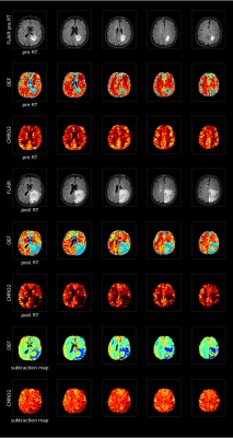 |
172 |
Effects of radiotherapy on tissue in patients with brain
metastases in terms of OEF and CMRO2
Jordi de Leeuw1,
Junghun Cho2,
Eva van Grinsven3,
Jeroen Siero4,
Marielle Philippens4,
and Alex Bhogal4
1Imaging Division, UMC Utrecht, Utrecht, Netherlands, 2SUNY Buffalo, Buffalo, NY, United States, 3Brain Department, Radiotherapy, UMC Utrecht, Utrecht, Netherlands, 4Radiology Department, UMC Utrecht, Utrecht, Netherlands Keywords: Tumors, Metabolism Radiotherapy might have an influence on the ratio of deoxygenated versus oxygenated blood in the venous blood vessels (OEF) in the brain. The relationship between delivered dose and the OEF may be a more direct measure of tissue damage or recovery. Preliminary results suggest that there is a marginal significant increase in OEF values after radiotherapy. This indicates that OEF can be of valuable use in determining the effects of radiotherapy. Additionally, CMRO2 maps show an identical increase or decrease in cerebral metabolic activity. Keywords: Oxygen extraction fraction Cerebral metabolic rate of oxygen extraction Radiotherapy Brain tumor |
|
3225.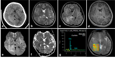 |
173 |
A preliminary study of radiological characteristics of diffuse
hemispheric gliomas, H3 G34-mutant
Yang Yu1,
Yan Ren1,
Qi Yue2,
and Zhenwei Yao1
1Radiology, Fudan University, Huashan Hospital, Shanghai, China, 2Neurosurgery, Fudan University, Huashan Hospital, Shanghai, China Keywords: Tumors, Tumor, H3 G34-mutant, glioma Diffuse hemispheric glioma, H3 G34-mutant (H3G34-DHG) is a rare newly proposed entity in 2021 WHO classification of central nervous system tumors. We retrospectively analysis characteristics of 10 cases of H3G34-DHG and found some certain characteristics in clinical and radiological manifestations in this entity. In most cases, absent or focally faint contrast enhancement initially suggested another diagnosis than a high-grade glioma. However, increased tumor vessels still suggested abundant tumor blood supply. Moreover, hyperintensity on CT and restricted diffusion reflected high density of tumor cells. MRS also showed extremely high metabolism active in the tumor. |
|
3226.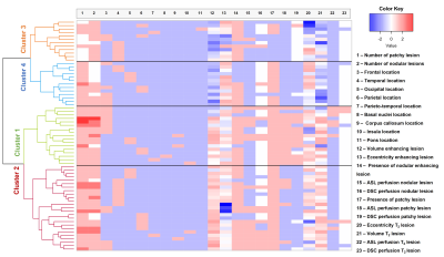 |
174 |
Brain MRI phenotypes of glioblastomas are suggestive of overall
patient survival
Bárbara Schmitz-Abecassis1,2,
Linda Dirven3,4,
Janey Jiang 5,
Jasmin A. Keller1,
Robert J.J. Croese3,4,
Daniëlle van Dorth1,
Ilse M. J. Kant 6,7,
Martin J.B. Taphoorn3,4,
Matthias J.P. van Osch1,2,
Johan A.F. Koekkoek3,4,
and Jeroen de Bresser1
1Department of Radiology, Leiden University Medical Center, Leiden, Netherlands, 2Medical Delta Cancer Diagnostics 3.0, South-Holland, Netherlands, 3Department of Neurology, Leiden University Medical Center, Leiden, Netherlands, 4Department of Neurology, Haaglanden Medical Center, The Hague, Netherlands, 5Department of Radiology, HagaZiekenhuis, The Hague, Netherlands, 6Clinical Artificial Intelligence Implementation and Research Lab (CAIRELab) and Department of Information Technology & Digital Innovation, Leiden University Medical Center, Leiden, Netherlands, 7Department of Digital Health, University Medical Center Utrecht, Utrecht, Netherlands Keywords: Tumors, Tumor, Perfusion Despite multimodal anti-tumor treatment, glioblastomas typically progress, yet identifying true tumor progression on MRI-scans is challenging. We aimed to establish brain MRI phenotypes of glioblastomas by combined analysis of radiological scoring of structural and perfusion tumor characteristics 3-months post-radiotherapy. Hierarchical clustering analysis method was applied to group patients by similar tumor characteristics and it was analyzed whether these groups showed differences in tumor progression and overall survival outcome. Four distinct MRI phenotypes of glioblastoma were established and showed between-group differences in median overall survival time. |
|
3227.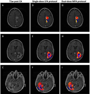 |
175 |
Identification of a single-dose, low-flip angle based CBV
threshold for fractional tumor burden (FTB) mapping in recurrent
glioblastoma
Aliya Anil1,
Ashley M Stokes1,
Lea Alhilali2,
John P Karis2,
Laura C Bell3,
Leland S Hu4,
Jerrold L Boxerman5,
Kathleen M Schmainda6,
and C Chad Quarles7
1Division of Neuroimaging Research and Barrow Neuroimaging Innovation Center, Barrow Neurological Institute, Phoenix, AZ, United States, 2Neuroradiology, Southwest Neuroimaging at Barrow Neurological Institute, Phoenix, AZ, United States, 3Early Clinical Development, Genentech, San Francisco, CA, United States, 4Department of Radiology, Division of Neuroradiology, Mayo Clinic Arizona, Phoenix, AZ, United States, 5Department of Neuroradiology, Rhode Island Hospital, Providence, RI, United States, 6Department of Biophysics, Medical College of Wisconsin, Milwaukee, WI, United States, 7The University of Texas MD Anderson Cancer Center, Houston, TX, United States Keywords: Tumors, DSC & DCE Perfusion Early differentiation of tumor recurrence from post treatment radiation effects (PTRE) in glioblastoma patients would improve patient management. The purpose of this study is to identify the optimal standardized relative cerebral blood volume (sRCBV) threshold for generating fractional tumor burden (FTB) maps derived from the new consensus protocol, single-dose, low-flip angle data. To establish the threshold, low-flip angle data was compared to well-validated, double-dose, moderate-flip angle data. In summary, with the optimized threshold, the single-dose, low-flip angle approach yielded FTB maps that strongly agreed with the reference standard, providing a compelling option for clinical use. |
|
3228. |
176 |
Differentiation of atypical high-grade glioma from primary
central nervous system lymphoma with mean apparent
propagator-MRI
Eryuan Gao1,
Guohua Zhao1,
Huiting Zhang2,
Xiaoyue Ma1,
Peipei Wang1,
Jie Bai1,
Xu Yan2,
Guang Yang3,
and Jingliang Cheng1
1Department of Magnetic Resonance, the First Affiliated Hospital of Zhengzhou University, Zhengzhou, China, 2MR Scientific Marketing, Siemens Healthcare, Shanghai, China, 3Shanghai Key Laboratory of Magnetic Resonance, East China Normal University, Shanghai, China Keywords: Tumors, Diffusion/other diffusion imaging techniques, mean apparent propagator Atypical high-grade glioma (HGG) (with no or little necrosis) and primary central nervous system lymphoma (PCNSL) are difficult to distinguish in routine MR images but their treatment strategies are totally different. Therefore, it’s important to distinguish between them before treatment. This study aimed to investigate the diagnostic efficiency of quantitative analysis based on mean apparent propagator-MRI in discriminating atypical HGG from PCNSL. Through quantitative analysis of MAP parameters, we found that MAP-MRI performed well in differentiating between atypical HGG and PCNSL. |
|
3229.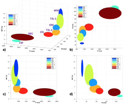 |
177 |
Segmentation of Contrast Enhancing Gliomas and Whole Brain
Multi-Parametric Mapping in a 6-minute Scan without Injecting
Contrast
Jing Liu1,
Angela Jakary1,
Duan Xu1,
Javier Villanueva-Meyer1,
Nicholas Butowski1,
Jennifer Clarke1,
Jennifer Taylor1,
Nancy Ann Oberheim Bush1,
and Janine Lupo1
1University of California San Francisco, San Francisco, CA, United States Keywords: Tumors, Cancer Multiple brain MRI sequences are routinely applied for tissue characterization and lesion detection, by acquiring multi-contrast anatomical images. We developed a single continuous data acquisition to generate multi-contrast images with high acquisition efficiency, which allows automatic brain tissue segmentation and lesion detection without contrast injection. Multi-compartment model-based dictionary searching method was developed to derive multi-parametric mapping, including T1, T2 and macromolecular proton fraction mapping. This short imaging acquisition (6 mins versus conventional ~30 mins scan time) without use of contrast injection and the efficient processing pipeline (automatic segmentation and quantitative mapping) can provide great potentials for imaging brain tumor patients. |
|
| 3230. | 178 |
Comparison Between Postcontrast T1-Weighted Thin-slice 2D Spin
Echo and 3D SPACE Sequences in Detection of Brain Metastases at
1.5T and 3T
Aaron Rulseh1,
Zuzana Ryznarova1,
and Josef Vymazal1
1Dept. of Radiology, Na Homolce Hospital, Prague, Czech Republic Keywords: Tumors, Tumor, Metastasis We compared standard T1-weighted thin-slice SE and T1-weighted SPACE sequences in the detection of metastatic brain lesions at 1.5 and 3 Tesla in 56 patients. Three raters evaluated the presence of lesions in 2 sessions minimally 6 weeks apart. Our results show that T1-weighted SPACE is not inferior to standard thin-slice SE sequences in the detection of brain metastases. All three experienced raters reached excellent consistency between SE and SPACE and agreement with ground truth. |
|
3231.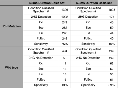 |
179 |
The Effect of RF Pulse Duration Settings in Basis Sets on
Sensitivity and Specificity of 2HG Spectroscopy
Sanghoon Kim1,
Changho Choi2,
Natasha Najam1,
and Alexander Lin1
1BWH, Boston, MA, United States, 2Vaderbilt University Institute of Imaging Science, Nashville, TN, United States Keywords: Tumors, Spectroscopy, 2HG detection 2HG is the specific biomarker for the IDH mutation in tumors. In this study, we utilized two different 2HG basis sets, one using a 4.8ms refocusing pulse duration and another with 5.8ms. Using the two different basis sets, we analyzed the same data and found large differences in the sensitivity and specificity results. |
|
3232. |
180 |
Discussion the WHO grading of brain invasion otherwise benign
meningiomas (BIOB) from the MR images perspective of 675
meningiomas
LUO XIAO1,
JIANG HONG2,
LIU XIAJING1,
LIN FAN1,
DENG KAN3,
JIANG JUN1,
ZHANG ZHU2,
WANG YULI1,
and YU JUAN1
1Medical Imaging Department, Shenzhen The Second People's Hospital, Shenzhen, China, 2Zhuhai People’s Hospital (Zhuhai hospital affiliated with Jinan University), Zhuhai, China, 3Philips Healthcare, Guangzhou, China Keywords: Tumors, PET/MR, WHO grade Should the BIOB meningiomas be classified as WHO grade 1 or WHO grade 2 atypical meningiomas, the core issue that scholars have questioned about the guidelines from an imaging perspective.In this study, we used BIOB as the entry point to comprehensively compare and analyze the MRI imaging characteristics of BIOB between WHO grade 1 and 2 meningiomas. Comparative analysis of multiple groups of MRI characteristics showed that BIOB and WHO grade 2 meningiomas were more similar in clinical and imaging features than grade 1 meningiomas. Therefore, it is reasonable to classify BIOB as WHO grade 2 meningiomas in the guidelines. |
|
 Back to Meeting Home
 Back to the Program-at-a-Glance
The International Society for Magnetic Resonance in Medicine is accredited by the Accreditation Council for Continuing Medical Education to provide continuing medical education for physicians.
 Back to Meeting Home
 Back to the Program-at-a-Glance
 View Presentation Video