Digital Poster
Brain Connectivity in Disease II
ISMRM & ISMRT Annual Meeting & Exhibition • 03-08 June 2023 • Toronto, ON, Canada

Digital Poster
Brain Connectivity in Disease II
| Computer # | |||
|---|---|---|---|
2843.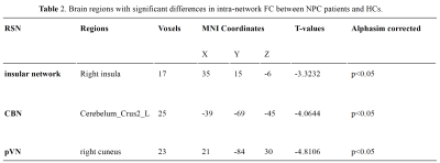 |
141 |
Altered resting-state connectivity in nasopharyngeal carcinoma
patients late stage post-radiotherapy: an Independent Component
Analysis
Xiaohan Song1 and
Lijun Wang1
1The First Affiliated Hospital of Dalian Medical University, Dalian, China Keywords: Head & Neck/ENT, fMRI (resting state) Radiotherapy is the primary treatment modality for patients with Nasopharyngeal carcinoma. However, radiotherapy in the treatment of nasopharyngeal carcinoma will also cause neurocognitive impairment. Previous studies mostly focused on the changes of brain structure in patients with nasopharyngeal carcinoma caused by radiotherapy, but it was reported that there were changes in brain network connectivity before the brain structure was abnormal. So, the purpose of thestudy was to investigate network-level functional connectivity alternations late stage post radiation therapy in Nasopharyngeal carcinoma patients using independent component analysis of rs-fMRI. |
|
2844.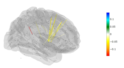 |
142 |
Therapy effects on the structural connectome of children treated
for medulloblastoma
John O Glass1,
Jia Liang2,
Ruitian Song1,
Yimei Li2,
Giles W Robinson3,
Amar Gajjar3,
Thomas E Merchant4,
and Wilburn E Reddick1
1Diagnostic Imaging, St. Jude Children's Research Hospital, Memphis, TN, United States, 2Biostatistics, St. Jude Children's Research Hospital, Memphis, TN, United States, 3Oncology, St. Jude Children's Research Hospital, Memphis, TN, United States, 4Radiation Oncology, St. Jude Children's Research Hospital, Memphis, TN, United States Keywords: Brain Connectivity, Cancer This study reports on the effects of pediatric medulloblastoma therapy in a population of 68 subjects. Structural connectivity was evaluated after surgery and again at the end of both radiation and chemotherapy. Linear models were written for the difference between these two time points controlling for age and risk arm. Fourteen significant edges were found connecting the basal ganglia to the dorsolateral prefrontal and prefrontal cortices as well as the cingulate. These regions are part of the executive function network and may point to the mechanisms of decreases in neurocognitive performance. |
|
2845.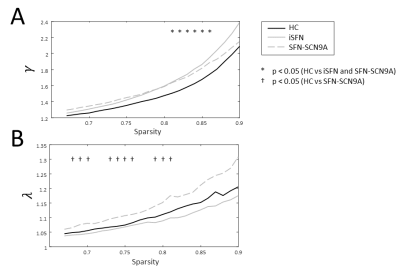 |
143 |
Altered response of the functional network during pain stimulus
in small fiber neuropathy
Gerhard Drenthen1,
Amir Far2,
Catharina Faber2,
Jaymin Upadhyay3,
David Linden4,
Raquel van Gool4,
Walter Backes1,
Janneke Hoeijmakers2,
and Jacobus Jansen1
1Department of Radiology & Nuclear Medicine, School for Mental Health and Neuroscience, Maastricht University Medical Center, Maastricht, Netherlands, 2Department of Neurology, Maastricht University Medical Center, Maastricht, Netherlands, 3Boston Children's Hospital, Boston, MA, United States, 4School for Mental Health & Neuroscience, Maastricht University Medical Center, Maastricht, Netherlands Keywords: Brain Connectivity, fMRI (task based), Chornic pain Small fiber neuropathy (SFN) is a neuropathic disorder that is associated with chronic pain, which may have an effect on the functional organization of the brain network. Here, we study the effect of pain on the functional network by applying a painful stimulus block-design during an fMRI acquisition. We show global changes in the functional network for patient with idiopathic and genetic SFN compared to controls. Moreover, self-reported pain scores correlated with nodal importance of the somatosensory cortex (postcentral gyrus) in the pain-evoked cerebral functional network, indicating that the functional network takes an important role in the perception of pain. |
|
2846. |
144 |
Disruption of default mode network and salience network dynamics
in acute traumatic pain states
Prasanna Karunanayaka1,
Sara L Mills-Huffnagle1,
Corinne M. Augusto1,
Aimee Cauffman1,
Joshua Hazelton1,
Sangam Kanekar1,
Andréa Hobkirk1,
and Jennifer E Nyland1
1Pennsylvania State University College of Medicine, Hershey, PA, United States Keywords: Brain Connectivity, Neuro, Pain Persistent and chronic pain following acute musculoskeletal injury is a significant contributor to diminished quality of life. We correlated self-reported pain using the McGill Pain Questionnaire with resting-state brain network connectivity in patients with blunt chest trauma. Results suggest that both inferior parietal and insular cortex connectivity were positively correlated to greater self-reported pain and a region within the salient network showed a negative correlation. These results support the hypothesis that aberrant functioning of brain circuits that assign salience values to stimuli may contribute to pain perception. Understanding abnormal activity/connectivity may identify targets to prevent persistent and chronic pain. |
|
2847.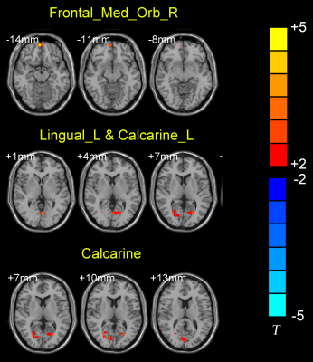 |
145 |
The abnormal functional connectivity of the locus coeruleus can
classify migraine without aura patients
Xiaozheng Liu1,
Jinming Chen2,
Keyang Chen1,
Kun Liu1,
Xi Zhang3,
Lipeng Dong2,
han lu4,
Yan Li1,
Zhihong Wang5,
and Yungang Cao1
1The Second Affiliated Hospital and Yuying Children’s Hospital, Wenzhou Medical University, Wenzhou, China, 2The Hebei General Hospital, Shijiazhuang, China, 3Xingtai people's Hospital, Xingtai, China, 4Philips Healthcare, Shanghai, China, Shanghai, China, 5The Second Affiliated hospital, Hebei Medical University, Shijiazhuang, China Keywords: Brain Connectivity, Nervous system, migraine Blood flow to the brainstem region is increased as a hypothesis for migraine. Locus coeruleus (LC) is the main noradrenergic nucleus in the brainstem. However, the functional characteristics of LC in migraine without aura (MwoA) patients are not currently known. Our results demonstrated that patients with MwoA exhibited significant LC FC differences in the brain areas associated with visual and cognitive function. Understanding the changes in the LC brain network in MwoA patients can provide us with new ideas to understand the pathological mechanisms of MwoA. |
|
2848.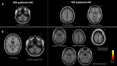 |
146 |
Time-Varying Functional Connectivity of the Hippocampus Is
Associated With Cognitive Performance in Multiple Sclerosis
Patients
Paola Valsasina1,
Olga Marchesi1,
Carmen Vizzino1,
Damiano Mistri1,
Maria Assunta Rocca1,2,3,
and Massimo Filippi1,2,3,4,5
1Neuroimaging Research Unit, Division of Neuroscience, IRCCS San Raffaele Scientific Institute, Milan, Italy, 2Neurology Unit, IRCCS San Raffaele Scientific Institute, Milan, Italy, 3Vita-Salute San Raffaele University, Milan, Italy, 4Neurorehabilitation Unit, IRCCS San Raffaele Scientific Institute, Milan, Italy, 5Neurophysiology Service, IRCCS San Raffaele Scientific Institute, Milan, Italy Keywords: Brain Connectivity, Multiple Sclerosis In this study, we explored hippocampal static and time-varying functional connectivity and its association with cognition in patients with multiple sclerosis. We found decreased static and time-varying connectivity of the hippocampus with temporo-parietal regions, and increased connectivity of the hippocampus with pre- and postcentral gyri, inferior temporal gyrus, precuneus and frontal regions. Better visuospatial and verbal memory were associated with higher hippocampal time-varying connectivity with precentral and inferior temporal gyri, while better attention scores were associated with higher hippocampal time-varying connectivity with the superior frontal cortex. |
|
2849.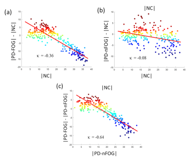 |
147 |
Disrupted topological organization of hubs in Parkinson’s
disease patients with freezing of gait.
Karthik R Sreenivasan1,
Ece Bayram2,
Xiaowei Zhuang1,
Jason Longhurst3,
Zhengshi Yang1,
Dietmar Cordes1,
Aaron Ritter4,
Jessica Caldwell1,
Jeffrey L Cummings5,
Zoltan Mari1,
Irene Litvan2,
Natividad Stover6,
Talene Yacoubian6,
Brent Bluett7,
and Virendra Mishra1,8
1Cleveland Clinic Lou Ruvo Center for Brain Health, Las Vegas, NV, United States, 2Department of Neurosciences, University of California San Diego, La Jolla, CA, United States, 3Department of Physical Therapy and Athletic Training, Saint Louis University, St. Louis, MO, United States, 4Memory & Cognitive Disorders Program Hoag, Pickup Family Neurosciences Institute, Newport Beach, CA, United States, 5Chambers-Grundy Center for Transformative Neuroscience, Department of Brain Health, School of Integrated Health Sciences, University of Nevada, Las Vegas, NV, United States, 6Department of Neurology, The University of Alabama at Birmingham, Birmingham, AL, United States, 7Central California Movement Disorders, Pismo Beach, CA, United States, 8Department of Radiology, The University of Alabama at Birmingham, Birmingham, AL, United States Keywords: Brain Connectivity, Parkinson's Disease Neuroimaging studies, including resting-state functional MRI (rs-fMRI), have implicated altered resting-state functional connectivity (rs-FC) in PD patients with freezing of gait (PD-FOG) and showed that the disruption of connectivity between the resting state networks (RSNs) was correlated with FOG. However, the network organization in PD-FOG remains poorly understood. In this study, we use rs-fMRI data and graph theoretical approaches to explore the reorganization of functional networks in PD-FOG. The results of our study suggest that there is a substantial reorganization of regional brain hubs and disruption in the higher-order functional network topology in PD-FOG participants compared to PD-nFOG. |
|
2850. |
148 |
Brain Micro-Structural and Functional Alterations for Cognitive
Function Prediction in the End-Stage Renal Disease Patients
Jiahui Zheng1,
Weiqiang Dou2,
Xiangxiang Wu1,
and Haifeng Shi1
1Department of Radiology, The Affiliated Changzhou NO.2 People’s Hospital of Nanjing Medical University, changzhou, China, 2GE Healthcare, MR Research China, Beijing, China Keywords: Brain Connectivity, Brain, DKI FC The goal of this study was to investigate the relationships of altered brain micro-structure and function, and cognitive function for ESRD patients undergoing maintenance hemodialysis. Specifically, diffusion kurtosis imaging (DKI), the resting-state functional connectivity (FC) algorithm, and the least squares support vector regression machine (LSSVRM) were utilized to conduct our study. Brain micro-structural and functional changes were found in ESRD patients, which may account for the onset of cognitive impairment in affected patients. These quantitative parameters combined with our optimized prediction model may be helpful to establish reliable imaging markers to detect and monitor cognitive impairment associated with ESRD. |
|
2851.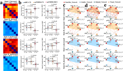 |
149 |
Dysregulated brain dynamics in visual-motor network of type 2
diabetes and its relation to cognitive impairment Ying Yu1,
Bo Hu1,
Wen Wang1,
Lin-Feng Yan1,
and Guang-Bin Cui1
1Department of Radiology & Functional and Molecular Imaging Key Lab of Shaanxi Province, Tangdu Hospital, Fourth Military Medical University (Air Force Medical University), Xi’an, China Keywords: Brain Connectivity, Diabetes To screen type 2 diabetes mellitus(T2DM)-specific effective connective (EC) network, the dynamic features of which may contribute to distinguishing T2DM patients with mild cognitive impairment (T2DM-MCI) from controls. Screening of resting-state functional MRI (rs-fMRI) data from early T2DM, T2DM-related static causality network mainly consisted of nodes in visual and sensorimotor network. In the visual-motor network, the fractional windows and mean dwell time of strong dEC state in T2DM-MCI patients were significantly higher than controls. The sum of dECs (sumdEC) could effective distinguish the mT2DM-MCI indicating sumdEC to be a promising biomarker for the early cognitive impairment in T2DM. |
|
2852.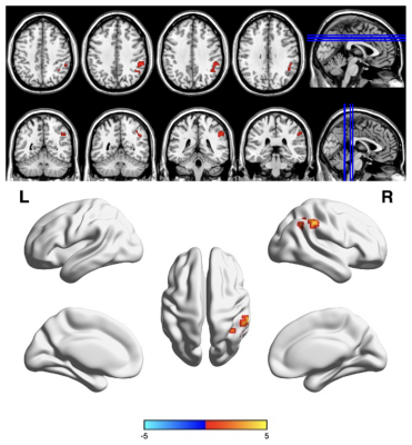 |
150 |
Increased regional homogeneity and correlation with clinical
indicators and scales in type 2 diabetes
Wei Du1,
Yangyingqiu Liu1,
Man Wang1,
Wanyao Li1,
Bingbing Gao1,
Weiwei Wang1,
Qingwei Song1,
and Yanwei Miao1
1Department of Radiology, the First Affiliated Hospital of Dalian Medical University, Dalian, China Keywords: Brain Connectivity, Diabetes, regional homogeneity To further investigate the changes in brain function of type 2 diabetes we prospectively enrolled 38 patients clinically diagnosed with T2DM and 24 age-matched healthy controls and underwent blood oxygen level-dependent fMRI scans. The image data was processed to obtain regional homogeneity values and compared by two sample t test. Results found that the local synchronization of spontaneous neural activity of the supramarginal gyrus in patients with T2DM was abnormal and correlated with cognitive function, which may be involved in the pathophysiological mechanism of T2DM. |
|
2853.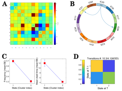 |
151 |
Altered dynamic effective connectivity of the default mode
network in T2DM
xu kun1,
wang jun2,
and zhang jing3
1medical, lanzhou unversity, lanzhou, China, 2lanzhou university, lanzhou, China, 3lanzhou university second hospital, lanzhou, China Keywords: Brain Connectivity, Brain Connectivity Type 2 diabetes (T2DM) as one of the risk factors for developing Alzheimer's, however, the underlying pathogenesis of cognitive impairment in patients with T2DM was still unknown. our study combined the method of sliding-window approach and Granger causality analysis and found decreased dynamic effective connectivity (DEC) in T2DM compared with healthy people, and the decrease was associated with abnormal blood glucose level. |
|
2854.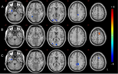 |
152 |
Altered functional hubs and connectivity in type 2 diabetes
mellitus with and without mild cognitive impairment
Yang Huang1,
Dongsheng Zhang1,
Xin Zhang1,
Kai Ai2,
Xiaoyan Lei1,
and Xiaoling Zhang1
1Shaanxi Provincial People’s Hospital, Xi’an, China, 2Philips Healthcare, Xi’an, China Keywords: Brain Connectivity, fMRI (resting state) To investigate the functional abnormalities of brain networks and hub nodes damaged in T2DM patients with mild cognitive impairment (DMCI). We applied degree centrality (DC) and seed-based functional connectivity (FC) analysis to identify the abnormal hub nodes and the FC patterns of these hubs in DMCI patients. Our results indicated that DMCI patients showed extensive alterations in the visual and memory pathways, and these alterations were correlated with clinical/cognitive variables. These results suggested that vision and memory-related brain regions dysfunctions may be involved in the neuropathology of visuospatial and memory function impairment in DMCI patients. |
|
2855. |
153 |
Aberrant brain gray matter and functional networks topology in
end stage renal disease patients with cognitive impairment
Jiahui Zheng1,
Jiankun Dai2,
Xiangxiang Wu1,
and Haifeng Shi1
1Department of Radiology, The Affiliated Changzhou NO.2 People’s Hospital of Nanjing Medical University, changzhou, China, 2GE Healthcare, MR Research China, Beijing, China Keywords: Brain Connectivity, fMRI (resting state) This study aimed to characterize the topological properties of gray matter (GM) and functional networks in end-stage renal disease (ESRD) patients undergoing maintenance hemodialysis to provide insights into the underlying mechanisms of cognitive impairment. Functional and GM networks were constructed based on resting-state functional magnetic resonance imaging (rs-fMRI) and diffusion kurtosis imaging (DKI), respectively. Disrupted topological organizations were observed, as indicated by significantly altered global measures, nodal efficiency, and degree centrality, which may account for the progression of cognitive dysfunction. And implementation of prediction models based on neuroimaging metrics may provide more objective information to promote early diagnosis and intervention. |
|
2856.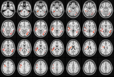 |
154 |
Altered Gray Matter Volume and Functional Connectivity in Type 2
Diabetes Mellitus
Xue Zhang1,
Wanjun Hu1,
and Jing Zhang1
1Department of Magnetic Resonance, Lanzhou University Second Hospital, LanZhou, China Keywords: Brain Connectivity, Diabetes, Neuroimaging, type 2 diabetes mellitus, VBM, resting-state fMRI, Cognition Keyword: Neuroimaging, type 2 diabetes mellitus, VBM, resting-state fMRI, Cognition The main purpose of this study was to evaluate the structural brain alterations and related functional abnormity of type 2 diabetes(T2DM), and its correlation with cognitive scale. Changes in brain volume were assessed using voxel-based morphology (VBM), while VBM Results were used to build ROI to analyse related functional connectivity (FC) changes in brain function. The result indicated that decreased gray matter volume and decreased functional connectivity may represent the neurobiological mechanism underlying cognitive dysfunction in patients with T2DM. |
|
2857.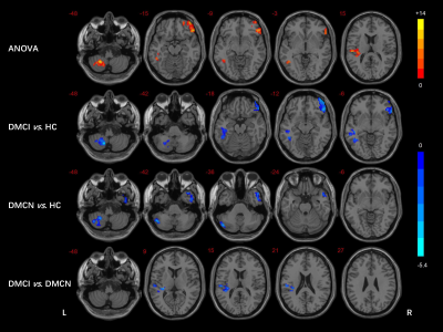 |
155 |
Aberrant functional connectivity density in different cognitive
states of T2DM patients
Dongsheng Zhang1,
Shasha Liu1,
Yang Huang1,
Weirui Liu1,
Wanting Liu1,
Kai Ai2,
Xiaoyan Lei1,
and Xiaoling Zhang1
1Shaanxi Provincial People’s Hospital, Xi’an, China, 2Philips Healthcare, Xi’an, China Keywords: Brain Connectivity, fMRI, T2DM To investigate the underlying neural mechanism of T2DM-related brain damage, the functional connectivity density (FCD) mapping method was used to examine the density distribution of whole-brain resting functional connectivity from 77 patients with T2DM under different cognitive states (with normal cognitive function, DMCN and mild cognitive impairment, DMCI) and 40 healthy controls (HCs). The decreased long-range FCD in the left superior temporal gyrus (STG) in DMCI patients and its correlation with Rey Auditory Verbal Learning Test (RAVLT) score in all T2DM patients, which suggested left STG may be involved in the neuropathology of auditory memory in T2DM patients. |
|
2858.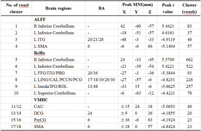 |
156 |
Evaluation of altered brain activity in type 2 diabetes by
multiple indices of brain function: a resting-state functional
MRI study
Ge Zhang1,
Xianchang Zhang2,
and Meiyun Wang1
1Radiology, Henan Provincial People's Hospital, Zhengzhou, China, 2MR Collaboration, Siemens Healthineers Ltd, Beijing, China Keywords: Brain Connectivity, fMRI (resting state) We combined multiple rs-fMRI indices to investigate the abnormal functional changes among T2DM patients. 52 T2DM patients and matched control participants were enrolled in this study. The functional abnormalities primarily involved motor and sensory function areas. More brain regions were revealed by the combination strategy than any single method and the indices of rs-fMRI showed significantly concordant alterations in most abnormal brain regions. Correlations of between clinical parameters and rs-fMRI indices were found. The current study demonstrated an enriched picture showing the brain functional alterations reflected by the combination of multiple indices in T2DM patients. |
|
2859.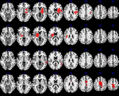 |
157 |
Microstructral and Functional Changes of brain in Pontine
Infarction: A Combined Arterial Spin Labeling and Diffusion
Tensor Imaging Study
Peipei Chang1,
Peng Sun2,
and Yanwei Miao1
1The First Affiliated Hospital of Dalian Medical University, Dalian, China, 2Philips Healthcare, BeiJing, China Keywords: Stroke, Perfusion, Arterial Spin Labeling Pontine infarction (PI) is very common in posterior circulation infarction, which can lead to behavioral dysfunctions, such as motor and cognition impairment. However, secondary changes in microstructure after PI in the whole brain remain unclear. This study aimed to explore the changes in cerebral blood flow (CBF) and fractional anisotropy (FA) in PI patients and to better understand the whole brain microstructure changes. We found PI could lead to extensive white matter tracts impairment in most of the whole brain regions and perfusion changes in some brain regions. |
|
2860.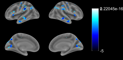 |
158 |
Voxel-mirrored homotopic connectivity is impaired in cortical
stroke but compensatory in subcortical stroke: a resting-state
fMRI study
Changjiang Zhao1,2,
Chengxin Yu1,2,
Bo Rao 3,
Junlong Pan1,
Li Zhu1,
Jiangjin Chen1,
Long Chen1,
Xiong Xiong 1,
Can Zhang 1,
Yong Ye1,
Zheng Wang 1,
Xiaoling Yang 1,
Lisi Xie 1,
Xiance Zhao4,
Chen Zhao5,
and Shan Huang4
1Yichang Central People's Hospital and The First College of Clinical Medical Science, China Three Gorges University, Yichang, China, 2Institute of Medcical Imaging, China Three Gorges University, Yichang, China, 3Department of Radiology, Zhongnan Hospital of Wuhan University, Wuhan University, Wuhan, China, 4Philips Healthcare, Shanghai, China, 5Philips Healthcare, Guangzhou, China Keywords: Stroke, fMRI (resting state) The alteration patterns of bi-hemispheric coordination between homologous areas in the whole brain of acute ischemic stroke patients with different infarct sites remains unclear. In the analysis of the voxel-mirrored homotopic connectivity (VMHC) among patients with frontoparietal lesion (G1), radiation coronal lesion (G2) and basal ganglia lesion (G3), VMHC was significantly decreased in G1 compared with G2 and G3. The impaired regions, such as precentral gyrus and postcentral gyrus, were part of the sensorimotor and default mode network. In contrast, VMHC mostly increased in patients with subcortical stroke, which indicates compensation rather than impairment of bi-hemispheric coordination. |
|
2861. |
159 |
Brain redundancy metric in cerebral small vessel disease:A
community-based study.
Lei Cui1 and
Minming Zang1
1Radiology, The second affiliated hospital of ZheJiang University, Hangzhou, China Keywords: Brain Connectivity, fMRI (resting state), redundancy We applied a dynamic whole-brain network metric, redundancy, to a community-based middle-aged and elderly population, revealed the potential mechanisms of SVD on cognitive impairment ; we also explored the relationship between cognitive reserve and brain redundancy metric.We found that aging, hypertension and lacunes could impair redundancy in brain networks, whereas better brain redundancy corresponds to better executive function, which partly explains the potential mechanism of cognitive impairment by SVD. Secondly, we found no significant correlation between redundancy and cognitive reserve. |
|
2862.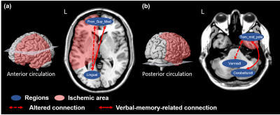 |
160 |
Association of brain connections in anterior and posterior
circulation with the side of asymptomatic internal carotid
stenosis and verbal memory
Jyun-Ru Chen1,
Chun-Jen Lin2,3,
I-Hui Lee2,3,4,
and Chia-Feng Lu1
1Department of Biomedical Imaging and Radiological Sciences, National Yang Ming Chiao Tung University, Taipei, Taiwan, 2School of Medicine, National Yang Ming Chiao Tung University, Taipei, Taiwan, 3Neurological Institute, Taipei Veterans General Hospital, Taipei, Taiwan, 4Institute of Brain Science, National Yang Ming Chiao Tung University, Taipei, Taiwan Keywords: Vessels, Ischemia Previous studies have reported that patients with asymptomatic internal carotid stenosis (aICS) had recall verbal memory impairment. However, the underlying mechanism to elucidate the altered functional connectivity (FC) associated with the anterior and posterior circulation was not proposed. Furthermore, whether the laterality of aICS is a factor in FC alterations and verbal memory impairment requires further investigation. In this study, we reported that the left and right aICS groups showed different patterns of FC changes related to secondary language regions in anterior and posterior circulation. These FCs could be served as imaging biomarkers for recall verbal memory. |
|
 Back to Meeting Home
 Back to the Program-at-a-Glance
The International Society for Magnetic Resonance in Medicine is accredited by the Accreditation Council for Continuing Medical Education to provide continuing medical education for physicians.
 Back to Meeting Home
 Back to the Program-at-a-Glance
 View Presentation Video