Digital Poster
Pediatric Cardiopulmonary MRI
ISMRM & ISMRT Annual Meeting & Exhibition • 03-08 June 2023 • Toronto, ON, Canada

| Computer # | |||
|---|---|---|---|
1977.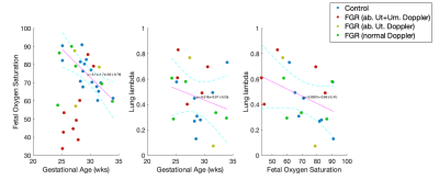 |
161 | Quantitative MRI Measurement of Lung Development in Early Onset Fetal Growth Restriction
Janina Schellenberg1, Paponrad Tontivuthikul2, Joanna Chappell1, Nada Mufti2, Dimitra Flouri1, Sebastien Ourselin1, Anna David2, Rosalind Aughwane2, and Andrew Melbourne1
1King's College London, London, United Kingdom, 2University College London, London, United Kingdom Keywords: Data Analysis, Fetus Fetal growth restriction (FGR) occurs when a fetus does not reach its full growth potential, predisposing the fetus to long-term impacts on lung function at birth and into childhood. We propose a novel model of fetal lung tissue based on combined diffusion and relaxation and apply this to a cohort of FGR and appropriately grown controls. We show differences in lung model parameters in FGR relative to controls and with gestational age. We also link these parameters to measurement of feto-placental oxygenation. This methodology could potentially provide a non-invasive method to assess fetal lung development. |
|
1978.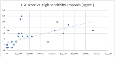 |
162 | The importance of cardiac MRI (CMR) in pediatric acute myocarditis, and it’s correlation with high sensitivity troponin
Maher Abadeer1, Munes Fares1, Jeanne Dillenbeck2, Maddy Artunduaga2, Sravani Avula1, Sanja Dzelebdzic1, Steven Philip1, Tarique Hussain1, and Gerald Greil1
1Pediatric Cardiology, UT Southwestern, Dallas, TX, United States, 2Pediatric Radiology, UT Southwestern, Dallas, TX, United States Keywords: Cardiovascular, Myocardium, Myocarditis, LGE, troponin Pediatric myocarditis is associated with major morbidity and mortality. Biomarkers are needed to predict clinical outcomes. High-sensitivity troponin (hs-TnT) and late gadolinium enhancement (LGE) have been shown to correlate with poor outcomes in adult populations. We found that LGE and hs-TnT linearly correlated with each other in our pediatric population, and that children with myocarditis were more likely to be male, have a higher LGE score, higher hs-TnT values, and increased T2 signal intensity than those who did not. A cut-off of 1200 ms for native T1 global mean was significantly associated with the need for inotropic support. |
|
1979.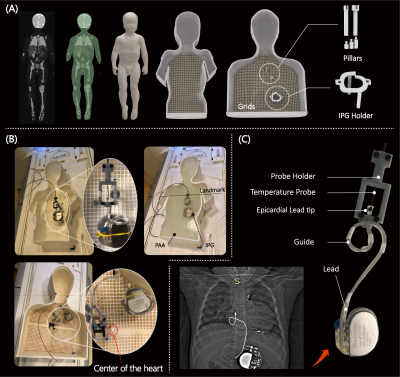 |
163 | Age matters: radiofrequency heating of epicardial and endocardial electronic devices in adults and children varies with body size and lead length
Fuchang Jiang1, Kaylee Henry1, Bhumi Bhusal2, Pia Sanpitak2, Gregory Webster3, Andrada Popescu4, Giorgio Bonmassar5, Christina Laternser6, Daniel Kim2, and Laleh Golestanirad1,2
1Department of Biomedical Engineering, Northwestern University, Evanston, IL, United States, 2Department of Radiology, Northwestern University, Chicago, IL, United States, 3Division of Cardiology, Ann and Robert H. Lurie Children's Hospital of Chicago, Chicago, IL, United States, 4Division of Medical Imaging, Ann and Robert H. Lurie Children's Hospital of Chicago, Chicago, IL, United States, 5A. A. Martinos Center for Biomedical Imaging, Massachusetts General Hospital, Boston, MA, United States, 6Center for Cardiovascular Innovation, Ann and Robert H. Lurie Children's Hospital of Chicago, Chicago, IL, United States Keywords: Heart, Safety, Implants Children with congenital heart defects often have life-sustaining indications for an epicardial cardiac implantable electronic device (CIED). However few data exist on endocardial systems in children. Additionally, the FDA has never approved an epicardial system as MRI-Conditional due to limited data on potential heating risk. To provide evidence-based knowledge on RF-induced heating of CIEDs in children and adults with epicardial and endocardial leads of different lengths, we recorded the temperature increase of 120 clinically relevant trajectories positioned into adult and pediatric phantoms. Our results highlight the need for age and device-specific assessments of MRI safety in children with CIEDs. |
|
1980.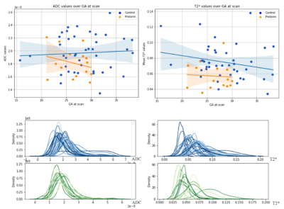 |
164 | Joint relaxometry-diffusion imaging and spectra of the fetal lungs.
Carla Avena-Zampieri1, Paddy J Slator2, Alena Uus1, Mary Rutherford1, Anne Greenough3,4,5, Jana Hutter1, and Lisa Story1,6
1Perinatal imaging, King's College London, London, United Kingdom, 2Centre for Medical Image Computing and Department of Computer Science, University College London, London, United Kingdom, 3Asthma UK Centre for Allergic Mechanisms in Asthma, King's College London, London, United Kingdom, 4Neonatal Unit, King’s College Hospital, London, United Kingdom, 5NIHR Biomedical Research Centre, Guy’s & St Thomas NHS Foundation Trusts and King’s College London, London, United Kingdom, 6Fetal Medicine Unit, Guy’s and St Thomas’ NHS Foundation Trust, London, United Kingdom Keywords: Data Analysis, Fetus, Prenatal lungs, non-invasive tool We scanned the fetal lungs of healthy controls and fetuses that subsequently delivered preterm, to characterise typical and atypical lung development in-utero, with a combined T2*-diffusion protocol. We analysed our data with a joint T2*-ADC model, yielding voxelwise maps of T2* and ADC, plus calculated whole-lung T2*-ADC spectra. A reduction in both T2* and ADC values was demonstrated in the preterm cohort, potentially reflecting alterations in pulmonary development commencing in-utero. Our study demonstrates that voxelwise T2* and ADC maps, and whole-lung T2*-ADC spectra have the potential to provide additional information on fetal lung microstructure and improve detection of atypical development. |
|
1981.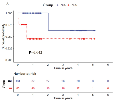 |
165 | Prognostic value of myocardial strain after chemotherapy in children with leukemia: a cardiac magnetic resonance study
Rong Xu1, Wei Huang2, Lini Liu1, Xiaoyong zhang3, and Yingkun Guo1
1West China Second University Hospital, Chengdu, China, 2West China Second Universityy Hospital, Chengdu, China, 3Clinical Science, Philips Healthcare, Chengdu, China Keywords: Cardiomyopathy, Cancer The stage of chemotherapy in children with ALL is a long process, and the myocardial damage caused by chemotherapy is still unknown. The different chemotherapy stages may have different degrees of myocardial damage. In this study, cardiac magnetic resonance was used to evaluate myocardial strain in ALL patients at different stages of treatment, and it was found that myocardial strain reduction was more pronounced in the early stages of treatment, and that global strain was a risk factor for adverse clinical outcomes. |
|
1982.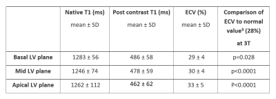 |
166 | Cardiac Magnetic Resonance Imaging at 3T Characterizes the Myocardium for Pediatric Cardiac Transplant Recipients: New Biomarker Development
Margaret M Samyn1, Adam M Gordon2, Ke Yan3, Jian Zhang4, Aspen M Duffin5, Jonathan H Soslow6, Bruce Damon7, Jennifer M Gerardin1, Benjamin M Goot8, Joseph R Cava1, and Steven J Kindel1
1Pediatrics, Medical College of Wisconsin/ Herma Heart Institute at Children's Wisconsin, Milwaukee, WI, United States, 2Medical College of Wisconsin, Milwaukee, WI, United States, 3Pediatrics, Medical College of Wisconsin/ Quantitative Health Sciences, Milwaukee, WI, United States, 4Quantitative Health Sciences, Medical College of Wisconsin/ Quantitative Health Sciences, Milwaukee, WI, United States, 5Herma Heart Institute, Children's Wisconsin, Milwaukee, WI, United States, 6Pediatrics, Vanderbilt Univesity Medical Center, Nashville, TN, United States, 7Bioengineering, Carle Foundation Hospital/University of Illinois, Urbana, IL, United States, 8Pediatrics (Cardiology), Medical College of Wisconsin/ Herma Heart Institute at Children's Wisconsin, Milwaukee, WI, United States Keywords: Cardiovascular, Tissue Characterization, Cardiac transplant; 3Tesla; Parametric Imaging Pediatric heart transplant recipients undergo frequent invasive cardiac catheterization with endomyocardial biopsy for routine allograft surveillance. Risks associated with catheterization and sampling error call into question this accepted “gold standard.” Cardiac magnetic resonance imaging (CMR), with parametric imaging, may advance myocardial surveillance in this population. Fifty-two pediatric patients (transplanted at ≤18 years old) underwent CMR (September 2016 to June 2019) at 3T to evaluate myocardial health using MOLLI sequence. Healthy transplant recipients had normal global biventricular function, native T1 higher than published values for healthy children at 3T with similar extracellular volume (ECV) despite history of treated rejection. |
|
1983.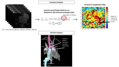 |
167 | Novel non-invasive cardiac MR oximetry and 4-dimensional flow in single ventricle hearts
Lajja Desai1, Elizabeth Weiss2, Juliet Varghese3, Rizwan Ahmad3, Orlando Simonetti3, Cynthia Rigsby1, and Michael Markl2
1Lurie Children's Hospital/Northwestern University Feinberg School of Medicine, Chicago, IL, United States, 2Northwestern University, Chicago, IL, United States, 3Ohio State University, Columbus, OH, United States Keywords: Cardiovascular, Translational Studies, 4D Flow Cardiac magnetic resonance imaging can provide novel non-invasive estimation of blood oxygen saturation and quantification of blood flow streaming in pediatric and adult patients with complex single ventricle physiology. Non-uniform distribution of inferior and superior caval blood flow and oxygenation to the left and right lungs may cause complications in patients with Fontan circulation. An individualized, comprehensive approach may identify patients at risk. |
|
1984. |
168 | Comprehensive 4D flow MRI analysis of aortic flow hemodynamics in pediatric/young adult patients with bicuspid aortic valve
Takashi Fujiwara1, LaDonna Malone1, Kelly Jarvis2, Kathryn C Chatfield3, Lorna P Browne1, and Alex J Barker1,4
1Department of Radiology, Children's Hospital Colorado, University of Colorado Anschutz Medical Campus, Aurora, CO, United States, 2Department of Radiology, Northwestern University, Chicago, IL, United States, 3Department of Pediatrics, University of Colorado Anschutz Medical Campus, Children’s Hospital Colorado, Aurora, CO, United States, 4Department of Bioengineering, University of Colorado Anschutz Medical Campus, Aurora, CO, United States Keywords: Flow, Velocity & Flow, 4D flow MRI Bicuspid aortic valve is associated with progressive aortic dilatation, which is seen both in pediatric and adult populations. Although several studies have found a connection between aortic hemodynamics, vessel wall architectural abnormalities, and progressive dilation, the role of hemodynamics in pediatric aortopathy remains unclear. We used 4D flow MRI data in pediatric bicuspid aortic valve patients to identify potential hemodynamic biomarkers associated with aortic diameter. Despite no clear association with aortic diameter, different flow features between BAV patients with/without coarctation were found. |
|
1985.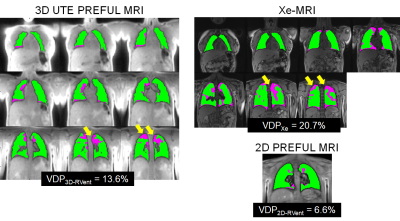 |
169 | Comparison of 3D Ultra-short Echo Time (UTE) Phase-Resolved Functional Lung (PREFUL) with Hyperpolarized 129Xe MRI in Pediatric Cystic Fibrosis
Samal Munidasa1,2, Brandon Zanette1,2, Marie-Pier Dumas3, Wallace Wee3, Jacky Au3, Sharon Braganza1, Daniel Li1, Felix Ratjen3, and Giles Santyr1,2
1Translational Medicine, The Hospital for Sick Children, Toronto, ON, Canada, 2Medical Biophysics, University of Toronto, Toronto, ON, Canada, 3Division of Respiratory Medicine, The Hospital for Sick Children, Toronto, ON, Canada Keywords: Data Processing, Lung 2D phase-resolved functional lung (PREFUL) MRI has been shown to correlate with hyperpolarized Xenon MRI (Xe-MRI) in pediatric Cystic Fibrosis (CF) lung disease but ventilation defects may be missed using the 2D approach. In this study we develop a 3D ultra-short echo time (UTE) PREFUL MRI method and compare to 2D PREFUL MRI and Xe-MRI in CF. 3D UTE PREFUL MRI showed a lower bias between Xe-MRI as compared to 2D PREFUL and was able to detect ventilation defects missed by the 2D approach, suggesting that the 3D approach may be a more comprehensive measure of ventilation in pediatric CF. |
|
1986. |
170 | Hemodynamics in children with partial anomalous pulmonary venous return and atrial septal defect using 4D-flow MRI.
Fraser Maurice Callaghan1,2, Barbara Burkhardt2,3, Emanuela Valsangiacomo Buechel2,3, Christian Kellenberger2,4, and Julia Geiger2,4
1Center for MR Research, University Children's Hospital Zurich, Zurich, Switzerland, 2Children’s Research Center, University Children's Hospital Zurich, Zurich, Switzerland, 3Division of Pediatric Cardiology, University Children's Hospital Zurich, Zurich, Switzerland, 4Department of Diagnostic Imaging, University Children's Hospital Zurich, Zurich, Switzerland Keywords: Cardiovascular, Cardiovascular, 4D flow, Congenital heart disease 4D flow MRI is used to assess flow dynamics in the pulmonary veins and left atrium of 10 patients (mean age 3.8 years) with partial anomalous pulmonary venous return and atrial septal defect compared with 13 age matched normal subjects. Anomalous veins do not have a higher proportion of total return flow compared to normal subjects. Flow across the atrial septal defect results in a higher proportion of flow during systole compared to normal subjects and results in lower atrial pressure and thus reduced left ventricle filling velocities. Atrial kick dynamics are not different compared with normal subjects. |
|
1987.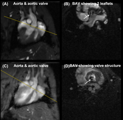 |
171 | Comprehensive evaluation of bicuspid aortic valve structure, hemodynamics, and tissue characterization in mice using MRI
El-Sayed H. Ibrahim1, John LaDisa1, and Joy Lincoln1
1Medical College of Wisconsin, Milwaukee, WI, United States Keywords: Cardiovascular, Valves, congenital heart disease Bicuspid Aortic Valve (BAV) is the most common congenital heart disease affecting 1-2% of all live births. Current clinical management includes periodic surveillance of aortic valve dysfunction, and only when the valve becomes stenotic is intervention recommended, which includes surgical procedures that come with insuperable long-term outcomes. We optimized a small-animal MRI protocol for comprehensive evaluation of the BAV structure, function, flow pattern, and tissue characterization. Children and adolescents with BAV would benefit from this comprehensive assessment of their risk profile during early stages of the disease to better predict outcomes and clinical management strategies. |
|
1988.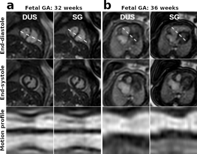 |
172 | Fetal Cardiovascular Magnetic Resonance: A Comparison of Doppler Ultrasound and Self-Gating Techniques
Paul Polak1, Henrik von Kleist2, Marcus Hott2, Erin K Englund1, Alex J Barker1, Bettina Cuneo3, Richard M Friesen3, Lorna P Browne1, and Mehdi Hedjazi Moghari1
1Radiology, University of Colorado Anschutz Medical Campus, Aurora, CO, United States, 2Technische Universitat Munchen, Munich, Germany, 3Cardiology, University of Colorado Anschutz Medical Campus, Aurora, CO, United States Keywords: Fetal, Cardiovascular Fetal cardiovascular magnetic resonance requires an accurate measure of the fetal heart rate for cine image acquisitions. In this work, we compared the use of Doppler ultrasound with a self-gating technique in 8 subjects. Images were acquired in the axial and short-axis orientations of the fetal heart, and the images were compared with qualitative scoring, heart rate analysis, and ventricular volumes. It was found that the techniques had only minor differences demonstrating the potential use of retrospective self-gating in the clinic, as it reduces workflow complexity and may lead to shorter exam times for pregnant women. |
|
1989.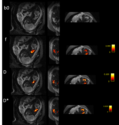 |
173 | Early Experience of Functional Fetal Lung Imaging using Low Field MRI
Kelly Payette1,2, Carla Avena Zampieri1, Jordina Aviles Verdera1,2, Lisa Story3, Raphael Tomi-Tricot4, Joseph V Hajnal1,2, Mary Rutherford1,2, and Jana Hutter1,2
1Centre for the Developing Brain, School of Biomedical Engineering and Imaging Sciences KCL, London, United Kingdom, 2Biomedical Engineering Department, School of Biomedical Engineering and Imaging Sciences, KCL, London, United Kingdom, 3Women’s Health, KCL, London, United Kingdom, 4MR Research Collaborations, Siemens Healthcare Limited, Frimley, United Kingdom Keywords: Fetal, Low-Field MRI Low field MRI for fetal imaging has the potential to be a powerful tool for prenatal diagnosis, with a favourable combination of scope for a larger bore (adding comfort and reducing claustrophobia), and lower SAR, susceptibility effects and receiver coil properties. We performed functional scans of the fetal lung to demonstrate the high image quality that can be acquired. The quantitative values of the fetal lungs (IVIM parameters, T1, T2*) calculated from the low field images match the expected values from higher field strengths reported in the literature. |
|
1990.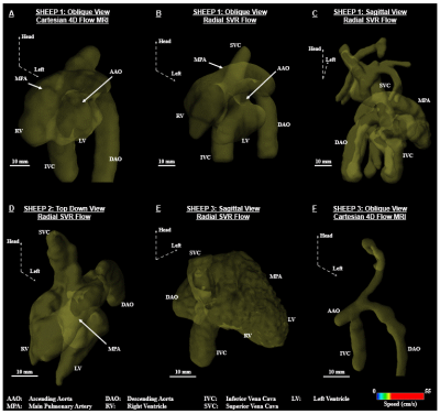 |
174 | Volumetric flow assessment in fetal sheep with Cartesian 4D flow MRI and radial slice-to-volume flow MRI with and without blood pool contrast
Datta Singh Goolaub1, Judd Storrs2, John Kelly3,4, Ramkumar Krishnamurthy2, Karen Texter5, James Strainic6, Ellie Ragsdale7, Christopher Breuer3, Aimee Armstrong4, Rajesh Krishnamurthy2, and Christopher K. Macgowan1,8
1Translational Medicine, The Hospital for Sick Children, Toronto, ON, Canada, 2Radiology, Nationwide Children’s Hospital, Columbus, OH, United States, 3Center for Regenerative Medicine, Nationwide Children’s Hospital, Columbus, OH, United States, 4Cardiology, Nationwide Children’s Hospital, Columbus, OH, United States, 5The Heart Center, Nationwide Children’s Hospital, Columbus, OH, United States, 6Pediatrics, University Hospitals Rainbow Babies & Children’s Hospital, Cleveland, OH, United States, 7OB/GYN-Maternal Fetal Medicine, University Hospitals Cleveland Medical Center, Cleveland, OH, United States, 8Medical Biophysics, University of Toronto, Toronto, ON, Canada Keywords: Fetal, Fetus We compare flow distributions in fetal sheep obtained with Cartesian 4D flow MRI and slice-to-volume multidimensional blood flow imaging using accelerated multislice radial phase contrast MRI. In the latter, stacks of 2D slices are imaged. Real-time reconstructions allow for in-plane motion correction. CINEs are combined into a dynamic flow sensitive volume using slice-to-volume reconstruction with interslice motion correction. Comparison is performed in the presence and absence of contrast agent. |
|
1991. |
175 | Accelerated 4D flow MRI in pediatric patients with congenital heart disease
Fatemeh Rastegar Jooybari1,2, Christopher Huynh2, Sharon Portnoy2, Jonathan Voutsas3, Diana Balmer-Minnes2, Ankavipar Saprungruang4, Shi-Joon Yoo5, Christopher Z Lam4, and Christopher K Macgowan1,2
1Medical Biophysics, University of Toronto, Toronto, ON, Canada, 2Translational Medicine, Hospital for Sick Children, Toronto, ON, Canada, 3Queen's University, Kingston, ON, Canada, 4Department of Diagnostic Imaging, Hospital for Sick Children, Toronto, ON, Canada, 5Departments of Medical Imaging and Paediatrics, University of Toronto, Toronto, ON, Canada Keywords: Cardiovascular, Quantitative Imaging, Flow, Heart, Cardiovascular, Congenital heart disease The application of conventional 4D flow to evaluate the hemodynamics of the heart and vessels is limited by long scan time. Recent advances aim to reduce scan time while providing sufficient accuracy. Here we investigate a 4D flow pipeline based on an undersampled 3D golden angle radial trajectory that offers reconstruction flexibility. We evaluated this technique in a cross-section of 11 pediatric patients with congenital heart disease and compared flows against conventional 2D phase contrast in target vessels. |
|
1992.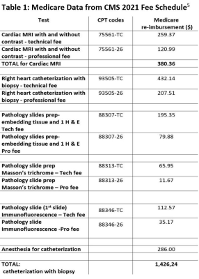 |
176 | Cost Analysis of Cardiac MRI for Cardiac Transplant Surveillance
Margaret M Samyn1, Ke Yan2, Justin Godown3, Steven J Kindel1, Bruce Damon4, Jonathan H Soslow5, and John Affleck-Graves6
1Pediatrics, Medical College of Wisconsin/ Herma Heart Institute at Children's Wisconsin, Milwaukee, WI, United States, 2Pediatrics, Medical College of Wisconsin, Milwaukee, WI, United States, 3Pediatrics, Vanderbilt University Medical Center, Nashville, TN, United States, 4Grainger College of Engineering, Carle Foundation Hospital/University of Illinois, Urbana, IL, United States, 5Pediatrics, Vanderbilt Univesity Medical Center, Nashville, TN, United States, 6University of Notre Dame Mendoza College of Business, Notre Dame, IN, United States Keywords: Cardiovascular, Health Care Economics, Heart Transplantation; Acute Rejection Current transplant surveillance relies on frequent cardiac catheterization with endomyocardial biopsy (cath EMB) to detect life threatening acute rejection. Cardiac MRI has emerged as a diagnostic tool which can characterize the myocardium. The PEACE study (R01-HL164995-01) aims to evaluate the potential for using cardiac MRI parametric mapping as a pre-cath EMB screening tool to alleviate both patient and family burden and the financial burden of cath EMB. Consistent with the recent emphasis on "Value MRI," we therefore sought to understand the potential Medicare savings, if MRI is used for transplant surveillance for healthy heart transplant recipients rather than cath EMB. |
|
1993.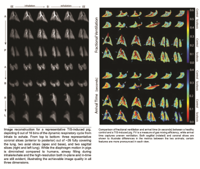 |
177 | Out-of-phase Ventiilation Observed With Dynamic Hyperpolarized Xenon-129 Mri In A Pig Thoracic Insu!ciency Syndrome Model
Hooman Hamedani1, Faraz Amzajerdian1, Stephen Kadlecek1, Kai Ruppert1, Mostafa Ismail1, Luis Loza1, Ian Duncan1, Madeline Boyes2, Klaus Hopster2, Rachel Hilliard2, Thomas Schaer2, Benjamin Sinder 3, Brian Snyder4, Axel Moore5, Dawn Elliott5, Kyle Meadows5, and Rahim Rizi1
1University of Pennsylvania, Philadelphia, PA, United States, 2University of Pennsylvania School of Veterinary Medicine, Kennett Square, PA, United States, 3Children’s Hospital of Philadelphia, Philadelphia, PA, United States, 4Boston Children’s Hospital, Boston, MA, United States, 5University of Delaware, Newark, DE, United States Keywords: Body, Skeletal In a pilot study of thoracic insufficiency syndrome in a pig model, we demonstrated dynamic hyperpolarized xenon-129 (HXe) MRI’s ability to noninvasively measure respiratory dysfunction in terms of gas distribution and mixing in the lungs, and specifically showed ‘out of phase’ ventilation in constricted regions of the lung. This marker of ventilation—i.e., the temporal characteristic of signal build-up during inhalation and its wash-out during exhalation—is inaccessible without our dynamic imaging technique. This technique can be implemented clinically to enhance our understanding of TIS pathophysiology and evaluate treatment strategies aimed at fostering age appropriate alveolarization. |
|
1994.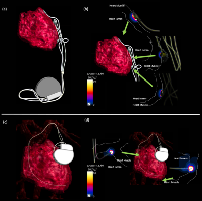 |
178 | Comparison of SAR Distributions around Epicardial and Endocardial Electrodes in Growing Pediatric Patients with Cardiac Pacemakers
Reina Oguchi1, Kyoko Fujimoto2, Kazuhiro Shiraga3, and Kagayaki Kuroda1
1Tokai University, Kanagawa, Japan, 2GE Healthcare, Waukesha, WI, United States, 3Department of Pediatrics, The University of Tokyo Hospital, Tokyo, Japan Keywords: Safety, Safety RF-induced SAR distributions around epicardial lead were compared with endocardial lead in the 29-month-old and 6-year-old pediatric anatomical models with cardiac pacemakers. Numerical simulations were performed with a 1.5-T birdcage coil and the pacemaker models segmented from the age-matching patient CT data. In both pediatric models, the simulation with the endocardial lead overall showed lower SAR values compared to that with the endocardial lead. The screw-like electrode attached to the endocardial lead may have resulted in much higher SAR values, compared to the tined-type electrode of the epicardial lead. |
|
1995.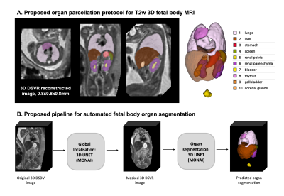 |
179 | Automated multi organ segmentation for 3D fetal body MRI: differences in the normal growth charts for different acquisition parameters
Alena U. Uus1, Lisa Story2,3, Megan Hall3,4,5, Vanessa Kyriakopoulou5, Alexia Egloff Collado5, Carla Avena Zampieri1,5, Jacqueline Matthew1,5, Irina Grigorescu1, Ayse Ceren Tanritanir5, Joseph V. Hajnal1,5, Jana Hutter1,5, Mary Rutherford5, and Maria Deprez1
1Department of Biomedical Engineering, King's College London, London, United Kingdom, 2Academic Women's Health Department, King's College London, London, United Kingdom, 3Fetal Medicine Department, GSTT, London, United Kingdom, 4Institute for Women’s and Children’s Health, King's College London, London, United Kingdom, 5Centre for the Developing Brain, King's College London, London, United Kingdom Keywords: Prenatal, Segmentation This work presents the first deep learning pipeline for segmentation of multiple body organs from motion corrected 3D ssTSE fetal MRI. It is used to compare growth charts of the normal body organ development during 21-37 week GA range for 254 fetal datasets with different acquisition protocols (field strength and TE). |
|
1996.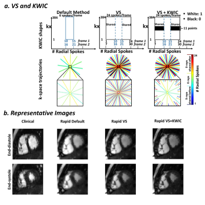 |
180 | View-Sharing and KWIC Filtering Achieves High Spatio-Temporal Resolution in 32-Fold Accelerated Real-Time Cardiac Cine with Radial Sampling
Lexiaozi Fan1,2, Kyungpyo D Hong2, Joshua D. Robinson2,3,4, Cynthia K. Rigsby2,3,5, and Daniel Kim1,2
1Department of Biomedical Engineering, Northwestern University, Evanston, IL, United States, 2Department of Radiology, Northwestern University Feinberg School of Medicine, Chicago, IL, United States, 3Department of Pediatric, Northwestern University Feinberg School of Medicine, Chicago, IL, United States, 4Division of Cardiology, Ann & Robert H. Lurie Children's Hospital of Chicago, Chicago, IL, United States, 5Department of Medical Imaging, Ann & Robert H. Lurie Children's Hospital of Chicago, Chicago, IL, United States Keywords: Cardiovascular, Image Reconstruction Highly-accelerated real-time cine MRI is a rapid technique that is particularly useful for patients with arrhythmia, dyspnea, and/or pediatric patients who may not be able to follow breath-hold instructions. Incorporating view-sharing (VS) and k-space weighted image contrast (KWIC) filtering into a compressed sensing (CS) reconstruction framework improves spatio-temporal resolution in our 32-fold accelerated real-time free-breathing cine MRI using radial k-space sampling. Resulting biventricular volumetric parameters are relatively accurate, which may eliminate the need for sedation/anesthesia in pediatric patients. |
|
 Back to Meeting Home
 Back to the Program-at-a-Glance
The International Society for Magnetic Resonance in Medicine is accredited by the Accreditation Council for Continuing Medical Education to provide continuing medical education for physicians.
 Back to Meeting Home
 Back to the Program-at-a-Glance
 View Presentation Video