Digital Poster
Pediatric Body & MSK MRI
ISMRM & ISMRT Annual Meeting & Exhibition • 03-08 June 2023 • Toronto, ON, Canada

| Computer # | |||
|---|---|---|---|
2135.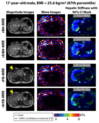 |
141 | Self-Navigated Rapid Radial Free-Breathing Liver MR Elastography: Assessment of Technical Performance in Children at 3T
Sevgi Gokce Kafali1,2, Bradley D. Bolster Jr.3, Shu-Fu Shih1,2, Timoteo I. Delgado1,4, Xiaodong Zhong5, Vibhas Deshpande6, Timothy R. Adamos7, Shahnaz Ghahremani1,7, Kara L. Calkins7, and Holden H. Wu1,2,4
1Radiological Sciences, David Geffen School of Medicine, University of California, Los Angeles, Los Angeles, CA, United States, 2Bioengineering, Henry Samueli School of Engineering, University of California, Los Angeles, Los Angeles, CA, United States, 3US MR R&D Collaborations, Siemens Medical Solutions USA, Inc., Salt Lake City, UT, United States, 4Physics and Biology in Medicine Interdepartmental Program, David Geffen School of Medicine, University of California, Los Angeles, Los Angeles, CA, United States, 5US MR Collaborations, Siemens Medical Solutions USA, Inc., Los Angeles, CA, United States, 6US MR Collaborations, Siemens Medical Solutions USA, Inc., Austin, TX, United States, 7Pediatrics, David Geffen School of Medicine, University of California, Los Angeles, Los Angeles, CA, United States Keywords: Elastography, Liver Hepatic stiffness measured by MR elastography (MRE) is a biomarker for fibrosis. Standard liver MRE requires breath-holding (BH) to avoid motion artifacts, but BH is challenging for children. Radial free-breathing (FB) MRE can overcome this challenge but requires longer scan times. Radial FB-MRE using rapid wave encoding decreases the scan time and self-navigation supports motion compensation. With the help of fractional encoding to reduce echo times, image quality can be further improved. This work demonstrated that self-navigated rapid and rapid fractional FB-MRE methods measure hepatic stiffness with close agreement and similar repeatability compared to corresponding BH-MRE in children at 3T. |
|
| 2136. | WITHDRAWN | ||
2137. |
142 | Free Breathing Simultaneous Multislice Diffusion Weighted Imaging of the Pediatric Abdomen Reduces Scan Time Without Sacrificing Image Quality
Ali B Syed1 and Praveen Jayapal2
1Department of Radiology, Stanford University, Redwood City, CA, United States, 2Department of Radiology, Stanford University, Palo Alto, CA, United States Keywords: Body, Diffusion/other diffusion imaging techniques Simultaneous Multislice (SMS) techniques can reduce scan time without significant impact on image quality. SMS techniques rely on coil geometry, which can be unfavorable in pediatric patients due to large variations in size. At the same time, pediatric imaging often demands minimizing scan time to increase patient compliance and reduce exposure to sedating medications. We evaluate the use of SMS diffusion weighted imaging applied to pediatric abdominal DWI and show that image quality is similar to standard DWI but with significant reduction in scan time. ADC values are not significantly affected. |
|
2138. |
143 | Evaluation of Pediatric Musculoskeletal Disease using CT-like MR of Pointwise Encoding Time Reduction with radial Acquisition sequence
Xiamei Zhuang1, Ke Jin1, Yan Yin 1, Junwei Li1, and Huiting Zhang2
1Hunan Children's Hospital, Changsha, China, 2MR Scientific Marketing, Siemens Healthineers, Wuhan, China Keywords: Body, Bone Cortical bone can be clearly displayed by CT-like MRI using ultrashort TE sequence. This study aimed to investigate the value of CT-like MRI using PETRA (ct-PETRA) sequence in the pediatric musculoskeletal diseases. Our results showed that ct-PETRA had no significant difference in measuring the C-E index and acetabulum index of children's hip joint compared with X-ray, and had no significant difference in visualization of lesions compared with X-ray and CT. Moreover, ct-PETRA has more advantages than X-ray in displaying trabecular fracture of fine bone. CT-like PETRA can be used as conventional complementary sequence for some assessments of pediatric musculoskeletal diseases. |
|
2139.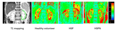 |
144 | Feasibility of Blood oxygen level dependent magnetic resonance imaging for distinguishing HSPN from HSP in children Synopsis
Hang Su1, Gang Zhang1, Jingjing Wu1, Wei Xing1, Yufu Hu1, Zhiwei Shen2, Ke Jiang2, Yuying Sun1, Xia Zhang1, and Xianqing Ren1
1The First Affiliated Hospital of Henan University of CM, Zhengzhou, China, 2Philips healthcare, Beijing, China Keywords: Adolescents, Kidney Blood oxygen level dependent magnetic resonance imaging (BOLD-MRI) could be used to detect renal tissue oxygen levels noninvasively, and the T2* time induced by the paramagnetic effect of deoxyhemoglobin can be obtained to indirectly assess blood oxygen utilization. Previous research has proven that BOLD-MRI can detect renal oxygenation levels with high sensitivity and the accuracy of the data has been validated by invasive probe measuring techniques. In this work, we investigated the clinical feasibility of T2* mapping for the detection of HSPN and HSP in children, since we predicted that HSPN would also result in aberrant renal blood oxygenation signals. |
|
2140.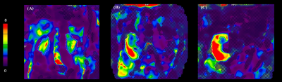 |
145 | Using MR Elastography in the Assessment of Low-Grade Fibrosis of Pediatric Kidney Transplant Recipients.
Suraj D Serai1,2, Hansel J Otero1,2, Tatiana Morales-Tisnes1, Mohamed M Elsingergy1, Tricia Bhatti1, and Bernarda Viteri1
1Children's Hospital of Philadelphia, Philadelphia, PA, United States, 2Radiology, University of Pennsylvania, Philadelphia, PA, United States Keywords: Kidney, Elastography, transplant MRE can propagate shear waves which can provide better estimates of stiffness of smaller and deeper structures. Here, we found a positive correlation between kidney elasticity and the presence of histological fibrosis where stiffness values tended to be higher in fibrosed allografts than stable or native kidneys. If clinically approved, elastography would not only be useful for early detection of interstitial fibrosis and tubular atrophy in renal transplants, which would individualize the need for immunosuppression therapy, but could also be used as a screening tool to evaluate fibrosis in potential donors to identify best candidates prior to the transplantation procedure. |
|
2141.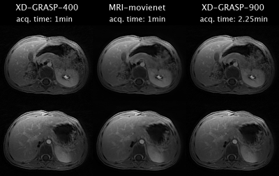 |
146 | Fast free-breathing pediatric MRI using accelerated radial acquisition and deep learning motion-resolved reconstruction
Victor Murray1, Syed Siddiq1, Gerald Behr2, and Ricardo Otazo1,2
1Department of Medical Physics, Memorial Sloan Kettering Cancer Center, New York, NY, United States, 2Department of Radiology, Memorial Sloan Kettering Cancer Center, New York, NY, United States Keywords: Body, Machine Learning/Artificial Intelligence Motion remains a significant challenge in pediatric MRI. Motion-resolved 4D MRI, such as XD-GRASP, is a promising alternative to motion correction. However, long scan times and particularly reconstruction times restricted routine clinical use. This work presents a deep learning approach called MRI-movienet for 4D reconstruction of radial data, which enables acceleration of both acquisition and reconstruction for free-breathing pediatric MRI with only 1 minute scan time and less than 2 seconds reconstruction time. The deep learning approach is demonstrated for free-breathing abdominal pediatric MRI without anesthesia using XD-GRASP as a reference for comparison. |
|
2142.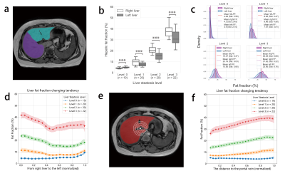 |
147 | Heterogeneous distribution of hepatic and pancreatic fat fraction in overweight or obese children and adolescents
Yang Cao1, Dingyi Lin1, Yufan Zhou1, Yi-Cheng Hsu2, Gaoyang Mi3, Hongxi Zhang3, and Min Wang1
1College of Biomedical Engneering & Instrument Science, Zhejiang University, Hangzhou, China, 2MR Collaboration, Siemens healthineers Itd, Shanghai, China, 3Department of Radiology, Zhejiang University School of Medicine Children's Hospital Binjiang Campus Pediatrics, Hangzhou, China Keywords: Data Analysis, Fat The ectopic fat is not evenly distributed in organs and can be a time dependent process that starts at early age. In this work, we focused on obese or overweight children and adolescents and calculated the voxel-wised liver and pancreas fat fraction. With our recently proposed deep learning method, we separately quantified the fat depositions in different compartments of liver and pancreas with high precisions. Our analysis revealed an unevenly distributed ectopic fat in both liver and pancreas at young age and suggested that hepatic and pancreatic fat depositions are weakly coupled but have different underlying mechanisms. |
|
2143.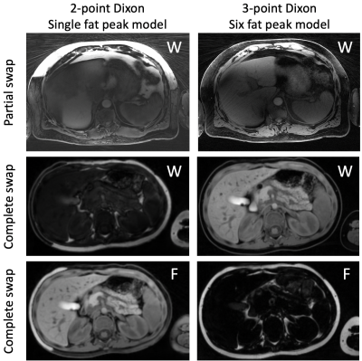 |
148 | High resolution, swap free, and motion compensated water/fat separation in free-breathing pediatric T1-weighted abdominal MRI
Reyhaneh Nosrati1, Onur Afacan1, Kristina Pelkola1, Sarah Bixby1, and Simon K. Warfield1
1Boston Children's Hospital, Harvard Medical School, Boston, MA, United States Keywords: Liver, Pediatric, Abdominal MRI T1-weighted GRE sequence with Dixon technique for water/fat separation is a routine component of abdominal MRI to detect lesions and characterize hemorrhage/fat content. The limitations of the routine free-breathing T1-weighted pediatric abdominal MRI and water/fat separation includes low-resolution to achieve high pixel-bandwidth and minimize chemical shift, respiratory motion blurring (despite radial acquisition) that degrades image sharpness, water/fat swapping, and off-resonance fat blurring due to multi-peak nature of fat spectrum. We have assessed a novel image acquisition and reconstruction pipeline based on 3-point Dixon method, multi-peak fat model, and residual motion correction to address all the limitations of the current practice. |
|
2144.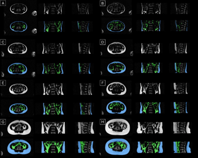 |
149 | Association of physical activity and screen time with DXA- and MRI-based body composition in adolescents, a population-based study
Tong Wu1, Junwen Yang-Huang2, Meike Vernooij1, María Rodriguez‑Ayllon3, Vincent Jaddoe4, Hein Raat2, Stefan Klein1, and Edwin Oei1
1Department of Radiology and Nuclear Medicine, Erasmus MC, Rotterdam, Netherlands, 2Department of Public Health, Erasmus MC, Rotterdam, Netherlands, 3Department of Epidemiology, Erasmus MC, Rotterdam, Netherlands, 4Department of Pediatrics, Erasmus MC, Rotterdam, Netherlands Keywords: Adolescents, Body, Body composition This study is embedded in the Generation R Study, a population-based prospective cohort study in the Netherlands. At 13 years, whole-body Dixon MRI was performed. A previously presented Competitive Dense Fully Convolutional Network (CDFNet) was retrained and used to quantify the abdominal subcutaneous and visceral fat. We found a higher physical activity and lower screen time are associated with lower levels of MRI-based abdominal visceral obesity in adolescents. Therefore, promoting physical activity and reducing screen time should be one of the goals of lifestyle intervention programs for tackling the childhood obesity. |
|
| 2145. | WITHDRAWN | ||
2146. |
150 | Exploring accelerated MRI based Bone Imaging in Pediatric Congenital Spinal Anomalies
Foram Bharat Gala1, Hirava Manek1, Indrajit Saha2, Narayana Krishna Rolla3, Pradeep Kumar4, and Rashmi Rao3
1Department of Radiology, Bai Jerbai Wadia hospital for children, Mumbai, India, 2Clinical Science, Philips India Limited, Delhi, India, 3Philips India Limited, Bangalore, India, 4Philips India Limited, Mumbai, India Keywords: MSK, Spinal Cord Congenital spinal anomalies are common in children resulting in spinal deformities and these could be due to isolated vertebral segmentation abnormalities or with added cord abnormalities referred to as spinal dysraphism. Early surgical intervention is necessary in these children which requires both MRI and CT scan evaluation for cord and bone details respectively. We utilized novel technique - Compressed Sense accelerated MRI based - Fast field echo Resembling A CT Using Restricted Echo-spacing (CS-FRACTURE) technique in pediatric patients which provides CT like contrast. We describe our experience in evaluation of congenital spinal anomalies |
|
2147.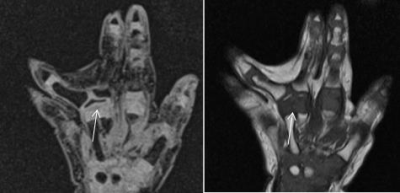 |
151 | The value of MRI 3D-DESS technique in preoperative evaluation of polydactyly in children
Li Jingling1
1Xijing Hospital, Air Force Military Medical University, Xi 'an, China Keywords: Joints, Pediatric Polydactyly is very common disease in pediatric orthopeadic. In this study, 18 patients with polydactyly were included. All the 3D-DESS image were evaluated by two senior diagnostic radiologists using a double-blind 3-point scale, the intraoperative results were used as the “gold standard” to analyze the consistency of image results and intraoperative results. The purpose of this study is to assess whether the 3D-DESS sequence can provide the growth and development information of cartilage epiphysis, tendon and bone tissue of polydactyly in children before surgery, which has value for preoperative evaluation and preparation of surgical treatment. |
|
2148.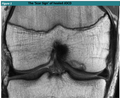 |
152 | On the MRI Signature of Healed Juvenile Osteochondritis Dissecans (JOCD) in the Knee – The ‘Scar Sign’
Jutta Ellermann1, Abdul Wahed Kajabi2, Marc Tompkins3, Joe Luchsinger1, Stefan Zbyn4, Shelly Marette1, Bradley Nelson3, and Takashi Takahashi1
1Radiology, University of Minnesota, Minneapolis, MN, United States, 2CMRR/Radiology, University of Minnesota, Minneapolis, MN, United States, 3Orthopedics, University of Minnesota, Minneapolis, MN, United States, 4University of Minnesota, Minneapolis, MN, United States Keywords: Bone, Adolescents, Epiphyseal Cartilage, Juvenile Osteochondritis Dissecans Healing of Juvenile Osteochondritis Dissecans requires progressive lesion ossification and osseous bridging with the parent bone. The ‘Scar Sign’ is an MR imaging finding that documents a healed knee JOCD lesion and is characterized by a distinct hypointense line at the prior lesion – parent bone interface, best identified on PD- or T1-weighted images w/o FS on routine clinical MRI. Knowledge of stages, timeline of healing and expected MR imaging findings of JOCD is important for the reporting Radiologist. It will facilitate prognostication and can serve as a reliable outcome measure for future research on evidence based Juvenile Osteochondritis Dissecans management. |
|
2149.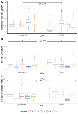 |
153 | Probing arm muscle structure underlying motor impairment in children with cerebral palsy using DTI tractography
Divya Joshi1,2, Alexandra Hruby1,2, Julius PA Dewald1,2,3, and Carson Ingo2,4
1Department of Biomedical Engineering, Northwestern University, Evanston, IL, United States, 2Department of Physical Therapy and Human Movement Sciences, Northwestern University Feinberg School of Medicine, Chicago, IL, United States, 3Department of Physical Medicine and Rehabilitation, Northwestern University Feinberg School of Medicine, Chicago, IL, United States, 4Department of Neurology, Northwestern University Feinberg School of Medicine, Chicago, IL, United States Keywords: Muscle, Diffusion Tensor Imaging, Cerebral Palsy Individuals with cerebral palsy (CP) exhibit musculoskeletal maladaptations that have a profound impact on wrist and hand function, yet there is little understanding regarding underlying arm muscle structural changes. We implemented DTI to estimate in vivo muscle architecture of a forearm flexor muscle in both paretic and non-paretic arms of children with hemiparetic CP (n=5). Bone length, muscle size, fascicle lengths, and MD decreased while fascicle curvature increased in the paretic limb. Interlimb differences in fascicle length and curvature were correlated with interlimb differences in MD. We show that DTI is effective at capturing forearm muscle architecture changes in CP. |
|
2150.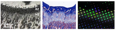 |
154 | Magnetic resonance and diffusion tensor imaging of the adolescent rabbit growth plate of the knee
Ola Kvist1, Peter Damberg1, Zelong Dou1, Johan Sanmartin Berglund2, Carl-erik Flodmark3, Ola Nilsson1,4, and Sandra Diaz Ruiz1,3
1Karolinska Institute, Stockholm, Sweden, 2Blekinge Institute of Technology, Stockholm, Sweden, 3Lunds University, Malmö, Sweden, 4Örebro University, Örebro, Sweden Keywords: Cartilage, Diffusion Tensor Imaging The hindlimbs of female rabbits aged 16, 20, and 24 weeks were studied in a 9.4T MRI scanner to assess the ability of MRI-DTI to evaluate growth plate morphology and activity compared with that of histomorphometry and micro-computed tomography (μCT) in rabbits. The MRI sequences included a multi-gradient echo 3D sequence and DTI in 14 directions (b-value= 984 s/mm2). The growth plate height and volume were similar for all modalities at each time point and age. Tract number and volume declined with age; however, tract length did not show any changes. MRI-DTI may be useful for evaluating the growth plates. |
|
2151.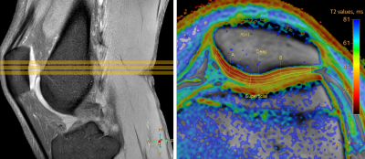 |
155 | T2 mapping of patellar cartilage after single and first-time traumatic lateral patellar dislocation episode
Elena Voronkova1,2, Petr Menshchikov3,4, Ilya Melnikov1, Olga Bozhko1, Andrei Manzhurtsev1,3,5, Maxim Ublinskiy1,3, Denis Vorobyev1, Dmitriy Kupriyanov4, and Tolib Akhadov1
1Radiodiagnosis, Clinical and Research Institute of Emergency Pediatric Surgery and Trauma, Moscow, Russian Federation, 2Institute of Biochemical Physics, Russian Academy of Sciences, Moscow, Russian Federation, 3Institute of Biochemical Physics, Russian Academy of Sciences, Moscow, Moscow, Russian Federation, 4LLC Philips, Moscow, Russian Federation, 5Moscow State University, Moscow, Russian Federation Keywords: MSK, Cartilage The purpose of the study was to examine short-term consequences of the single and first-time lateral patellar displacement on patellar cartilage quality in teenagers using T2 mapping. The research showed a principal difference in T2 changes between medial and lateral cartilage facet. The medial part usually suffering first after LPD, the absence of T2 changes for the mild group may indicate completed reparative processes, while the decrease in T2 denotes that the reparation is still ongoing. T2 increase in lateral patellar regardless chondromalacia severity indicates secondary damage. |
|
2152.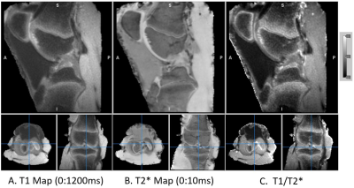 |
156 | T1 and T2* Relaxometry MRI of Knee Growth Plate Using Ultra-Short TE MRI: Feasibility Study
Kwan-Jin Jung1 and Elahe Ganji2
1Biomedical Imaging Center, Beckman Institute, University of Illinois at Urbana-Champaign, Urbana, IL, United States, 2Beckman Institute, University of Illinois at Urbana-Champaign, Urbana, IL, United States Keywords: Cartilage, Relaxometry, growth plate, UTE Growth plates are one of the most common sites of injury in the young skeleton. The growth plate is an interwoven and multi-layered interfacial tissue that transitions from cartilaginous to mineral morphology as it extends into metaphysis of long bones. We used Ultrashort Echo-time (UTE) MR imaging as a promising sensitive technique to detect the growth plate transition that has short T2. The UTE sequence was extended to measure T2* and T1 using multi-echoes and variable flip angles. The contrast of the transitional zone was further enhanced by a ratio of T1 over T2*. |
|
2153. |
157 | The Performance of 3D-UTE MRI in Children with Pneumonia: Comparison with Standard 3D T1-GRE and T2-FSE Sequences
yan sun1, yujie chen1, xuesheng li1, yi liao1, xijian chen1, yu song1, xinyue liang2, yongming dai2, and gang ning1
1Key Laboratory of Birth Defects and Related Diseases of Women and Children(Sichuan University), Ministry of Education,West China Second university Hospital, Sichuan University, Chengdu, China, 2Central Research Institute, United Imaging Healthcare, shanghai, shanghai, China Keywords: Lung, Pediatric, pediatric pneumonia As a promising MRI technique, ultrashort echo‑time (UTE) sequence has been widely used in the evaluation of pulmonary diseases, allowing for increased contrast-to-noise ratio and the comparable image quality with CT. Literature on the use of the UTE in pneumonia is broadly available in older children and adults, whereas the data in younger children group is limited. This study illustrated the ability of UTE lung imaging for assessing pneumonia patients under 3 years old and has good diagnostic utility in detecting the typical morphological findings of pediatric pneumonia. |
|
2154.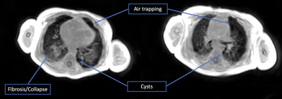 |
158 | Validating UTE-based Assessment of Bronchopulmonary Dysplasia at 3T
Narayan Iyer1,2, Matthew Borzage1,2, and Eamon Doyle2,3
1Pediatrics, Children's Hospital of Los Angeles, Los Angeles, CA, United States, 2Keck School of Medicine, University of Southern California, Los Angeles, CA, United States, 3Radiology, Children's Hospital of Los Angeles, Los Angeles, CA, United States Keywords: Lung, Pediatric, BPD, UTE Bronchopulmonary dysplasia (BPD) is the most common serious complication of prematurity affecting approximately 30,000 infants annually in the USA alone. Decisions regarding treatment course as well as prognostication require an understanding of regional lung changes. However, chest radiographs poorly distinguish the severity of BPD. Advances in ultra-short echo time imaging have made lung imaging feasible at clinically relevant field strengths. We have implemented a UTE-based imaging protocol at our institution and started validation in pre-term infants. Initial results demonstrate that clinically interpretable images can be acquired in an infant with BPD. |
|
 Back to Meeting Home
 Back to the Program-at-a-Glance
The International Society for Magnetic Resonance in Medicine is accredited by the Accreditation Council for Continuing Medical Education to provide continuing medical education for physicians.
 Back to Meeting Home
 Back to the Program-at-a-Glance
 View Presentation Video