Digital Poster
Fetal MRI
ISMRM & ISMRT Annual Meeting & Exhibition • 03-08 June 2023 • Toronto, ON, Canada

| Computer # | |||
|---|---|---|---|
 |
2332.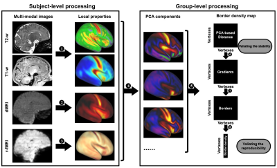 |
161 | Multi-modal multi-resolution atlas of the human neonatal cerebral cortex based on microstructural similarity
Mingyang Li1, Xinyi Xu1, Zuozhen Cao1, Ruike Chen1, Ruoke Zhao1, Zhiyong Zhao1, and Dan Wu1
1College of Biomedical Engineering & Instrument Science, Zhejiang University, Zhejiang, China Keywords: Gray Matter, Multimodal, parcellation, morphometric similarity, atlas In this work, we aimed to generate a comprehensive parcellation of the human neonatal cortex based on multi-modal MRI features. We collected the dataset from the developing human connectome project and estimated ten different MRI features to calculate the similarity between different locations in the neonatal cortex. We developed an automated algorithm based on gradient of the integrated similarity map to generate parcellations at different resolutions. We also provided a manual parcellation based on the multimodal similarity for higher anatomical interpretability. The present work may facilitate structural-functional connectome analysis in early brain development. |
2333.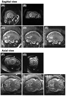 |
162 | High-resolution fetal brain anatomical imaging using a reduced field-of-view with outer volume suppression
MinJung Jang1,2 and Zungho Zun1
1Department of Radiology, Weill Cornell Medicine, New York, NY, United States, 2Department of Biomedical Engineering, Ulsan National Institute of Science and Techonology, Ulsan, Korea, Republic of Keywords: Fetal, Fetus Reliable fetal brain anatomical imaging is critical to identify structural brain abnormalities early in utero. Fetal brain imaging with increased spatial resolution may provide more details but is limited by a large acquisition matrix size with a long readout time. Here, we demonstrate high-resolution fetal brain imaging using a reduced field-of-view with outer volume suppression. The performance of outer volume suppression pulse was evaluated in simulations and phantom/in vivo experiments. High-resolution imaging with reduced field-of-view demonstrated improved image quality than conventional imaging without visible blurring or aliasing artifacts. |
|
2334.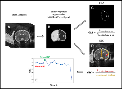 |
163 | Quantitative Assessment of Gyrification Patterns and Symmetry of the Fetal Brain based on Routine Fetal 2D MRI
Bossmat Yehuda1,2, Aviad Rabinowich, MD1,3,4, Dafi Surani1, Yair Wexler5, Netanell avisdris1,6, Ori Ben-Zvi1, Bella Spector1,6, Leo Joskowicz6, Liat Ben-Sira, MD1,3, Elka Miller, MD7, and Dafna Ben Bashat1,2,4
1Sagol Brain Institute, Tel Aviv Sourasky Medical Center, Tel Aviv, Israel, 2Sagol School of Neuroscience, Tel Aviv University, Tel Aviv, Israel, 3Department of Radiology, Tel Aviv Sourasky Medical Center, Tel Aviv, Israel, 4Sackler Faculty of Medicine, Tel Aviv University, Tel Aviv, Israel, 5School of Neurobiology, Biochemistry and Biophysics, Faculty of Life Sciences, Tel Aviv University, Tel Aviv, Israel, 6School of Computer Science and Engineering, The Hebrew University of Jerusalem, Jerusalem, Israel, 7CHEO Medical Imaging, University of Ottawa, Ottawa, ON, Canada Keywords: Fetal, Neuro, gyrification Current clinical assessment of fetal brain gyrification is performed manually and subjectively. Previous studies that suggested methods for quantitative assessment of fetal brain gyrification were based on 3D reconstruction. In this study we propose an automatic method for quantifying the gyrification from routinely acquired 2D clinical data. We calculate five different gyrification indices for both the right and left hemispheres. We present developmental curves of 134 control fetuses in a wide range of GA (18-37 weeks) and demonstrate differences between control fetuses and fetuses with malformation of cortical development: five with lissencephaly and 13 with polymicrogyria. |
|
2335.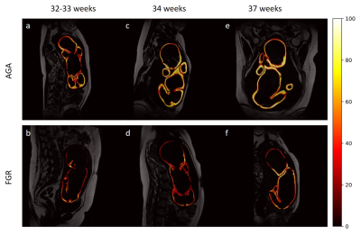 |
164 | Reduced adiposity in growth-restricted fetuses using quantitative two-point Dixon analysis
Aviad Rabinowich1,2,3, Netanell Avisdris1,4, Ayala Zilberman2,5, Sapir Lazar2,6, Daphna Link-Sourani1, Jacky Herzlich2,7, Bella Specktor-Fadida4, Leo Joskowicz4, Gustavo Malinger2,5, Liat Ben Sira2,6, Liran Hiersch2,5, and Dafna Ben Bashat1,2,8
1Sagol Brain Institute, Tel Aviv Sourasky Medical Center, Tel Aviv, Israel, 2Sackler Faculty of Medicine, Tel Aviv University, Tel Aviv, Israel, 3Department of Radiology, Tel Aviv Soursaky Medical Center, Tel Aviv, Israel, 4School of Computer Science and Engineering, The Hebrew University of Jerusalem, Jerusalem, Israel, 5Department of Obstetrics and Gynecology, Lis Hospital for Women, Tel Aviv Sourasky Medical Center, Tel Aviv, Israel, 6Department of Radiology, Tel Aviv Sourasky Medical Center, Tel Aviv, Israel, 7Neonatal Intensive Care Unit, Dana Dwek Children's Hospital, Tel Aviv Sourasky Medical Center, Tel Aviv, Israel, 8Sagol School of Neuroscience, Tel Aviv University, Tel Aviv, Israel Keywords: Fetal, Fetus, Fetal growth restriction Adipose tissue (AT, i.e., fat) deposition is reduced in undernourished growth-restricted fetuses (FGR) and may correlate with disease severity. Previous studies showed reduced AT using mainly linear sonographic measurements of the subcutaneous fat. We demonstrate the use of a two-point Dixon sequence for whole-body subcutaneous AT. The fat content of 64 fetuses (37 appropriate for gestation age [AGA], 18 FGR, and 9 small for gestational age) was measured using three adiposity parameters. We demonstrated third-trimester AT accretion in AGA fetuses and reduced AT in FGR. This study is the first to demonstrate whole-fetus subcutaneous AT changes in FGR. |
|
2336.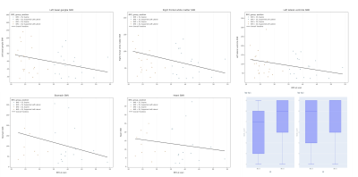 |
165 | Exploring the effect of elevated maternal BMI on image quality in fetal MRI
Kathleen Elizabeth Colford1, Daniel Cromb1, Zoe Hesketh1, Tom Finck1,2, Ayse Ceren-Tanritanir1,3, Serena Counsell1, and Mary Rutherford1,4
1Centre for the Developing Brain, School of Biomedical Engineering & Imaging Sciences, Kings College London, London, United Kingdom, 2Department of Diagnostic and Interventional Neuroradiology, Klinikum rechts der Isar der Technischen Universität München, Munich, Germany, 3Guys and St. Thomas’s NHS Foundation Trust, London, United Kingdom, 4MRC Centre for Neurodevelopmental Disorders, King’s College London, London, United Kingdom Keywords: Fetal, Data Analysis, Maternal BMI, Signal to Noise Ratio We assessed SNR (signal-to-noise-ratio) against elevated maternal BMI in fetal brain/body MRI imaging to examine if SNR decreases both objectively/subjectively, and if MRI could be an alternative imaging modality in prenatal care. Spearman’s Correlation-Rank coefficient was used to identify relationships between SNR measurements in ROI’s and maternal BMI. There’s a negative downward trend between maternal BMI and SNR seen for all fetal ROIs (particularly in regions more susceptible to SNR); subjective assessment didn’t identify any association between BMI and image quality. Our results suggest that MRI is a viable alternative to ultrasound in pregnant women with elevated BMI. |
|
2337.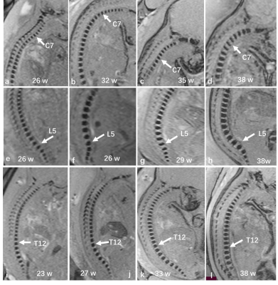 |
166 | A quantitative study on the growth patterns of vertebrae in utero fetuses in second-third trimester:a prospective study based “black-bone” MRI
Cai Xianyun1, Chen Xin1, Jinxia Zhu2, and Guangbin Wang1
1Shandong Provincial Hospital Affiliated to Shandong First Medical University, jinan, China, 2MR Collaboration, Siemens Healthineers Ltd, Beijing, China. Email: jinxia.zhu@siemens-healthineers.com, beijing, China Keywords: Fetal, Normal development Imaging the anatomy of the spine and its relevant pathology is clinically important for the early identification of spinal malformations and anomalous osseous development. our study applied MRI-based “black-bone” sequence to elucidate the growth dynamics of vertebrae ossification center in utero fetuses in second-third trimester. We found that the fetal vertebrae were correlated with gestational age and follow a certain formula, providing the existing literature with completely novel quantitative data. |
|
2338.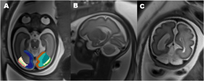 |
167 | Volumetric MRI analysis of occipital gyrus in fetuses with isolated ventriculomegaly
Xin Zhang1, Fan Wu1, Zhaoji Chen1, Yuchao Li1, Zhenqing Liu1, Chenxin Xie1, and Hongsheng Liu1
1Department of Diagnostic Radiology, Guangzhou Women and Children's Medical Center, Guangzhou Medical University,Guangdong Provincial Clinical Research Center for Child Health, 510623, China, Guangzhou, China Keywords: Fetal, Brain, Fetal MRI; Isolated ventriculomegaly The lateral ventricle is closely related to the occipital lobe anatomically. We found changes in cortical volume and white matter volume in isolated fetuses ventriculomegaly. This study will address whether occipital and occipital gyrus volumes differ in IVM and investigate the developmental pattern of the occipital lobe with increasing gestational age investigate. This study will help to provide the research basis for the study the neurodevelopment in IVM fetuses. |
|
2339.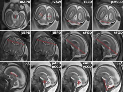 |
168 | Fetal brain Super-Resolution Reconstruction reliability from clinical MRI data
Tommaso Ciceri1,2, Letizia Squarcina3, Adele Ferro4, Florian Montano5, Alessandra Bertoldo2,6, Nicola Persico7, Simona Boito7, Fabio Maria Triulzi3,8, Giorgio Conte8, Paolo Brambilla3,4, and Denis Peruzzo5
1NeuroImaging, IRCCS Eugenio Medea, Bosisio Parini (LC), Italy, 2Department of Information Engineering, University of Padua, Padova, Italy, 3Pathophysiology and Transplantation, University of Milan, Milano, Italy, 4Neurosciences and Mental Health, Fondazione IRCCS Ca’ Granda Ospedale Maggiore Policlinico, Milano, Italy, 5NeuroImaging Lab, IRCCS Eugenio Medea, Bosisio Parini (LC), Italy, 6Neuroscience Center, University of Padua, Padova, Italy, 7Department of Woman, Child and Newborn, Fondazione IRCCS Ca’ Granda Ospedale Maggiore Policlinico, Milano, Italy, 8Services and Preventive Medicine, Fondazione IRCCS Ca’ Granda Ospedale Maggiore Policlinico, Milano, Italy Keywords: Fetal, Brain, Neurodevelopment, Biometry, Image Reconstruction In this study, we characterized the geometric reliability of the Super-Resolution (SR) images reconstructed via NiftyMIC and MIALSRTK toolkits over a common clinical fetal MR dataset (20-21 weeks of gestation). We compare 15 biometric measures derived from the acquired 2D images with those derived from the SR brain volumes. Furthermore, we examined two different acquisition sequences (TSE Vs. b-FFE) to evaluate which of them lead to more reliable measures and high-resolution reconstructions. Our findings strengthen the adoption of SR toolkits for fetal brain reconstructions to perform biometry evaluations, suggesting to use TSE sequences. |
|
2340.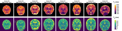 |
169 | T1 and T2 measurements from Neonates at 7 Tesla
Shaihan J Malik1,2,3, Raphael Tomi-Tricot2,4, Philippa Bridgen2,5, Anthony N Price3,5, Megan Quirke5, Daniel V Cromb3, Paul Cawley3,5, Enrico De Vita2,3, Jonathan O’Muircheartaigh3,6, Serena J Counsell3, Sharon Giles2,5, Mary Rutherford3, A. David Edwards3,5,6, Joseph V Hajnal1,2,3, and Tomoki Arichi3,5,6
1Biomedical Engineering Department, School of Biomedical Engineering and Imaging Sciences, King's College London, London, United Kingdom, 2London Collaborative Ultrahigh field System (LoCUS), King's College London, London, United Kingdom, 3Centre for the Developing Brain, School of Biomedical Engineering and Imaging Sciences, King's College London, London, United Kingdom, 4MR Research Collaborations, Siemens Healthcare Limited, Frimley, United Kingdom, 5Guy's and St. Thomas' NHS Foundation Trust, London, United Kingdom, 6Centre for Neurodevelopmental Disorders, King's College London, London, United Kingdom Keywords: Neonatal, Relaxometry T1 and T2 measurements were made in 8 neonates (median age 42+1 weeks) scanned on a 7 Tesla MRI system, using inversion recovery and spin echo methods respectively. The aim of this study is to help to establish expected ranges, to prepare for systematic relaxometry studies, and to identify optimal operating points for imaging neonates at ultra-high field. Region of interest measurements show strong age dependence of T1, and some variation in T2 (particularly in frontal white matter). The measured T1 times are longer than in adults at 7T, and also longer than in neonates imaged on lower field systems. |
|
2341.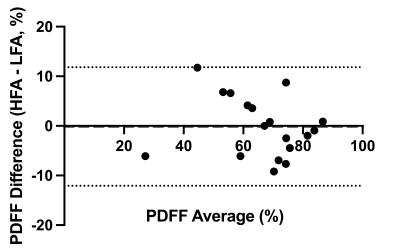 |
170 | High Flip Angle Chemical-Shift Encoded MRI for Imaging Fetal Adipose Tissue
Stephanie A Giza1, Simran Sethi1, Genevieve Eastabrook2,3, Barbra de Vrijer2,3, and Charles A McKenzie1,3
1Medical Biophysics, University of Western Ontario, London, ON, Canada, 2Obstetrics and Gynaecology, University of Western Ontario, London, ON, Canada, 3Maternal, Fetal and Newborn Health, Children's Health Research Institute, London, ON, Canada Keywords: Fetal, Fat Fat fractions in fetal adipose tissue vary across gestation and across different regions of the body, and can be measured by chemical shift encoded MRI which provides accurate measurements of fat fraction above 10%. The assessment of lower fat fractions can be improved by increasing flip angle, but this introduces T1 bias. We demonstrated that this T1 bias can be corrected using fetal adipose tissue water and lipid T1 values, preserving accuracy of the fat fraction measurement while simultaneously improving its precision in fetal adipose tissue.
|
|
2342.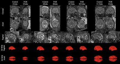 |
171 | Feto-placental oxygenation and brain development in fetal growth restriction
Emily Peacock1, Paponrad Tontivuthikul2, Joanna Chappell1, Nada Mufti1,2, Janina Schellenberg2, Michael Ebner1, Sebastien Ourselin1,3, Anna David2,4,5, Rosalind Aughwane2, and Andrew Melbourne1,2,3
1School of Biomedical Engineering and Imaging Sciences, King's College London, London, United Kingdom, 2Elizabeth Garrett Anderson Institute for Women's Health, University College London, London, United Kingdom, 3Department of Medical Physics and Biomedical Engineering, University College London, London, United Kingdom, 4University Hospital KU Leuven, Leuven, Belgium, 55NIHR University College London Hospitals Biomedical Research Centre, London, United Kingdom Keywords: Image Reconstruction, Brain, Fetus, Machine Learning State-of-the-art machine learning algorithms were applied to create MRI 3D super-resolution reconstructions of fetal brains to analyse brain development in fetal growth restriction. Reconstructions were segmented into the grey matter, white matter, deep grey matter, cerebellum and brainstem, and volumetric and cortical surface data was extracted. We found that the five brain regions segmented were significantly smaller in the FGR cohort than in controls, and that this effect was linked to feto-placental blood oxygen saturation. FGR fetuses showed evidence of brain sparing from MRI and ultrasound measurements and preservation of tissue relationships, such as the grey:white matter ratio. |
|
2343.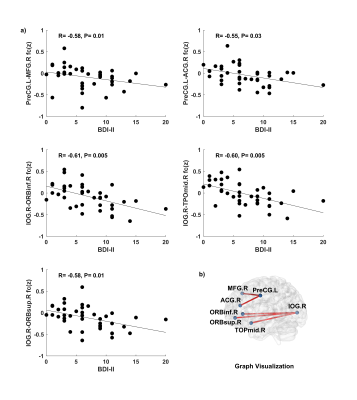 |
172 | Associations between Maternal Depression during Pregnancy and Neonatal Brain Resting-State Functional Connectivity
Xiaoxu Na1, Aline Andres2,3,4, Charles M. Glasier1, Jayne Bellando2, Haitao Chen5,6,7, Wei Gao5,6, and Xiawei Ou1,2,3,4
1Radiology, University of Arkansas for Medical Sciences, Little Rock, AR, United States, 2Pediatrics, University of Arkansas for Medical Sciences, Little Rock, AR, United States, 3Arkansas Children’s Nutrition Center, Little Rock, AR, United States, 4Arkansas Children’s Research Institute, Little Rock, AR, United States, 5Biomedical Sciences and Imaging, Cedars Sinai Medical Center, Los Angeles, CA, United States, 6Biomedical Imaging Research Institute, Cedars Sinai Medical Center, Los Angeles, CA, United States, 7Bioengineering, University of California at Los Angeles, Los Angeles, CA, United States Keywords: Neonatal, fMRI (resting state) This study reports associations between maternal depression symptoms during pregnancy and neonatal brain functional connectivity. The mothers self-rated their depression symptoms during pregnancy using Beck Depression Inventory-II, and their newborns underwent a brain MRI examination including structural 3D T1-weighted images and resting-state fMRI for functional connectivity measurements. Most participants scored in the minimal range for depressive symptoms. Significant negative associations between maternal depression symptoms at ~36 weeks of pregnancy and newborn functional connectivity were observed in multiple brain regions/networks, indicating a negative influence of antenatal depression symptoms on neonatal brain functional development even in women with low symptoms of depression. |
|
2344.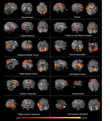 |
173 | Ultra-high field characterisation of resting state networks in the neonatal brain
Tomoki Arichi1,2,3, Philippa Bridgen2,4, Raphael Tomi-Tricot1,4,5, Daniel Cromb1, Paul Cawley1,2, Megan Quirke1,2, Anthony N Price1,2, Enrico De Vita1,4, Jonathan O'Muircheartaigh1,3, Serena J Counsell1, A David Edwards1,3, Joseph V Hajnal1,4, and Shaihan Malik1,4
1Department of Perinatal Imaging, Centre for the Developing Brain, School of Biomedical Engineering and Imaging Sciences, King's College London, London, United Kingdom, 2Guy's and St Thomas' NHS Foundation Trust, London, United Kingdom, 3MRC Centre for Neurodevelopmental Disorders, King's College London, London, United Kingdom, 4London Collaborative Ultra high field System (LoCUS), Kings College London, London, United Kingdom, 5MR Research Collaborations, Siemens Healthcare Limited, Frimley, United Kingdom Keywords: Neonatal, fMRI (resting state) Acquiring BOLD fMRI data at ultra-high field offers marked gains in sensitivity and spatial specificity including within cortical layers and distinct subcortical nuclei. We describe the first pilot data demonstrating feasibility of characterizing resting state networks in the neonatal brain using a 7 Tesla system. In 3 neonates imaged at full term, we show that in addition to the canonical networks seen at standard field strengths, ultra-high field fMRI enables network delineation with higher spatial specificity, including better localization to the cortex and with definition of individual networks corresponding to distinct anatomical regions and tissues. |
|
2345.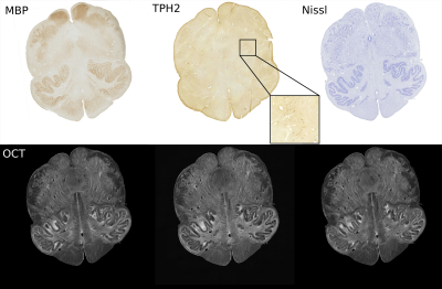 |
174 | Integration of imaging measurements at micro-, meso and macro-scale of the caudal medulla on a postmortem infant subject
Caroline Magnain1, Robin Haynes2, Jean Augustinack1, Hannah Kinney2, and Lilla Zollei1
1MGH, Boston, MA, United States, 2BCH, Boston, MA, United States Keywords: Neonatal, Multimodal, postmortem, brain development The core lesion of SIDS is a set of medullar nuclei with abnormalities correlated with sudden infant death syndrome (SIDS). We performed ex vivo whole brain and brainstem MRI, optical coherence tomography and histology at an unprecedented spatial resolution on a postmortem infant brain to investigate the structural properties of the caudal medulla. In our image processing pipeline, we register all image modalities into the same coordinate system along with a rich set of segmentation labels. Such multimodal construction helps us validate imaging findings at various resolution levels and will serve as prior information in automated segmentation solutions. |
|
2346.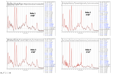 |
175 | Ultra-high-field 7T Neonatal Proton MRS and Metabolite Relaxation Times
Enrico De Vita1,2,3, Maria Yanez-Lopez4, Anthony N Price2,5, Philippa Bridgen3,6, Shaihan Malik2,3, Megan Quirke2,6,7, Raphael Tomi-Tricot3,7,8, Mary A Rutherford2, Joseph V Hajnal2,3,7, and Tomoki Arichi2,6,9
1MR Physics group, Radiology Department, Great Ormond Street Hospital, London, United Kingdom, 2Centre for the Developing Brain, School of Biomedical Engineering and Imaging Sciences, King’s College London, London, United Kingdom, 3London Collaborative Ultra high field System (LoCUS), King’s College London, London, United Kingdom, 4MR Physics Group, Department of Medical Physics and Clinical Engineering, Swansea Bay University Health Board, Swansea, United Kingdom, 5MR Physics, Guys and St Thomas’ NHS Foundation Trust, London, United Kingdom, 6Guys and St Thomas’ NHS Foundation Trust, London, United Kingdom, 7Biomedical Engineering Department, School of Biomedical Engineering and Imaging Sciences, King’s College London, London, United Kingdom, 8MR Research Collaborations, Siemens Healthcare Limited, Frimley, United Kingdom, 9MRC Centre for Neurodevelopmental Disorders, King’s College London, London, United Kingdom Keywords: Neonatal, Spectroscopy, 7T, ultra-high field We present 1H-MRS results from a pilot cohort of newborn infants, acquired on a 7T system. Whilst a customised approach is required for patient preparation and sequence calibrations, our preliminary data points to highly improved sensitivity and data quality at ultra-high field compared to typical spectra from adults at the same field or newborn infants at lower field strengths. We also report initial estimates of T1 and T2 for the main metabolites in the neonatal brain at 7T. |
|
2347.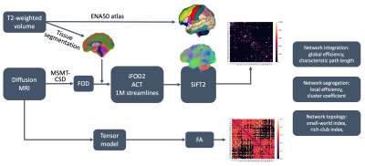 |
176 | Assessing the development of brain's structural connectivity in the perinatal stage with diffusion MRI
Yihan Wu1, Lana Vasung1, Davood Karimi1, and Ali Gholipour1
1Harvard Medical School & Boston Children's Hospital, Boston, MA, United States Keywords: Neonatal, Data Analysis This study analyzed the development of brain's structural connectivity during the perinatal period using the diffusion MRI data of 263 subjects from the dHCP dataset. We used anatomically constrained probabilistic tractography to reconstruct structural connectomes. We computed connectivity metrics to assess network integration and segregation and tested for the effects of postmenstrual age (PMA), premature birth, and gender. Results showed increasing network integration and segregation with PMA in the range 29 to 45 weeks. Premature birth and male gender were associated with significantly lower rates of increase in global efficiency. |
|
2348.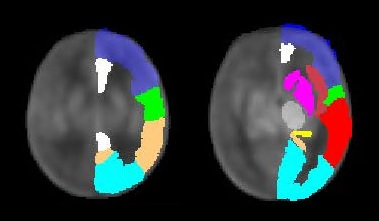 |
177 | Cerebral perfusion in neonates with severe congenital heart disease
Alexandra De Silvestro1,2,3, Giancarlo Natalucci4, Maria Feldmann3,5, Cornelia Hagmann3,6, Thi Dao Nguyen4, Seline Coraj4, Beatrice Latal3,5, Walter Knirsch1,3, and Ruth Tuura2,3
1Pediatric Cardiology, Pediatric Heart Center, Department of Surgery, University Children's Hospital Zurich, Zurich, Switzerland, 2Center for MR-Research, University Children's Hospital Zurich, Zurich, Switzerland, 3Children's Research Center, University Children's Hospital Zurich, Zurich, Switzerland, 4Department of Neonatology, University Hospital Zurich, Zurich, Switzerland, 5Child Development Center, University Children's Hospital Zurich, Zurich, Switzerland, 6Department of Neonatology and Pediatric Intensive Care, University Children's Hospital Zurich, Zurich, Switzerland Keywords: Neonatal, Arterial spin labelling We aimed to compare cerebral perfusion in neonates with severe congenital heart disease (CHD) and healthy controls using arterial spin labeling magnetic resonance imaging. Sixty-seven scans of CHD patients and 23 scans of controls were analyzed (postmenstrual age at scan 41.6±1.8 and 41.9±2.1 weeks, respectively). As in healthy neonates, cerebral perfusion in CHD patients increased with age. Whereas age-adjusted whole brain perfusion was similar to controls, redistribution of regional perfusion was detected in CHD patients. Furthermore, the presence of a systemic-to-pulmonary shunt was associated with cerebral hypoperfusion. Effects of cerebral perfusion alterations on neurodevelopment need to be further assessed. |
|
2349. |
178 | Distance-dependent changes in functional connectivity in neonatal brain
Yihan Wu1, Ruolin Li1, Yifan Shuai1, Zhiyong Zhao1, and Dan Wu1
1Key Laboratory for Biomedical Engineering of Ministry of Education, Department of Biomedical Engineering, College of Biomedical Engineering & Instrument Science, Zhejiang University, Hangzhou, China Keywords: Neonatal, fMRI (resting state) The present study aimed to explore functional connectivity changes with anatomical distance in neonatal brain by using the second dHCP resting-state fMRI dataset. We calculated distance-dependent functional connectivity density (FCD) maps to test for the effect of age, preterm-birth, and gender. Results found that the FCD in neonatal brains showed distance-dependent changes with PMA, in which the short- and long-range FCD changes were associated with maternal environment and postnatal experience, respectively. Moreover, the preterm- and term-born infants showed different patterns in short- and long-range FCD. These findings suggested distance-dependent changes in functional connectivity in neonatal brain during early development. |
|
2350.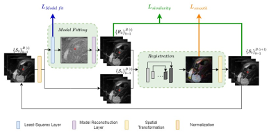 |
179 | qDWI-Morph: Motion-compensated quantitative Diffusion-Weighted MRI analysis for fetal lung maturity assessment
Yael Zaffrani-Reznikov1, Onur Afacan2, Sila Kurugol2, Simon Warfield2, and Moti Freiman1
1Technion, Haifa, Israel, 2Boston Children's Hospital, Boston, MA, United States Keywords: Fetal, Diffusion/other diffusion imaging techniques, fetal imaging Quantitative analysis of fetal lung Diffusion Weighted MRI (DWI) data shows potential in providing quantitative imaging biomarkers for assessing fetal lung maturation. However, fetal motion during the acquisition impairs the accuracy and robustness of the analysis. We introduce qDWI-morph, an unsupervised deep-neural network architecture for motion correction and quantitative DWI (qDWI) analysis. We simultaneously estimate the qDWI parameters and the motion model by minimizing a bio-physically-informed loss. The qDWI-morph achieved an improved correlation between qDWI parameters in the fetal lung with the gestational age (R-squared=0.32) over baseline analysis without motion correction (R-squared=0.13) and our network with registration loss solely (R-squared=0.28). |
|
2351.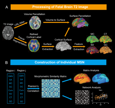 |
180 | Developmental pattern of individual morphometric similarity network in the human fetal brain
Ruoke Zhao1, Xinyi Xu1, Zhiyong Zhao1, Mingyang Li1, Ruike Chen1, Yao Shen1, Cong Sun2, Guangbin Wang3, and Dan Wu1
1College of Biomedical Engineering & Instrument Science, Zhejiang University, Hangzhou, China, 2Department of Radiology, Beijing Hospital, National Center of Gerontology, Institute of Geriatric Medicine, Chinese Academy of Medical Sciences, Beijing, China, 3Department of Radiology, Shandong Provincial Hospital Affiliated to Shandong First Medical University, Jinan, China Keywords: Fetal, Brain Connectivity The fetal brain morphological network is important for us to understand early brain organization but has not characterized previously. In the study, we constructed fetal individual morphometric similarity networks using multiple cortical features based on in utero MRI data. The results demonstrated the decline of morphological symmetry between hemispheres during development. The morphological similarity between the limbic regions and other regions significantly increased while the similarity between the parieto-occipital regions and other regions significantly decreased. Small world organizations appeared as early as 22 weeks, and the network topology developed towards more integrated and less segregated during fetal period. |
|
 Back to Meeting Home
 Back to the Program-at-a-Glance
The International Society for Magnetic Resonance in Medicine is accredited by the Accreditation Council for Continuing Medical Education to provide continuing medical education for physicians.
 Back to Meeting Home
 Back to the Program-at-a-Glance
 View Presentation Video