Digital Poster
Pediatric Neuroimaging
ISMRM & ISMRT Annual Meeting & Exhibition • 03-08 June 2023 • Toronto, ON, Canada

| Computer # | |||
|---|---|---|---|
2509.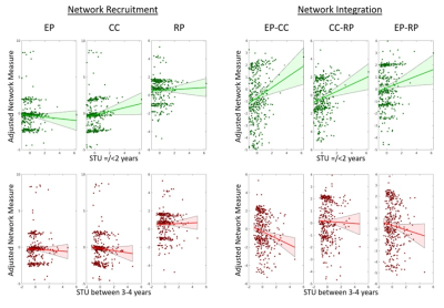 |
161 | Brain Network Topology: The Missing Link Between Early Childhood Screen Time Utilization and Executive Function Performance
Ai Peng Tan1,2,3, Pei Huang4, Shi Yu Chan4, Zhen Ming Ngoh4, Zi Yan Ong4, Xi Zhen Low5, Evelyn C. Law4,6,7, Peter D. Gluckman4,8, Michelle Z.L. Kee4, Yap Seng Chong4,6,7, Juan H. Zhou6, and Michael J. Meaney4
1Diagnostic Imaging, National University Hospital, Singapore, Singapore, 2Diagnostic Imaging, National University of Singapore, Singapore, Singapore, 3Translational Neuroscience, Singapore Institute for Clinical Sciences, Singapore, Singapore, 4Singapore Institute for Clinical Sciences (SICS), Singapore, Singapore, 5Diagnostic Imaging, National University Hospital, SIngapore, Singapore, 6National University of Singapore, Singapore, Singapore, 7National University Hospital, Singapore, Singapore, 8Liggins Institute, Auckland, New Zealand Keywords: Normal development, Brain Connectivity Excessive screen time utilization in childhood has been linked to executive dysfunction. Our primary aim was to investigate the potential influence of screen time in early childhood on brain network topology and later executive function (EF). We collected data on screen time (between ages 0-4 years) (n = 950), followed by MRI brain at age 6 (n = 414) and assessment of executive function at age 7 (n = 620). Screen time in early childhood was significantly associated with emotion processing-cognitive control network integration which mediates the effect of screen time on EF performance. |
|
2510.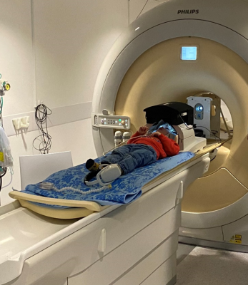 |
162 | A novel VR based and motion tolerant capability for MR imaging of awake young children
Kun Qian1, Joseph V Hajnal1, Lucilio Cordero-Grande1,2, Jonathan O'Muircheartaigh1, A David Edwards1,3,4, and Tomoki Arichi1,3,4
1Department of Perinatal Imaging, Centre for the Developing Brain, School of Biomedical Engineering and Imaging Sciences, King's College London, London, United Kingdom, 2Biomedical Image Technologies, ETSI Telecomunicación, Universidad Politécnica de Madrid and CIBER-BBN, ISCIII, Madrid, Spain, 3Guy's and St Thomas' NHS Foundation Trust, London, United Kingdom, 4MRC Centre for Neurodevelopmental Disorders, King's College London, London, United Kingdom Keywords: Normal development, New Devices MRI examinations in young children are typically performed during natural or induced sleep to reduce distress and movement artefacts. However, those approaches have high failure rate and/or carry risks. We describe a system for MR imaging in awake children which combines immersive and interactive MR compatible virtual reality, eye tracking, and robust post-acquisition motion correction. We demonstrate effectiveness in a pilot study with a 2 year old child who used the system on 3 occasions for average 19.5 minutes. The described approach opens new possibilities for awake MR studies in young children for both clinical and research purposes. |
|
2511.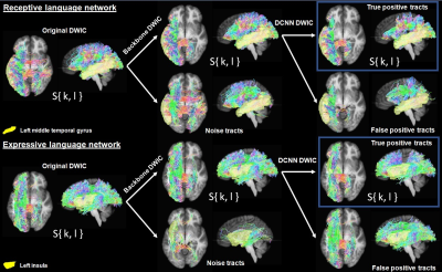 |
163 | Deep learning-based DWI connectome analysis to improve the prediction of postoperative language improvement in pediatric epilepsy
Min-Hee Lee1,2, Nathan Sim3, Marie Papamarcos3, Masaki Sonoda4, Csaba Juhász1,2, Eishi Asano1,5, and Jeong-Won Jeong1,2
1Pediatrics, Wayne State University, Detroit, MI, United States, 2the Translational Imaging Laboratory, Children's Hospital of Michigan, Detroit, MI, United States, 3Medical Doctor Program, Wayne State University, Detroit, MI, United States, 4Neurosurgery, Yokohama City University, Yokohama, Japan, 5Neurology, Children's Hospital of Michigan, Detroit, MI, United States Keywords: Neuro, Epilepsy, Prediction of postoperative language improvement in children with epilepsy We present a novel deep learning-based tract classification to effectively remove false positive tract streamlines from preoperative DWI connectome data of children with medically intractable epilepsy. Compared to the prediction model without the presented classification where uncontrollable false positive tracts significantly limit the accurate prediction of postoperative language improvement using local efficiency values of key hub regions in the receptive and expressive language networks, the prediction model with the presented classification enhanced the accuracy of about 34% up to 100%/88% for the prediction of receptive/expressive language improvement, especially when the local efficiency values were combined with the clinical variables. |
|
2512. |
164 | The evaluation of Synthetic MRI and machine learning in differentiating autism and developmental language disorders
Yanyong Shen1, Xin Zhao1, Kaiyu Wang2, Qingna Xing1, Honglei Shang1, Hongrui Ren1, Yongbing Sun3, and Xiaoan Zhang1
1Department of Radiology, the Third Affiliated Hospital of Zhengzhou University, Zhengzhou, China, 2MR Research China, GE Healthcare, Beijing 100000, PR China, Beijing, China, 3Department of Medical Imaging of Henan Provincial People’s Hospital, Zhengzhou University People’s Hospital, Zhengzhou, China Keywords: Neuro, Quantitative Imaging, Autism Spectrum Disorder Distinguishing early Autism Spectrum Disorder (ASD) from Developmental language disorder (DLD) in clinical practice is challenging as they are usually diagnose by behavioral tests and subjective observation. The emerging technique Synthetic MRI can be used to quantify the changed in biological tissues. Machine learning is also widely used for improvement of diagnostic performance. This study was aimed to identify ASD from DLD by using Synthetic MRI in combination with machine learning methods. Results show that T1 mapping in Synthetic MRI can be used for differentiation of the two diseases and the SVM model with linear kernel have the best performance. |
|
2513.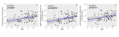 |
165 | Association of socio-economic status and perceived parental stress effects with infant cortical development
Jessica Hyland1, Kay Sindabizera 1, Minhui Ouyang1,2, Tianjia Zhu1,3, Juri Kim1,3, and Hao Huang1,2
1Department of Radiology, Children's Hospital of Philadelphia, Philadelphia, PA, United States, 2Department of Radiology, Perelman School of Medicine, University of Pennsylvania, Philadelphia, PA, United States, 3Department of Biomedical Engineering, School of Engineering and Applied Science, University of Pennsylvania, Philadelphia, PA, United States Keywords: Normal development, Gray Matter Environmental factors such as socio-economic status (SES) and parental stress have significant impacts on cognitive performance, but their links to brain development during infancy are not known. We collected high-resolution structural MRI, SES and perceived stress scales (PSS) of 70 infants and their caregivers aged 0-20 months to investigate the association of these environmental factors and brain development. The total brain, gray matter, and white matter volumes all increase rapidly with age. Higher SES is significantly correlated with greater cortical volume, particularly in the right hemisphere. Lower PSS tends to be associated with higher cortical volume, though not significantly. |
|
2514.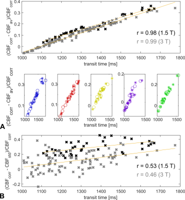 |
166 | Vascular territorial analysis of cerebral blood flow in pediatric Moyamoya to exclude arterial transit time bias
Rahel Heule1,2, Raimund Kottke3, and Ruth Tuura1,2
1Center for MR Research, University Children's Hospital, Zurich, Switzerland, 2Children's Research Center, University Children's Hospital, University of Zurich, Zurich, Switzerland, 3Department of Diagnostic Imaging, University Children’s Hospital, Zurich, Switzerland Keywords: Neuro, Arterial spin labelling Delayed perfusion in patients with Moyamoya vasculopathy can lead to a bias in CBF quantification, especially for single-delay ASL acquisitions. In this work, we investigate the clinical utility of arterial transit time corrected CBF derived from a 7-delay prototype ASL sequence for assessment of hemodynamically mediated perfusion failure in pediatric Moyamoya patients. The multi-delay ASL acquisition proved successful in capturing the whole distribution of transit times in the investigated patient cohort and may thus be a valid contrast agent-free alternative to DSC-enhanced perfusion imaging. |
|
2515.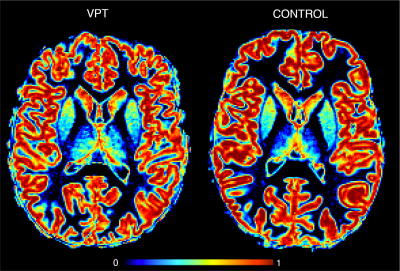 |
167 | Cortical alterations after very preterm birth and the association with socio-emotional abilities from childhood to early adolescence
Elda Fischi-Gomez1, Vanesssa Siffredi2,3,4, Maria Chiara Liverani5, Cristina Borradori-Tolsa2,5, Russia Hà-Vinh Leuchter6, and Petra Susan Hüppi6
1Computer Imaging and Machine Learning, CIBM CHUV-EPFL SP Section, Lausanne, Switzerland, 2Division of Development and Growth, Department of Paediatrics, Gynaecology and Obstetrics, Geneva University Hospitals, Geneva, Switzerland, Geneva, Switzerland, 3Institute of Bioengineering, Center for Neuroprosthetics, Ecole Polytechnique Fédérale de Lausanne, Switzerland, Lausanne, Switzerland, 4Department of Radiology and Medical Informatics, Faculty of Medicine, University of Geneva, Switzerland, Geneva, Switzerland, 5SensoriMotor, Affective and Social Development Laboratory, Faculty of Psychology and Educational Sciences, University of Geneva, Geneva, Switzerland, Geneva, Switzerland, 6Division of Development and Growth, Department of Paediatrics, Gynaecology and Obstetrics, Geneva University Hospitals, Geneva, Switzerland Keywords: Adolescents, Pediatric This study compared GM concentration and its developmental trajectory in vey preterm (VPT) and full-term (FT) children aged 6 to 14 years. Widespread abnormal GM concentration was found with complicated patterns of increases/decreases of GM concentration across cortical and subcortical regions. Socio-emotional abilities were association with GM concentration in regions known to be involved in such process for both VPTand FT peers. Our findings suggest the trajectory of brain development following VPT birth may be fundamentally distinctive with impact on socio-emotional abilities. |
|
2516.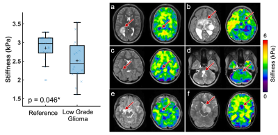 |
168 | Mechanical Properties of Pediatric Central Nervous System Tumors in Children With and Without Neurofibromatosis Type 1
Grace McIlvain1, Abdulhafeez M Khair2, Vinay Kandula2, Gurcharanjeet Kaur2, Andrew Walter2, Lauren Averill2, Arabinda K Choudhary2, Curtis L Johnson1,2, and Rahul M Nikam2
1University of Delaware, Newark, DE, United States, 2Neuroradiology, Nemours Children's Health, Wilmington, DE, United States Keywords: Neuro, Tumor Pediatric brain tumors have developmental consequences even when prognoses are favorable. Mechanical characterization using magnetic resonance elastography (MRE) has previously revealed adult gliomas are softer than brain tissue. However, pediatric tumors are known to differ behaviorally and cytoarchitecturally from adult tumors. Neurofibromatosis type 1 (NF1) imparts genetic predisposition to gliomas, but also results in spongiform regions in white matter tissue. Here we use MRE for the first time in NF1 and in pediatric low-grade gliomas and we find that pediatric gliomas are softer than reference tissue, while spongiforms are stiffer. |
|
2517.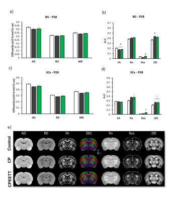 |
169 | Effects of Intensive sensorimotor training following experimental cerebral palsy assessed by advanced diffusion MRI
Yohan van de Looij1,2, Eduardo Sanches1, Ho Dini1, Audrey Toulotte1, Laetitia Baud3, Quentin Barraud3, Rodrigo Araneda4, Yannick Bleyenheuft4, Sylvain Brochard5,6, Gregoire Courtine3, and Stéphane Sizonenko1
1Department of Paediatrics and Gynaecology-Obstetrics, Division of Development and Growth, University of Geneva, Geneva, Switzerland, 2Center for Biomedical Imaging, Animal Imaging Technology section, Federal Institute of Technology of Lausanne, Lausanne, Switzerland, 3Center for Biomedical Imaging, Federal Institute of Technology of Lausanne, Lausanne, Switzerland, 4Institute of Neuroscience, Université catholique de Louvain, Brussels, Belgium, 5Physical and medical rehabilitation department, CHRU Brest, Brest, France, 6Paediatric physical and medical rehabilitation department, Fondation ILDYS, Brest, France Keywords: Neonatal, Brain, Cerebral palsy, preclinical animal model Injury to the developing brain is a major cause of Cerebral Palsy (CP) leading to motor and cognitive disabilities. HABIT-ILE is a 2-week intensive sensorimotor rehabilitation program with proven effects decreasing motor impairments in infants with CP. Here, we combined early environmental enrichment (EE) and treadmill motor training (TT) to model HABIT-ILE (EETT) for treating experimental CP in rats assessing then histological and microstructural parameters (diffusion MRI at 9.4T). Exvivo DTI/NODDI showed altered brain microstructure in CP rats not reversed by HABIT-ILE. HABIT-ILE modulated BDNF signaling and decreased the over-expression of proteins involved in excitatory function induced by CP. |
|
2518.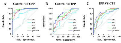 |
170 | Diagnostic value of adenohypophyseal magnetic resonance imaging features in girls with precocious puberty
Dong Liu1, Weiyin Vivian Liu2, Yuanyuan Qin1, Guojun Ding1, and Wenzhen Zhu1
1Department of Radiology, Tongji Hosptial of Tongji Medical College of Huazhong University of Science and Technology, Wuhan, China, 2MR Research, GE Healthcare, Beijing, China Keywords: Normal development, Endocrine, pituitary gland It is difficult to distinguish IPP from CPP in the clinical diagnosis. GnRH stimulation test has long been considered the gold standard for the diagnosis of CPP and evaluation of hypothalamic-pituitary-gonadal axis activation, but it is a high cost and an invasive approach. To simplify examination and dig out an alternative method is necessary. In our study, analysis of clinical data, adenohypophyseal MRI features and laboratory characteristics of PP girls help us better comprehensive PP. A combined model of adenohypophyseal MRI features and clinical characteristics improved the diagnostic efficacy of PP and provided us a non-invasive and reliable diagnostic method. |
|
2519.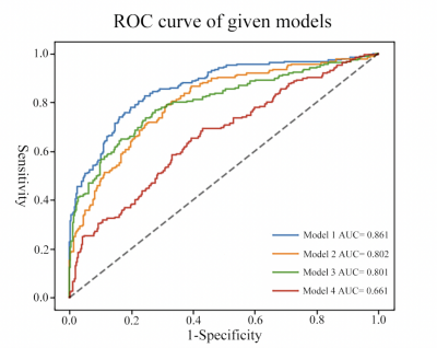 |
171 | A machine learning model for identifying idiopathic central precocious puberty in girls based on medical images and clinical multi-parameters
Yi Lu1, PinFa Zou1, LingFeng Zhang1, lu han2, and Zhihan Yan1
1Radiology, The Second Affiliated Hospital and Yuying Children’s Hospital of Wenzhou Medical University, Wenzhou, China, 2Philips Healthcare, Shanghai, China Keywords: Adolescents, Endocrine Early identification of precocious puberty (PP) is important to guarantee the growth and development of children. The aim of this study was to propose a robust machine learning model that incorporate information from pituitary MRI images, carpal bone age, gonadal ultrasound, baseline sex hormone tests, and clinical information to identify idiopathic central precocious puberty (ICPP). The experiments show that the AUCs are 0.860, 0.862, and 0.866 respectively for the training set, internal validation sets, and external validation sets. The performance suggests that the presented model could be an alternative clinical approach. |
|
2520.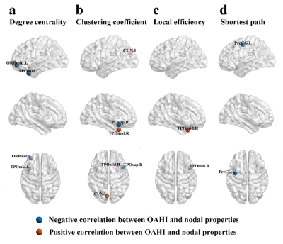 |
172 | Disturbed cerebral white matter network topological mediate the symptom severity and cognition in children with obstructive sleep apnea
Yi Lu1, Fangfang Chen1, Chenyi Yu2, Tao Chen3, lu han4, Zhihan Yan1, and Yuchuan Fu1
1Radiology, The Second Affiliated Hospital and Yuying Children’s Hospital of Wenzhou Medical University, Wenzhou, China, 2Pediatrics, The Second Affiliated Hospital and Yuying Children’s Hospital of Wenzhou Medical University, Wenzhou, China, 3Radiology, The First Affiliated Hospital of Zhejiang University, Hangzhou, China, 4Philips Healthcare, Shanghai, China Keywords: Adolescents, Brain Connectivity, obstructive sleep apnea Obstructive sleep apnea (OSA) in children can cause deficits in cognition. The purpose of this study was to explore the underlying mechanisms of OSA-related cognitive impairment by investigating the altered topology of brain white matter networks in children with OSA. Based on graph theory and mediating effects analysis, we discovered that OSA had significantly higher global assortativity than control group, and that the nodal clustering coefficients of temporal lobes mediated the relationship between the symptom severity of OSA and the verbal comprehension index. Our findings provided a further neuroimaging evidence of impaired cognitive function in children with OSA. |
|
2521. |
173 | Cortical morphology-based prediction of communication disorder in children with bilateral spastic cerebral palsy
jie hu1,2, yanli yang1, haifeng ran1, jingjing zhang3, cheng he4, heng liu1, and tijiang zhang1
1Department of Radiology, the Affiliated Hospital of Zunyi Medical University, zunyi, China, 2Department of Radiology and Nuclear Medicine, Xuanwu Hospital, Capital Medical University, beijing, China, 3Department of Radiology, Mianyang Hospital of T.C.M, mianyang, China, 4Department of Radiology, Chongqing University Central Hospital,, chongqing, China Keywords: Neuro, Brain This study were preliminarily established an individualized diagnosis model of communication dysfunction in children with bilateral spastic cerebral palsy based on cortical morphological parameters extracted from structure magnetic resonance images by using Support Vector Machines classification algorithm. This may provide a novel idea for the diagnose of communication disorder in children with BSCP. |
|
2522.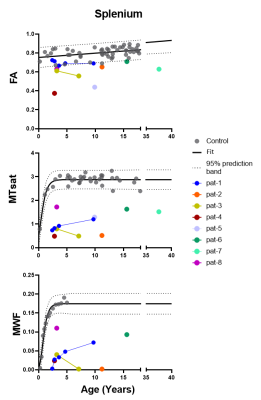 |
174 | Multimodal quantitative MRI for the assessment of different disease processes in a childhood hypomyelinating leukodystrophy
Prativa Sahoo1, Caroline Köhler2, Irini Gkalimani1, Paul Kunte2, Peter Dechent3, Gunter Helms4, Sean Deoni5, Hagen H Kitzler2, and Steffi Dreha Kulaczewski1
1Department of Pediatrics and Adolescent Medicine, University Medical Center Göttingen, Göttingen, Germany, 2Department of Diagnostic and Interventional Neuroradiology, University Hospital Carl Gustav Carus Dresden, Dresden, Germany, 3Department of Cognitive Neurology, University Medical Center Göttingen, Göttingen, Germany, 4Lund University, Lund, Sweden, 5Bill and Medlinda Gates Foundation, Seattle, WA, United States Keywords: Neuro, White Matter, DTI , MTI X-linked Pelizaeus Merzbacher disease is characterized by failure of myelin formation and subsequent axonal damage. Severity of disease courses varies depending on the underlying gene mutation. Eight male patients were recruited for the study. MR-imaging protocol included MTI, MWI for evaluation of myelin related parameters and DTI for assessment of axonal integrity. Decreased values in quantitative MTI and MWI parameter maps and ROI analyses indicated severe myelin deficit in all patients. However, FA reflected various degrees of axonal involvement among patients with deferent genotypes. Multimodal MRI can facilitate assessment of simultaneous disease processes in childhood hypomyelinating leukodystrophies. |
|
2523. |
175 | the value of ASL-based cerebral blood flow and network metrics in evaluating children with MR-negative epilepsy
Yi Zhuang1, Chenchen Hua2, Lu Qiu1, and Haoxiang Jiang1
1Department of Radiology, The Affiliated Wuxi Children's Hospital of Nanjing Medical University, Wuxi, China, 2Department of Radiology, Wuxi People's Hospital Affiliated to Nanjing Medical University, Wuxi, China Keywords: Neuro, Epilepsy Epilepsy is the most common chronic neurological disorder with significant morbidity and mortality. Frontal lobe epilepsy is a common type of seizure after temporal lobe epilepsy, which makes the diagnosis of frontal lobe epilepsy and its treatment difficult due to its presence of not only motor symptoms but also other autonomic symptoms during seizures. Arterial spin labeling can be used to identify brain regions and brain network lesions associated with the propagation of epileptiform activity, and is of higher investigative value especially in children with negative MRI. keyword: Epilepsy, brain network, arterial spin labeling, pediatrics |
|
2524.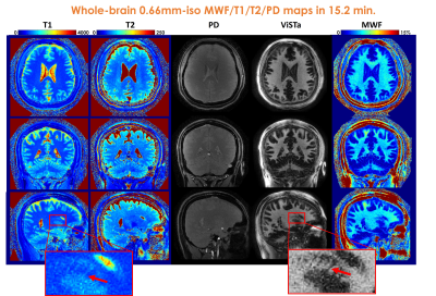 |
176 | Mesoscale myelin-water fraction and T1/T2/PD mapping using optimized 3D ViSTa-MR Fingerprinting
Congyu Liao1,2, Xiaozhi Cao1,2, Siddharth Srinivasan Iyer1,3, Sophie Schauman1,2, Zihan Zhou4, Xiaoqian Yan5, Quan Chen1,2, Ting Gong6, Zhe Wu7, Hongjian He4, Jianhui Zhong4,8, Adam B Kerr2,9, Kalanit Grill-Spector5, and Kawin Setsompop1,2
1Department of Radiology, Stanford University, Stanford, CA, United States, 2Department of Electrical Engineering, Stanford University, Stanford, CA, United States, 3Department of Electrical Engineering and Computer Science, Massachusetts Institute of Technology, Cambridge, MA, United States, 4Center for Brain Imaging Science and Technology, College of Biomedical Engineering & Instrument Science, Zhejiang University, Hangzhou, China, 5Department of Psychology, Stanford University, Stanford, CA, United States, 6Athinoula A. Martinos Center for Biomedical Imaging, Massachusetts General Hospital and Harvard Medical School, Charlestown, MA, United States, 7Techna Institute, University Health Network, Toronto, ON, Canada, 8Department of Imaging Sciences, University of Rochester, Rochester, NY, United States, 9Stanford Center for Cognitive and Neurobiological Imaging, Stanford University, Stanford, CA, United States Keywords: Normal development, Microstructure In this work, we developed an optimized ViSTa-MRF method, which combined Visualization of Short Transverse relaxation time component (ViSTa) technique with MR Fingerprinting (MRF), to achieve high-fidelity whole-brain myelin-water fraction (MWF) and T1/T2/PD mapping at sub-millimeter isotropic resolution. To achieve high image quality, fast acquisition, and memory-efficient reconstruction, the proposed ViSTa-MRF sequence leverages a CRLB-optimized flip-angle (FA) protocol, SNR-efficient 3D spiral-projection sampling scheme and a GPU-based subspace reconstruction. We also applied the proposed method to enable high-resolution assessment of MWF/T1/T2 for infant brain development as well as for post-mortem brain sample. |
|
2525.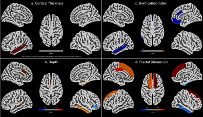 |
177 | Efficacy of virtual reality therapy using functional connectivity and structural measures in children with cerebral palsy
S Senthil Kumaran1, N M Shruthi2, Priyanka Bhat3, Sheffali Gulati2, and Tapan Kumar Gandhi4
1Department of NMR, All India Institute of Medical Sciences, New Delhi, India, 2Department of Pediatrics, All India Institute of Medical Sciences, New Delhi, India, 3Center for BioMedical Engineering, Indian Institute of Technology Delhi, New Delhi, India, 4Electrical Engineering, Indian Institute of Technology, Delhi, New Delhi, India Keywords: Neuro, fMRI (task based) Hemiparetic cerebral palsy patients were administered Virtual reality therapy (VRT) in addition to constraint induced movement therapy (CIMT) in VRT group, and CIMT alone in CIMT group in a randomised trial. Task based functional MRI of left and right fist clenching and structural data were acquired in a 3T MR scanner. Cortical thickness, gyrification (using cat12 toolbox) and functional connectivity (using conn toolbox) results revealed presence of salience and motor networks in the VRT group, highlighting better recovery mechanism stimulated by virtual reality based training. |
|
2526.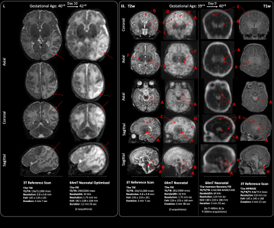 |
178 | Clinical Utility of Optimised Ultra-Low Field Structural MR Brain imaging in Neonates
Paul Cawley1,2,3, Francesco Padormo1,4, Daniel Cromb1,5, Alessandra Maggioni5, Jennifer Almalbis1, Miguel De La Fuente Botell5, Massimo Marenzana1, Rui Teixeira4, UNITY Consortium6, Steve Williams6, Serena Counsell1, Tomoki Arichi1,3, Mary Rutherford1,3, Joseph V Hajnal1, and A David Edwards1,3,5
1Centre for the Developing Brain, School of Biomedical Engineering and Imaging Sciences, King's College London, London, United Kingdom, 2Neonatal Intensive Care Unit, Evelina Children's hospital, London, United Kingdom, 3MRC Centre for Neurodevelopmental Disorders, King's College London, London, United Kingdom, 4Hyperfine, Inc., Connecticut, CT, United States, 5Neonatal Intensive Care Unit, Evelina Children’s Hospital, London, United Kingdom, 6Centre for Neuroimaging Sciences, King's College London, London, United Kingdom Keywords: Neuro, Low-Field MRI Perinatal brain injury and congenital brain abnormalities are common. Access to definitive neuro imaging is limited globally, particularly within resource constrained settings, or in infants too sick to transfer to scanning departments. New devices utilising ultra-low field magnets may herald a revolution in lower-cost high-access MRI, but preliminary results using manufacturer standard adult-optimised sequences has produced suboptimal image quality in neonates. We performed optimisation of low field MRI pulse sequence design and demonstrate enhanced visualization of key brain tissues and neuroanatomical structures. Neonatal-specific sequences show promising performance across a range of gestational maturities and perinatal brain abnormalities. |
|
2527.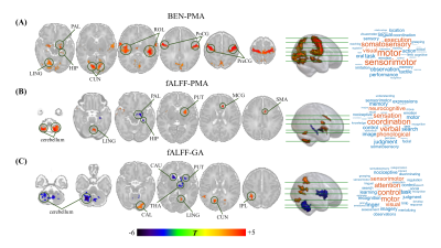 |
179 | Functional plasticity development of visual-motor cortex in neonatal brain is modulated by postnatal experience
Yifan Shuai1, Ruolin Li1, Yihan Wu1, Zhiyong Zhao1, and Dan Wu1
1Key Laboratory for Biomedical Engineering of Ministry of Education, Department of Biomedical Engineering, College of Biomedical Engineering & Instrument Science, Zhejiang University, Hangzhou, China Keywords: Neonatal, Normal development, functional plasticity The present study aimed to investigate how the brain entropy (BEN) changes with age during early development and whether it exists differences between term- and preterm-born neonates. The results found that the BEN in visual-motor cortex was positively correlated with PMA but had no significant correlation with GA, which showed an increase in preterm- than term-born neonates with matched PMA. Moreover, both age and preterm have a significant effect on the functional connectivity between visual-motor cortex and other regions. These findings suggest the functional plasticity development of visual-motor cortex is modulated by early postnatal experience in human neonates. |
|
2528.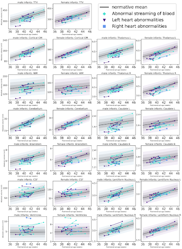 |
180 | Individualised perioperative brain growth in infants with congenital heart disease (CHD): correlation with clinical risk factors
Daniel Cromb1,2, Alexandra Bonthrone1,2, Alessandra Maggioni1, Paul Cawley1,3, Ralica Dimitrova1,4, Christopher Kelly1, Lucilio Cordero-Grande1,5, Olivia Carney1, Alexia Egloff-Collado1, Emer Hughes1, Joseph V Hajnal1,2, John Simpson1,6, Kuberan Pushparajah1,6, Mary Rutherford1,3, A David Edwards1, Jonathan O'Muircheartaigh1,3,4, and Serena J Counsell1,2
1Centre for the Developing Brain, School of Biomedical Engineering and Imaging Sciences, King's College London, London, United Kingdom, 2Biomedical Engineering Department, School of Biomedical Engineering and Imaging Sciences, King's College London, London, United Kingdom, 3MRC Centre for Neurodevelopmental Disorders, King's College London, London, United Kingdom, 4Department for Forensic and Neurodevelopmental Sciences, King's College London, London, United Kingdom, 5Biomedical Image Technologies, ETSI Telecomunicación, Universidad Politécnica de Madrid and CIBER-BBN, Madrid, Spain, 6Paediatric Cardiology, Evelina London Children's Hospital, London, United Kingdom Keywords: Neuro, Brain, Cardiovascular Infants with congenital heart disease (CHD) are at risk of neurodevelopmental impairments, which may be associated with impaired brain growth. We mapped brain volumes from pre- and postoperative brain MRI in 36 infants with CHD undergoing cardiac surgery or intervention to normative curves derived from 219 healthy infants. Perioperative brain growth was impaired, and was associated with clinical and surgical risk factors, including higher preoperative serum creatinine levels, older postnatal age at surgery, longer cardiopulmonary bypass duration and longer postoperative intensive care stay. Brainstem and deep grey matter growth appear particularly vulnerable to clinical factors. |
|
 Back to Meeting Home
 Back to the Program-at-a-Glance
The International Society for Magnetic Resonance in Medicine is accredited by the Accreditation Council for Continuing Medical Education to provide continuing medical education for physicians.
 Back to Meeting Home
 Back to the Program-at-a-Glance
 View Presentation Video