Digital Poster
Device & Multimodal II
ISMRM & ISMRT Annual Meeting & Exhibition • 03-08 June 2023 • Toronto, ON, Canada

| Computer # | |||
|---|---|---|---|
4542.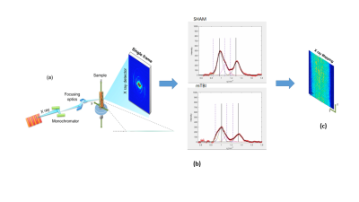 |
121 |
A multiscale approach based on the combination of DTI, XRD and
histology to study the myeloarchitecture in a rat model of mTBI
Michela Fratini1,2,
Isabel San Martín Molina3,
Manfred Burghammer4,
Tilman Grünewald4,5,
Raimo A. Salo3,
Omar Narvaez3,
Maria Guidi6,
Federico Giove6,
Manisha Aggarwal7,
Jussi Tohka3,
Alejandra Sierra3,
and Gaetano Campi8
1Nanotec, CNR, Roma, Italy, 2IRCSS Santa Lucia Foundation, Roma, Italy, 3University of Eastern Finland,, Kuopio, Finland, 4European Synchrotron Radiation Facility, Grenoble, France, 5Aix-Marseille Université, CNRS, Centrale Marseille, Institut Fresnel, Marseille, France, 6Museo Storico della Fisica e Centro Studi e Ricerche Enrico Fermi, Rome, Italy, 7Russell H. Morgan Department of Radiology and Radiological Science, , John Hopkins University School of Medicine, Baltimore, MD, United States, 8Institute of Crystallography, CNR, Rome, Italy Keywords: Multimodal, Tissue Characterization, x ray scanning diffraction We have combined high-resolution structural imaging techniques, such as scanning micro-X-ray diffraction (SμXRD), DTI and histology to study the myeloarchitecture in both healthy and pathological rat brains. For the latter, we focused on an animal model of mild traumatic brain injury (mTBI) that is the most common form of acquired brain injury caused by an external force into the brain. We have performed a quantitative morphometrical analysis of myelin, mapping its relevant morphological and topological parameters (period, orientation, and integrated intensity fluctuation) to assess tissue microstructural damage 35 days after mTBI and sham-operation |
|
4543.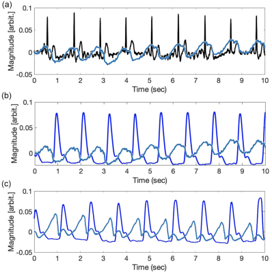 |
122 |
Continuous wave radar for carotid pulse sensing in Magnetic
Resonance Imaging
Renesmee Kuo1,
John Pauly1,
Fraser Robb2,
Shreyas Vasanawala1,
and Greig Scott1
1Stanford University, Stanford, CA, United States, 2GE HealthCare, Aurora, OH, United States Keywords: Hybrid & Novel Systems Technology, Image Reconstruction Non-contact synchronization MRI with heart motion is still an open challenge. Inaccurate cardiac gating often complicates image reconstruction and leads to poor or non-diagnostic images. Continuous wave doppler radar is a non-contact, noninvasive novel systems technology that could monitor cardiac motions without distortion from the MRI electromagnetic environment while offering an alternative to state-of-the-art (ECG) triggering. |
|
4544.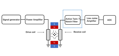 |
123 |
Design and Implement Active Twin-T Notch Filter for Signal
Reception in Magnetic Particle Imaging
Kangjian Huang1,
Congcong Liu1,
Ye Li1,
Yanjie Zhu1,
Hairong Zheng1,
Dong Liang 1,
and Haifeng Wang1
1Shenzhen Institute of Advanced Technology,Chinese Academy of Sciences, shenzhen, China Keywords: Hybrid & Novel Systems Technology, Hybrid & Novel Systems Technology Because of the imaging coding mode of Magnetic particle imaging (MPI), the drive-field signal directly couples into the receiving coil and covers the magnetic particle signal, which one aims to detect. Therefore, some method is needed to remove the drive-field signal and retain the magnetic particle signal. One of the methods is to use a passive band-stop filter to filter the excitation signal, but the general passive band-reject filter has a large stopband, which filters part of the magnetic particle signal while filtering the drive-field signal. In this study, we design an active twin-T notch filter with narrow stopband, which better restore the magnetic particle signal and improve the signal-to-noise in MPI. |
|
4545. |
124 |
AI inspired engineering of future MRI machines and components
J. Coupling1,
E. Current1,
and Line SP. Litting1
1Division of Medical Physics, Department of Diagnostic and Interventional Radiology, University Medical Center Freiburg, Faculty of Medicine, University of Freiburg, Freiburg, Germany Keywords: Hybrid & Novel Systems Technology, New Devices The topic of this contribution is how artificial intelligence (AI) can be used to engineer future magnetic resonance imaging (MRI) machines and components. MRI machines are vital tools in the medical field, used for everything from diagnosing injuries to detecting cancer. However, they are also extremely complex devices, made up of many different parts that must work together seamlessly. |
|
4546.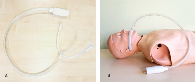 |
125 |
Feasibility of high-frequency shear wave MR elastography of the
parotid glands
Vitaliy Atamaniuk1,
Jun Chen 2,
Kevin Glaser2,
Andrii Pozaruk1,
Marzanna Obrzut3,
Bogdan Obrzut4,
Wojciech Domka5,
Krzysztof Gutkowski6,
Richard L Ehman2,
and Marian Cholewa1
1Department of Biophysics, Institute of Physics, College of Natural Sciences, University of Rzeszow, Rzeszow, Poland, 2Department of Radiology, Mayo Clinic, Rochester, MN, United States, 3Center for Diagnostic Medical Sonography, Rzeszow, Poland, 4Department of Obstetrics and Gynecology, Institute of Medical Sciences, Medical College, University of Rzeszow, Rzeszow, Poland, 5Department of Otorhinolaryngology, Institute of Medical Sciences, Medical College, University of Rzeszow, Rzeszow, Poland, 6Institute of Medical Sciences, Medical College, University of Rzeszow, Rzeszow, Poland Keywords: New Devices, Elastography A specialized driver was developed to enable magnetic resonance elastography of the parotid and other salivary glands using high shear wave frequencies to provide improved spatial resolution and optimized ergonomics. The performance of this new device was tested in volunteer studies. Preliminary results showed that suitable shear wave illumination of the parotid gland is possible at frequencies as high as 120 Hz and that high-frequency shear wave MR elastography of the parotid gland is feasible. |
|
4547. |
126 |
Monitoring performance of a GaN HEMT switch across different
magnetic field strengths for implementation of On-Coil Tx
Amplifiers
Brett Setera1,2,
Aristos Christou2,
and Natalia Gudino1
1Laboratory of Functional and Molecular Imaging, National Institutes of Health, Bethesda, MD, United States, 2Department of Materials Science & Engineering, University of Maryland, College Park, MD, United States Keywords: New Devices, New Devices, Radiofrequency Technology We present a method for monitoring multiple GaN HEMT device signals at different orientations with B0 during RF switching to determine possible magnetic field impacts on device performance and assist the development of an optimized GaN based device for on-coil amplification. Reduction in output current with increased magnetic field strength were not detected and device orientation did not impact signal amplitude. |
|
4548. |
127 |
Postmortem Imaging with Reusable 3D Printed Ex Vivo Brain
Enclosures/Cutting Guide for MRI Registration with Gross Anatomy
Photographs at 7T
Jacob Patrick Berardinelli1,
Julia Kofler2,
Jinghang Li3,
Owen Flaugh3,
Nadim Farhat3,
Tales Santini3,
Andrea Sajewski3,
Noah Schweitzer3,
Joseph Mettenburg2,3,
Milos Ikonomovic2,
Howard J. Aizenstein3,
and Tamer S. Ibrahim3
1Bioengineering, University of Pittsburgh, Pittsburgh, PA, United States, 2UPMC, Pittsburgh, PA, United States, 3University of Pittsburgh, Pittsburgh, PA, United States Keywords: New Devices, Ex-Vivo Applications The ability to continuously prototype and 3D print new containers as well as using an improved transmit coil has led to major improvements in ex-vivo image quality. This has also allowed for a more automated registration process of the MR images to the gross anatomy
|
|
4549.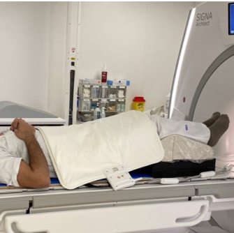 |
128 |
MRI IN RADIATION THERAPY: VALIDATION OF THE EFFECT FROM BLANKET
MRI-COIL ON THE PELVIS REGION OF HEALTHY VOLUNTEERS
Yosef Al-Abasse1,
Tova Roman Rung1,
Peter Larsson1,
Dan Josefsson1,
Lena Lundkvist2,
Kristina Redelius2,
Kristina Hultman2,
Maria Nilsson2,
Jan Rzepecki2,
and Peter Lundberg1,3
1Department of Health, Medicine and Care, Linköping University, Linköping, Sweden, 2Department of Oncology, and Department of Biomedical and Clinical Sciences, Linköping University, Linköping, Sweden, 3Center for Medical Imaging and Visualization (CMIV), Linköping University, Linköping, Sweden Keywords: Multimodal, Radiotherapy, Synthetic CT, T2-w, software Conventional coils for dose planning purposes cannot be placed on the patient as the outer body contour then will be deformed. Coils are therefore placed on a special holder which creates a distance between coil and patient, but this leads to less SNR. The blanket-coils are significantly lighter and can be placed directly on the patient. Three similarity comparison methods were used here, Hausdorff distance, Dice similarity coefficient (DSC) and Surface DSC. Eight of eleven comparisons were within 4 mm difference for Hausdorff distance; Surface DSC was >99%, at a 3 mm tolerance, and DSC >98.5%.
|
|
4550.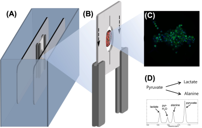 |
129 |
Benchtop NMR for lab-on-a-chip
Marc Azagra1,
Alejandro Portela1,
Hetal Patel2,
Dian Weerakonda2,
Jose Yeste1,
Alba Herrero-Gómez1,
Matthew Fallon2,
Megdouda Benamara3,
Marc Dubois3,
Tryfon Antonakakis3,
Javier Ramon1,
and Irene Marco-Rius1
1Institute for Bioengineering of Catalonia, Barcelona, Spain, 2Oxford Instruments, Abingdon, United Kingdom, 3Multiwave Imaging, Marseille, France Keywords: New Devices, Hyperpolarized MR (Non-Gas), Molecular Imaging A benchtop NMR spectrometer has been designed and optimized to monitor real-time metabolism of 3D tissue engineered on microfluidic platforms using hyperpolarization by dynamic nuclear polarisation. We show the design process, which included the modification of a commercial benchtop NMR spectrometer, the design and fabrication of a microfluidic platform for a reliable injection of a hyperpolarized substrate and a constant delivery and renewal of the cell media and its integration with a RF coil for transmit/receive, and the design and fabrication of a sample carrier. We also present our preliminary NMR data using this system. |
|
| 4551. | WITHDRAWN | ||
4552. |
130 |
Development of a MR-compatible bioreactor for 3D cell structure
metabolic analysis in real-time for a 60 MHz benchtop NMR
spectrometer.
Lluís Mangas-Florencio1,
Alba Herrero-Gómez1,
Marc Azagra1,
James Ellis1,
and Irene Marco-Rius1
1Institute for Bioengineering of Catalonia, Barcelona, Spain Keywords: New Devices, Cancer, Metabolism Bioreactor platforms have been used in HP-NMR applications to analyze cellular metabolism in real-time, however they require complex set-ups and automated controlled systems, limiting their use. We present a system that overcomes these limitations consisting of a 3D printed platform, an NMR compatible 3D cell model and a recirculation circuit for single-shot and longitudinal hyperpolarization-enhanced NMR experiments. The platform presents no signal interference, a media system for biological support, and a straightforward and simplified manufacturing and assembly process. As a proof-of-concept, we present preliminary metabolic data of a 3D HeLa cell model analyzed with our bioreactor. |
|
4553.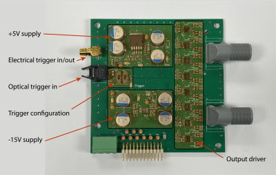 |
131 |
An in-bore PIN diode driver based on a single chip with
nanoseconds rise/fall time
Christoph Michael Schildknecht1,
Russell L Lagore2,
Matt Waks2,
Gregor Adriany2,
and Klaas Paul Pruessmann1
1Institute for Biomedical Engineering, ETH Zurich and University of Zurich, Zürich, Switzerland, 2Center for Magnetic Resonance Research, University of Minnesota, Minneapolis, MN, United States Keywords: New Devices, New Devices An in-bore PIN diode driver with eight output channels is presented here. The driver is based on a single-chip solution making the implementation simple and of high performance. The rise and fall times of the driver's output are in the single-digit nanosecond range with a peak output current of up to 5.7A per channel. Triggering the driver can be done either with an optical or electrical signal. The driver was tested on the bench and in-bore of a 10.5T MR scanner. |
|
4554.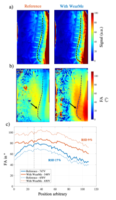 |
132 |
Ultra-thin metasurface pads for spine MRI
Marc Dubois1,
Amira Trabelsi1,2,3,
Tania Vergara Gomez1,2,3,
Pierre Jomin3,
Djamel Berrahou1,
David Bendahan2,4,
Frank Kober2,4,
Virginie Callot2,4,
Stefan Enoch3,
and Redha Abdeddaim3
1Multiwave Imaging, Marseille, France, 2Aix Marseille Univ, CNRS, CRMBM, Marseille, France, 3Aix Marseille Univ, CNRS, Centrale Marseille, Institut Fresnel, Marseille, France, 4APHM, Hopital Universitaire Timone, CEMEREM, Marseille, France Keywords: New Devices, Spinal Cord, Metamaterial, Metasurface Spine imaging is usually conducted at 3T given the need of high spatial resolution. Unfortunately, the lumbar and thoracic spine regions of interest will lie in areas of weak transmit efficiency. Recent studies showed that high permittivity ceramic blocks can improve image quality. Here we present a new approach based on ultra-thin, lightweight, flexible metasurface pads yielding improved efficiency and homogeneity of the B1+ field along the spine in clinical 3T MRI settings. Our design offers seamless integration to regular equipment leading to more efficient and safer spine imaging protocols. |
|
4555.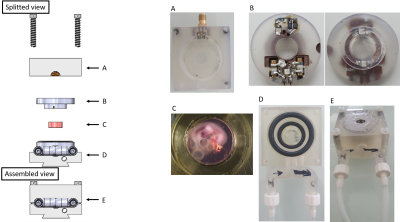 |
133 |
The MR-Bioreactor : Micro-MRI of thick living tissues to
characterize 4-days old ex-ovo chick embryo morphology.
Jean-Lynce Gnanago1,
Sophie Calvet2,
Tony Gerges1,
Valernst Gilmus1,
Camila Pereira Sousa1,
Michel Cabrera1,
Julien Falk2,
and Simon Auguste Lambert1
1Université de Lyon, INSA Lyon, Université Claude Bernard Lyon 1, Ecole Centrale de Lyon, CNRS, Ampère UMR5005, Villeurbanne, France, Lyon, France, 2MeLis, CNRS UMR 5284-INSERM U1314-Université Claude Bernard Lyon 1 Institut NeuroMyoGène, Lyon, France, Lyon, France Keywords: New Devices, Animals Thanks to their accessibility for experimental manipulations, their availability and their low cost, chick embryos have been used as animal models for developmental biology for long. In these applications, non-invasive 3D imaging of the embryos is needed to monitor the embryos development. Morphological characterization of ex-ovo chick embryo generally require tissue fixing which can alter MR characterizations. In this work, we use our MR-Bioreactor to perform a high resolution morphological characterization of a chick embryo maintained in living conditions. |
|
4556.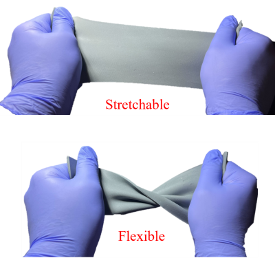 |
134 |
Silicon Carbide (SiC) Based Flexible and Stretchable Dielectric
Pad for B1 Field Enhancement
Seyedamin Hashemi1,
Kezia Sharon Christopher1,
Sri Kirthi Kandala1,
Kwanjoon Song2,
Benjamin Bartelle1,
and Sung-Min Sohn1
1School of Biological and Health Systems Engineering, Arizona State University, Tempe, AZ, United States, 2Department of Biomedical engineering, Yonsei University, Wonju, Korea, Republic of Keywords: New Devices, RF Arrays & Systems, Invisible Dielectric pad, Silicon Carbide, SNR enhancement We propose a stretchable, flexible, and MRI invisible dielectric pad to enhance the quality of acquired MR images. A silicon-based elastomer is mixed with Silicon Carbide (SiC) to develop this proposed dielectric pad. Preclinical in-vivo imaging of a mouse head was conducted at 9.4T to validate the performance of the dielectric pad. 2.54% (in dB) increase in the central SNR of the brain was observed after using the dielectric pad and peripheral brain locations have shown improved contrast. The flexible and stretchable dielectric pad could be used as a substitute for conventional dielectric pads, especially in irregular sample shapes. |
|
4557.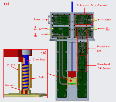 |
135 |
A Design of 10-100 MHz Broadband NMR Transmission-line Probe
Shouquan Yao1,
Juncheng Xu1,
Yiqiao Song2,
Ming Shen1,
Bingwen Hu1,
and Yu Jiang1
1Shanghai Key Laboratory of Magnetic Resonance, School of Physics and Electronic Science, East China Normal University, Shanghai, China, 2Department of Radiology, Massachusetts General Hospital, Charlestown, MA, United States Keywords: New Devices, New Devices, Probe This work demonstrated a broadband NMR probe with a bandwidth of 10-100 MHz. Based on transmission-line structure, broadband transmission was achieved. As for broadband reception, high-speed active T/R switches and low-noise differential amplifiers were used for high-sensitivity RF reception. The SNR of FID signal detected by broadband probe was 74% of that of conventional resonance probe, which means the feasibility of the broadband NMR transmission-line probe was verified. |
|
4558.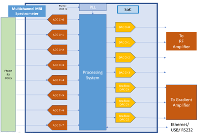 |
136 |
State of Art, Powerful, System On Chip, Design,Development and
Implementation of Multi Channel Transceiver for MRI Spectrometer
Rajesh Harsh1,
Dharmesh Verma1,
Aparna Gunvant Raut1,
and Mahadev Kashinath Dipnaik1
1Medical Systems Division, S.A.M.E.E.R. Mumbai, Mumbai, India Keywords: New Devices, Spectroscopy, System -on-chip,MIMO Based smart MRI Spectrometer The increasing complexity of modern MRI hardware design makes system challenging. MRI systems equipped with multi-channel exciter and multi-channel data-acquisition modules, particularly high-channel-counts often requires a huge cost. This abstract summaries the design and development of a low cost, small form factor, system on chip approach to realize a complete 1.5 T MRI spectrometer on a small chip of 28 nanometer fabric. The developed system executes parallel image processing algorithms and compatible instrumentation for accurate spatial and temporal requirements. |
|
4559.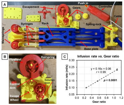 |
137 |
Developing an In-Bore MRI-Compatible Syringe Pump
Martin D Holland1,
Seth N Lee2,
and Harrison D Kim2
1Interdisciplinary Engineering, University of Alabama at Birmingham, Birmingham, AL, United States, 2Radiology, University of Alabama at Birmingham, Birmingham, AL, United States Keywords: New Devices, New Devices, Syringe pump, MRI Compatibility, 3D Printing Delivering a contrast (or therapeutic) agent to a subject inside an MRI bore requires an expensive electronically programmed syringe pump and a long tube filled with the agent. We developed an inexpensive 3D-printed MRI-compatible syringe pump that can be used inside an MRI bore. This syringe pump yields a highly reproducible infusion rate, which can be readily adjusted using gears with different gear ratios. |
|
4560.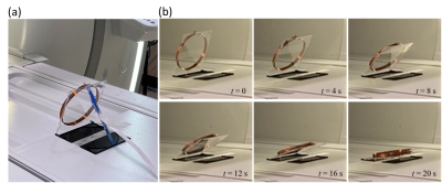 |
138 |
Falling conductive loop: A simple analytical model for magnetic
damping
Seung-Kyun Lee1
1GE Global Research, Niskayuna, NY, United States Keywords: Magnets (B0), Magnets (B0), magnetic damping The motion of a conductive object is profoundly affected by the presence of a strong magnetic field. A conductive loop falling inside an MRI magnet can serve as an analytically solvable model to explore magnetic damping. We present a pair of magneto-mechanical coupled equations to describe its motion and verify their solution with experimental data. A simple formula for the loop's falling time was obtained, which indicates that magnetic damping is proportional to B02. |
|
4561.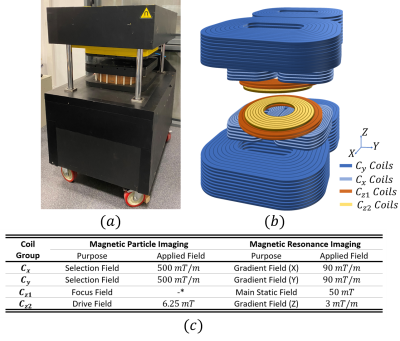 |
139 |
Open-Bore Hybrid MPI-MRI Scanner: A Proof-of-Concept Simulation
Study
Sefa Karaca1,2,3,
Damla Alptekin Soydan1,
Emine Ulku Saritas2,3,
and Can Barış Top1
1Aselsan Research Center, ASELSAN A.Ş., Ankara, Turkey, 2Electrical and Electronics Engineering, Bilkent University, Ankara, Turkey, 3National Magnetic Resonance Research Center (UMRAM), Bilkent University, Ankara, Turkey Keywords: Hybrid & Novel Systems Technology, Low-Field MRI, Magnetic Particle Imaging Magnetic particle imaging (MPI) is a novel technique that images background-free spatial distribution of magnetic nanoparticles inside the body. The lack of signal from the background tissue provides high sensitivity to MPI. However, anatomical information is also required in many applications. Here, we present an open-bore hybrid preclinical MPI-MRI system, in which the coils can be utilized interchangeably between MPI and MRI data acquisitions. We analyze the imaging performance for both modalities using a simulation model based on our in-house prototype MPI system that can generate 0.5 T/m selection field gradients for MPI and 50 mT B0 for low-field MRI. |
|
 Back to Meeting Home
 Back to the Program-at-a-Glance
The International Society for Magnetic Resonance in Medicine is accredited by the Accreditation Council for Continuing Medical Education to provide continuing medical education for physicians.
 Back to Meeting Home
 Back to the Program-at-a-Glance
 View Presentation Video