Digital Poster
DTI & DWI I
ISMRM & ISMRT Annual Meeting & Exhibition • 03-08 June 2023 • Toronto, ON, Canada

| Computer # | |||
|---|---|---|---|
3605.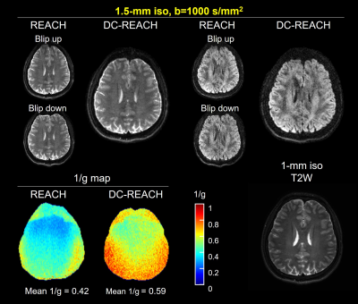 |
21 |
Hybrid-space reconstruction with add-on distortion correction
for simultaneous multi-slab DWI
Jieying Zhang1,
Simin Liu1,
Yishi Wang2,
and Hua Guo1
1Center for Biomedical Imaging Research, Department of Biomedical Engineering, School of Medicine, Tsinghua University, Beijing, China, 2Philips Healthcare, Beijing, China Keywords: Image Reconstruction, Diffusion Tensor Imaging 3D simultaneous multi-slab imaging (SMSlab) can achieve high-resolution DWI with high SNR efficiency. Recently, we integrated SMSlab DWI with blipped-CAIPI gradients (blipped-SMSlab) and proposed a hybrid-space reconstruction algorithm, REACH. In this study, REACH is extended for distortion correction, which is called DC-REACH. It can correct the image distortions and the phase interferences introduced by the blipped-CAIPI gradients simultaneously. It also distinctly reduces the g-factor penalty via the joint reconstruction of the blip-up/down data. |
|
3606.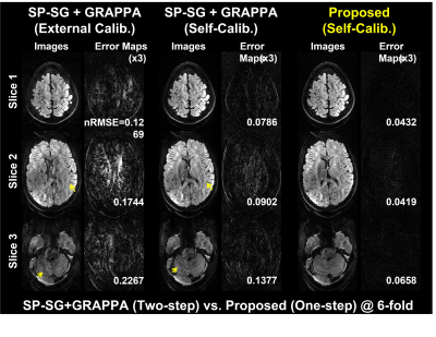 |
22 |
Self-Calibrating Aliasing-Controlled Simultaneous Multi-Slice
Reconstruction for Diffusion MRI
Eun Ji Lim1,
Hyunkyung Maeng1,
and Jaeseok Park1
1Department of Intelligent Precision Healthcare Convergence, Sungkyunkwan University, Suwon, Korea, Republic of Keywords: Image Reconstruction, Diffusion Tensor Imaging To jointly resolve inter-slice leakages and in-plane aliasing for combined in-plane- and slice-accelerated diffusion MRI data, we proposed a novel, one-step solution for SMS-reconstruction optimally exploiting self-calibrating data from generalized 3D Fourier encoding perspective. To this end, we propose a generalized SMS forward signal model with an extended controlled aliasing and an extended self-calibration. Aliasing artifacts are jointly resolved in ky-and kz-directions by balancing null space consistency with a low rank prior while enforcing data fidelity in 3D k-space. We demonstrated the proposed method outperforms competing methods for diffusion MRI at SMS=3, R=2. |
|
3607.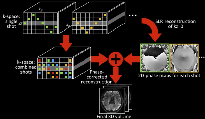 |
23 |
Self-navigated high-resolution 3D diffusion MRI using an
extended blipped-CAIPI sampling and structured low-rank
reconstruction
Ziyu Li1,
Xi Chen1,
Mark Chiew1,2,3,
Karla L. Miller1,
and Wenchuan Wu1
1Wellcome Centre for Integrative Neuroimaging, FMRIB, Nuffield Department of Clinical Neurosciences, University of Oxford, Oxford, United Kingdom, 2Physical Sciences, Sunnybrook Research Institute, Toronto, ON, Canada, 3Department of Medical Biophysics, University of Toronto, Toronto, ON, Canada Keywords: Image Reconstruction, Diffusion/other diffusion imaging techniques, Diffusion Acquisition & Reconstruction 3D multi-shot multi-slab imaging can provide superior SNR efficiency for high-resolution dMRI but suffers from motion-induced shot-to-shot phase variations. We propose a highly-efficient, self-navigated method to correct for phase variations in 3D multi-slab dMRI. The sampling of each shot is designed to intersect with the kz=0 plane. These intersections are used to reconstruct a 2D phase map for each shot using a structured low-rank reconstruction that leverages the redundancy in shot and coil dimensions. The phase maps are used to eliminate the phase inconsistency in the final 3D multi-shot reconstruction. We demonstrate the method’s efficacy using highly realistic simulations. |
|
3608.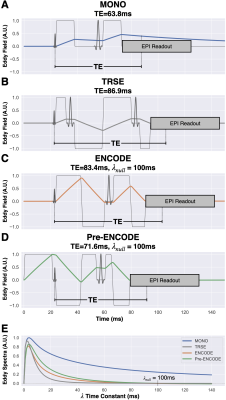 |
24 |
Pre-Excitation Gradients for Eddy Current-Nulled Convex
Optimized Diffusion Encoding to Mitigate Distortion in 2D
Diffusion Weighted Imaging
Matthew J. Middione1,
Michael Loecher1,
Xiaozhi Cao1,
Kawin Setsompop1,2,
and Daniel B. Ennis1,3
1Department of Radiology, Stanford University, Stanford, CA, United States, 2Department of Electrical Engineering, Stanford University, Stanford, CA, United States, 3Cardiovascular Institute, Stanford University, Stanford, CA, United States Keywords: Pulse Sequence Design, Diffusion/other diffusion imaging techniques, Eddy Currents Diffusion encoding gradients produce eddy currents that cause image distortions in DWI. Twice refocused spin-echo (TRSE) and eddy current-nulled convex optimized diffusion encoding (ENCODE) mitigate eddy current-induced image distortions in DWI, but at the expense of extending the TE. Herein, we revise the original ENCODE method by playing additional pre-excitation gradient lobes (Pre-ENCODE). Using simulations, phantom experiments, and in vivo imaging we demonstrate that Pre-ENCODE mitigates eddy current-induced image distortions in DWI with a shorter TE than TRSE and ENCODE. |
|
3609.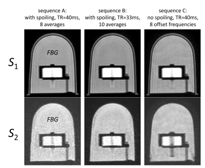 |
25 |
Impact of gradient spoiling for diffusion-weighted Double-Echo
Steady-State sequences
Ulrich Katscher1,
Jakob Meineke1,
and Jochen Keupp1
1Philips Research Europe, Hamburg, Germany Keywords: Pulse Sequence Design, Diffusion/other diffusion imaging techniques Double-Echo Steady-State (DESS) sequences are a promising candidate for diffusion weighted imaging (DWI) free of geometric distortions. While diffusion weighted DESS (dwDESS) sequences were originally introduced with a unipolar diffusion weighting gradient GD, a bipolar GD is required to obtain a motion robust fully balanced sequence. The inevitable banding artefacts occurring for bipolar GD can be handled via different techniques like gradient spoiling (thus deviating from the fully balanced sequence) or phase cycling. This study compares these techniques to optimize SNR for a given scan time and given diffusion weighting. |
|
3610.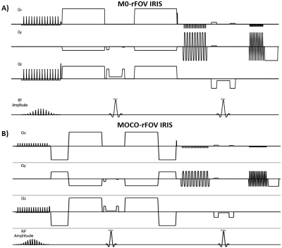 |
26 |
Motion robust and high resolution DWI utilizing the combination
of Multi-shot Reduced FOV imaging with motion-compensated
diffusion gradients
Zhigang Wu1,
Yajing Zhang2,
Guillaume Gilbert3,
Wengu Su4,
Yan Zhao5,
and Jiazheng Wang6
1Philips Healthcare, Shenzhen, Ltd., Shenzhen, China, 2Philips Health Technology, Suzhou, China, 3MR Clinical Science, Philips Healthcare, Mississauga, ON, Canada, 4BU MR Application, Philips Health Technology, Suzhou, China, 5BU MR R&D, Philips Health Technology, Suzhou, China, 6Philips Healthcare, Beijing, China Keywords: Pulse Sequence Design, Diffusion/other diffusion imaging techniques, Diffusion, Motion compensated diffusion gradients, Reduced FOV imaging, Multi-shot DWI Reduced FOV imaging (rFOV) and multi-shot DWI both are very useful techniques to improve spatial resolution for detection of pancreatic lesion. However, respiratory and cardiovascular motion will introduce severe artifacts and ADC bias. Motion compensated diffusion gradients (MOCO) could be used to improve the image quality. In this study, a new sequence named MOCO-rFOV IRIS was developed, which combines the advantages of rFOV, MOCO and image reconstruction using image-space sampling function based multi-shot DWI (IRIS). Results from in vivo data demonstrated that the proposed method could be used to realize motion robust and high resolution DWI for pancreas |
|
3611.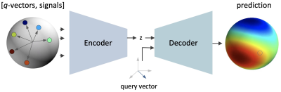 |
27 |
DISCUS: Diffusion MRI Signal Reconstruction with Continuous
Sampling
Christian Ewert1,
David Kügler1,
Anastasia Yendiki2,3,
and Martin Reuter1,2,3
1AI in Medical Imaging, German Center for Neurodegenerative Diseases (DZNE), Bonn, Germany, 2Athinoula A. Martinos Center for Biomedical Imaging, Massachusetts General Hospital, Boston, MA, United States, 3Department of Radiology, Harvard Medical School, Boston, MA, United States Keywords: Image Reconstruction, Diffusion/other diffusion imaging techniques, q-space, denoising DISCUS addresses two challenges currently limiting the analysis potential of diffusion MRI: sparsity of measurements and variability in q-space sampling schemes. Our method combines the advantages of model-fit approaches with continuous sampling (spherical harmonics, SHORE) and rigid, discrete learning-based methods. DISCUS can be initialized from any acquisition scheme and permits signal prediction for an arbitrary q-vector. Despite the added flexibility, DISCUS performs on par with other, far less flexible learning methods, while outperforming model-fit methods. DISCUS-derived signals translate to higher-quality FA estimates promising accurate analyses even from very short acquisitions. |
|
3612.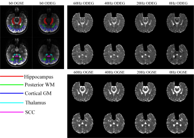 |
28 |
Orthogonal diffusion encoding gradient sequence (ODEG) improves
time-dependency measurements in the human brain
Qinfeng Zhu1,
Haotian Li1,
Yi-Cheng Hsu2,
Yi Sun2,
and Dan Wu1
1zhejiang University, Hangzhou, China, 2Siemens Healthcare China, Shanghai, China Keywords: Pulse Sequence Design, Diffusion/other diffusion imaging techniques Oscillatory gradient dMRI is used to access restricted diffusion at short diffusion times (td). But its clinical application is limited due to the limited gradient strength, leading low b-value and low resolution, and thus is subject to contamination from microcirculation and CSF partial volume. Here we proposed an orthogonal diffusion encoding gradient (ODEG) sequence to improve td–dependency measurements in human brain, by applying a pulse gradient orthogonal to the oscillating gradient to suppress the fast diffusion from microcirculation and free water. The results showed that td-dependency was significantly improved by the ODEG sequence in the hippocampus and cortical gray matter. |
|
3613.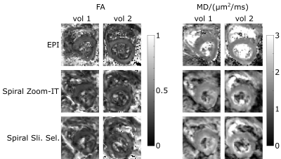 |
29 |
Cardiac DTI with spiral readouts with third order k-space
correction but without inner volume excitation
Lars Mueller1,
Sam Coveney1,
Maryam Afzali1,2,
Irvin Teh1,
Fabrizio Fasano3,4,
Filip Szczepankiewicz5,
Erica Dall’Armellina1,
Christopher Nguyen6,
Derek K Jones2,
and Jurgen E Schneider1
1Leeds Institute of Cardiovascular and Metabolic Medicine, University of Leeds, Leeds, United Kingdom, 2Cardiff University Brain Research Imaging Centre (CUBRIC), Cardiff University, Cardiff, United Kingdom, 3Siemens Healthcare Ltd, Camberly, United Kingdom, 4Siemens Healthcare GmbH, Erlangen, Germany, 5Medical Radiation Physics, Clinical Sciences Lund, Lund University, Lund, Sweden, 6Cleveland Clinic, Cleveland, OH, United States Keywords: Pulse Sequence Design, Diffusion Tensor Imaging, Spiral Using spiral trajectories instead of EPI can reduce the echo time in diffusion weighted MRI. The feasibility of spiral trajectories for cardiac DTI has recently been demonstrated, but the larger object size (i.e torso vs. for example skull) may require an inner volume excitation to keep the readout at an useable duration. Here we examine the use of an undersampled spiral with SENSE reconstruction and standard slice selective pulses. We show that MD and FA derived from the slice selective and inner volume excitation yield comparable values with the MD being higher than the one measured with EPI. |
|
3614.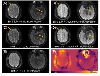 |
30 |
Feasibility of diffusion imaging using SMS-spiral acquisition
with corrections on gradient waveform and field inhomogeneities
Zhe Wu1,
Alexander Jaffray2,
Lars Kasper1,
and Kamil Uludag1,3
1Techna Institute, University Health Network, Toronto, ON, Canada, 2University of British Columbia, Vancouver, BC, Canada, 3Department of Medical Biophysics, University of Toronto, Toronto, ON, Canada Keywords: Pulse Sequence Design, Diffusion/other diffusion imaging techniques, Spiral Imaging, field inhomogeneity correction, gradient correction We propose a short-TE signal-to-noise ratio (SNR) enhanced diffusion imaging method using simultaneous multi-slice (SMS) accelerated spiral acquisition. The correction of field inhomogeneity and gradient waveforms are introduced in the reconstruction without any assistance of external hardware (e.g. field camera). Results showing the feasibility of this method on both phantom and human subjects, and the corrections of B0 and gradient waveforms are essential to improve the image quality. |
|
3615.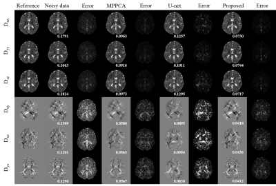 |
31 |
A deep learning method for diffusion tensor imaging using
spherical harmonic representation
Yunwei Chen1 and
Xinyuan Zhang1
1School of Biomedical Engineering, Southern Medical University, Guangzhou, China Keywords: Machine Learning/Artificial Intelligence, Diffusion Tensor Imaging Deep learning methods have been demonstrated state-of-the-art performance in Diffusion Magnetic Resonance Imaging (dMRI) denoising and parameter estimation. However, existing deep learning methods for dMRI are limited to the specific acquisition scheme. To solve the limitation, we proposed to use spherical harmonic coefficients as the deep learning network’s input. Our results have shown that the proposed method has a high performance in denoising and parameter estimation for DTI with a strong generalization ability. |
|
3616. |
32 |
Gauge equivariant convolutional neural networks for diffusion
MRI
Uzair Hussain1 and
Ali Khan1,2,3
1Robarts Research Institute, Centre for Functional and Metabolic Mapping, London, ON, Canada, 2Department of Medical Biophysics, Western University, London, ON, Canada, 3Western Institute for Neuroscience, Western University, London, ON, Canada Keywords: Machine Learning/Artificial Intelligence, Diffusion Tensor Imaging One shortcoming of diffusion MRI (dMRI) is long scan times as numerous images have to be acquired to achieve a reliable angular resolution of diffusion gradient directions. In this work we introduce gauge equivariant convolutional neural network (gCNN) layers that overcome the challenges associated with the dMRI signal being acquired on a sphere instead of a rectangular grid. We apply this method to upsample angular resolution to predict diffusion tensor imaging (DTI) parameters from just six diffusion gradient directions. Additionally, gCNNs are able to train with fewer subjects and are general enough to be applied to other dMRI related problems. |
|
3617.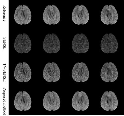 |
33 |
Accelerating High Resolution Diffusion Tensor Imaging Using
Intra- and Inter-image Correlation
Zhongbiao Xu1,
Rongli Zhang2,
Wei Huang1,
Junying Cheng3,
Yingjie Mei4,
Yihao Guo5,
Hengwen Sun1,
Yaohui Wang6,
and Zhifeng Chen7
1Department of Radiotherapy, Guangdong Provincial People's Hospital, Guangzhou, China, 2Department of Imaging and Interventional radiology, The Chinese University of Hong Kong, HongKong, China, 3Department of MRI, The first Affiliated Hospital of Zhengzhou University, zhengzhou, China, 4School of Biomedical Engineering, Southern Medical University, guangzhou, China, 5Hainan General Hospital, hainan, China, 6Institute of Electrical Engineering, Chinese Academy of Sciences, beijing, China, 7Monash Biomedical Imaging, Department of Data Science and AI, Monash University, Clayton, Australia Keywords: Image Reconstruction, Diffusion Tensor Imaging DTI is challenged by the prolonged scan time in frontier studies and clinical applications. Parallel imaging can reduce the scan time, but with the SNR loss and the limitation of acceleration factor. In this work, we combined SENSE with self-supervised BM4D reconstruction model to improve image quality. The in vivo experiments demonstrated that the proposed method can obtain greatly improved image quality even with high acceleration factor of 5, compared to conventional methods. |
|
3618.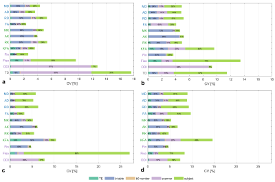 |
34 |
Evaluation of the impact of protocol settings on the variability
of model estimations in multicenter diffusion MRI studies
Qiqi Tong1,
Jinsong Li1,2,
Jianhui Zhong3,4,
and Hongjian He4,5
1Research Center for Healthcare Data Science, Zhejiang Lab, Hangzhou, China, 2College of Biomedical Engineering and Instrument Science, Zhejiang University, Hangzhou, China, 3Department of Imaging Sciences, University of Rochester, Rochester, NY, United States, 4Center for Brain Imaging Science and Technology, College of Biomedical Engineering and Instrument Science, Zhejiang University, Hangzhou, China, 5School of physics, Zhejiang University, Hangzhou, China Keywords: Data Analysis, Diffusion Tensor Imaging, multicenter A multicenter diffusion magnetic resonance imaging (dMRI) study was designed with different b-table schemes, non-diffusion b0 number, and varied echo time (TE) in two 3T scanners of different vendors. Global sensitivity analysis of 6 traveling subjects was conducted to evaluate the impact of imaging protocol setting on the observed cross-scan variability of diffusion metrics. |
|
3619.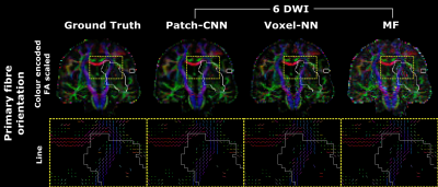 |
35 |
Patch-CNN provides high-fidelity directional & scalar parameter
estimation from 6-directional DWI robust to pathology unseen
during training
Tobias Goodwin-Allcock1,
Guglielmo Genovese2,3,4,
Belen Zaid3,5,
Stéphane Lehericy2,3,
Charlotte Rosso3,5,
Ting Gong1,
Robert Gray6,
Parashkev Nachev6,
Marco Palombo7,8,
and Hui Zhang1
1Department of Computer Science and Centre for Medical Image Computing, UCL, London, United Kingdom, 2Centre de NeuroImagerie de Recherche - CENIR, Paris Brain Institute - ICM, Paris, France, 3UMR S 1127, Inserm U 1127, CNRS UMR 7225, ICM, F-75013, Sorbonne Université, Paris, France, 4Center for Magnetic Resonance Research, Department of Radiology, University of Minnesota, Minneapolis, MN, United States, 5Paris Brain Institute - ICM, Centre de NeuroImagerie de Recherche - CENIR, Paris, France, 6University College London Queen Square Institute of Neurology, London, United Kingdom, 7Cardiff University Brain Research Imaging Centre (CUBRIC), School of Psychology, Cardiff University, Cardiff, United Kingdom, 8School of Computer Science and Informatics, Cardiff University, Cardiff, United Kingdom Keywords: Data Processing, Diffusion Tensor Imaging, Machine Learning This work evaluates the clinical viability of Patch-CNN for estimating diffusion MRI (dMRI) parameters from only 6 diffusion-weighted images (DWIs). Machine learning (ML) has been proposed to improve fitting from 6-directional DWIs. However, directional measures, e.g. primary fibre orientation, have only been estimated using CNNs. CNNs have not yet been validated on pathology that is not contained within the training dataset. As pathological diversity is difficult to capture in typical applications, ML methods are clinically viable only if they can generalise to unseen pathology. We show that Patch-CNN may generalise to unseen pathology and estimate directional measures. |
|
3620.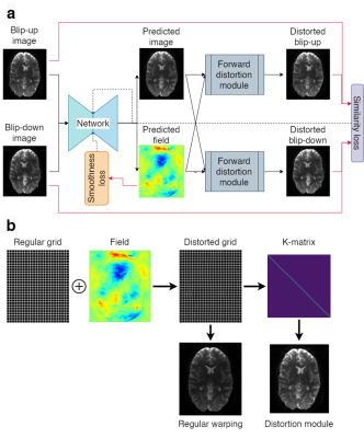 |
36 |
Unsupervised Susceptibility Artifact Correction in DTI Using a
Deep Learning Forward-Distortion Model
Abdallah Zaid Alkilani1,2,
Tolga Çukur1,2,3,
and Emine Ulku Saritas1,2,3
1Deparment of Electrical and Electronics Engineering, Bilkent University, Ankara, Turkey, 2National Magnetic Resonance Research Center (UMRAM), Bilkent University, Ankara, Turkey, 3Neuroscience Graduate Program, Bilkent University, Ankara, Turkey Keywords: Data Analysis, Diffusion Tensor Imaging, Susceptibility, Machine Learning/Artificial Intelligence, Brain, Artifacts Diffusion weighted imaging (DWI) requires correction of susceptibility artifacts before conducting quantitative analyses. Correction is typically performed by acquiring DWI images in reversed phase-encode directions, which are used to estimate and correct for the effects of susceptibility-induced field. In this work, we propose a Forward-Distortion Network (FD-Net) for correcting susceptibility artifacts at multiple b-values. We evaluate the quality of the corrected DWI images and Diffusion Tensor Imaging (DTI) metrics, using FSL’s TOPUP as a reference classical method. In addition to rapid execution times, FD-Net exhibits high-fidelity performance for both DWI images and DTI metrics.
|
|
3621.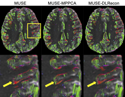 |
37 |
High-resolution Diffusion Tensor Imaging with Deep Learning
Reconstruction: Preliminary Results in Sub-cortical Fiber
Tracking
Zhangxuan Hu1,
Xiaocheng Wei1,
Jie Lu2,
and Bing Wu1
1GE Healthcare, Beijing, China, 2Department of Radiology and Nuclear Medicine, Xuanwu Hospital, Capital Medical University, Beijing, China Keywords: Machine Learning/Artificial Intelligence, Diffusion Tensor Imaging Diffusion tensor imaging (DTI) is a well-established tool for providing insights into brain structural connectivity and detecting brain microstructure. High spatial resolution diffusion MRI can provide improved resolvability of fibers with high-curvature (u-fibers). Segmented k-space methods such as Multiplexed sensitivity-encoding (MUSE) are often used to achieve high resolution diffusion images, however the shortcomings, such as prolonged scan time and low signal-noise-ratio (SNR), still exist. In this study, we aim to further improve the image quality of high-resolution diffusion images acquired with MUSE by combing with a deep learning based reconstruction method and thus to improve the sub-cortical fiber tracking accuracy. |
|
3622.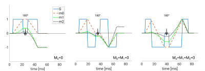 |
38 |
Segmented thick-slab 3D DWI with first and second order
motion-compensated diffusion gradients
Jens Johansson1,
Kerstin Lagerstrand2,3,
Hanna Hebelka1,4,
and Stephan E Maier1,5
1Radiology, Clinical Sciences, Sahlgrenska Academy, University of Gothenburg, Gothenburg, Sweden, 2Medical Radiation Sciences, Clinical Sciences, Sahlgrenska Academy, University of Gothenburg, Gothenburg, Sweden, 3Medical Physics and Biomedical Engineering, Sahlgrenska University Hospital, Gothenburg, Sweden, 4Radiology, Sahlgrenska University Hospital, Gothenburg, Sweden, 5Radiology, Brigham and women's hospital, Boston, MA, United States Keywords: Data Acquisition, Diffusion/other diffusion imaging techniques, Motion correction, 3D imaging Routine clinical diffusion imaging is generally performed with 2D echo planar sequences. A single thick-slab 3D approach could offer higher signal-to-noise ratio and better slice resolution, but has not been adopted due to the difficulty to avoid motion-induced phase errors that interfere with multi-shot spatial encoding. A new approach to enable 3D DWI is introduced here: rather than relying on navigator echoes for phase correction, first and second order motion-compensated diffusion encoding gradients are used to minimize phase variations at the source. |
|
3623.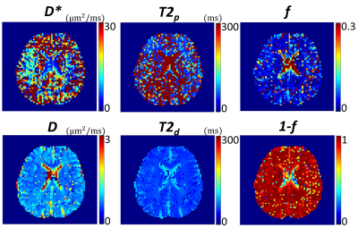 |
39 |
Time-efficient Relaxation-Incorporated IVIM Diffusion Imaging
Kaibao Sun1,
Guangyu Dan1,2,
Qingfei Luo1,
and Xiaohong Joe Zhou1,2,3
1Center for MR Research, University of Illinois at Chicago, Chicago, IL, United States, 2Department of Biomedical Engineering, University of Illinois at Chicago, Chicago, IL, United States, 3Departments of Radiology and Neurosurgery, University of Illinois at Chicago, Chicago, IL, United States Keywords: Data Acquisition, Diffusion/other diffusion imaging techniques Overestimation of perfusion volume fraction has been reported in conventional IVIM imaging. In a model known as extended T2-IVIM, compartmentalized relaxation times are incorporated to improve quantification of perfusion volume fraction, which requires acquisition of additional images at different TEs. A main challenge is that the scan time is lengthened, decreasing the efficiency while increasing vulnerability to motion. We herein introduce a novel sequence for time-efficient, relaxation-incorporated (TERI) IVIM imaging to address the aforementioned issues. TERI IVIM imaging uses multiple EPI readouts at different TEs in a single shot. The proposed technique has been demonstrated in the human brain. |
|
3624.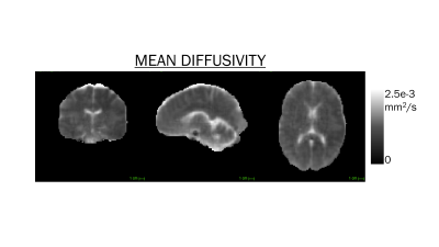 |
40 |
Super-resolution diffusion tensor imaging at 64 mT
Alix Plumley1,
Mara Cercignani1,
Álvaro Planchuelo-Gómez1,2,
James Gholam1,
and Derek K Jones1
1Cardiff University, Cardiff, United Kingdom, 2University of Valladolid, Valladolid, Spain Keywords: Software Tools, Diffusion Tensor Imaging, Low-field A super-resolution approach was used to create 2mm isotropic diffusion tensor images (DTI) from diffusion-weighted imaging data acquired on a low field, portable system. Mean diffusivity, fractional anisotropy and principal eigenvector orientation maps are shown. This work extends the very recently implemented capability of performing DTI on a 64mT system, and shows substantial improvement due to the increased through-plane resolution achieved with super-resolution. |
|
 Back to Meeting Home
 Back to the Program-at-a-Glance
The International Society for Magnetic Resonance in Medicine is accredited by the Accreditation Council for Continuing Medical Education to provide continuing medical education for physicians.
 Back to Meeting Home
 Back to the Program-at-a-Glance
 View Presentation Video