Digital Poster
Low-Field & Point-of-Care/Portable MRI I
ISMRM & ISMRT Annual Meeting & Exhibition • 03-08 June 2023 • Toronto, ON, Canada

Digital Poster
Low-Field & Point-of-Care/Portable MRI I
| Computer # | |||
|---|---|---|---|
1585.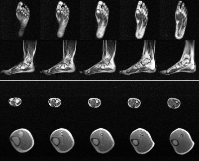 |
121 | 3D Balanced Steady-State Free Precession (bSSFP) Imaging on an Ultra-Low-Field 0.055T MRI System
Ye Ding1,2, Shi Su1,2, Linfang Xiao1,2, Yujiao Zhao1,2, Jiahao Hu1,2, Junhao Zhang1,2, Vick Lau1,2, Christopher Man1,2, Alex T.L. Leong1,2, and Ed X. Wu1,2
1Laboratory of Biomedical Imaging and Signal Processing, The University of Hong Kong, Hong Kong, China, 2Department of Electrical and Electronic Engineering, The University of Hong Kong, Hong Kong, China Keywords: Low-Field MRI, Body Recently, there has been an impetus to develop ultra-low-field (ULF) MRI technologies, which present opportunities for low-cost and portable imaging in point-of-care scenarios. Balanced steady-state free precession (bSSFP) is a time-efficient imaging sequence yet its feasibility at ULF remains unexplored. In this study, we implemented bSSFP sequence at 0.055T, and successfully demonstrated phantom, brain and extremity imaging. |
|
1586.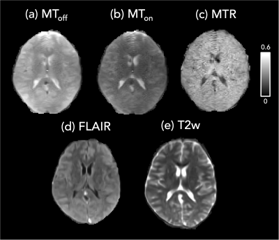 |
122 | Magnetization Transfer Imaging using non-balanced SSFP at Ultra-Low Field
Sharada Balaji1, Adam Dvorak1, Francesco Padormo2, Rui Pedro A.G Teixeira2, Megan E. Poorman2, Alex L. MacKay1,3, Tobias C. Wood4, Steven C.R. Williams4, Sean C.L. Deoni5, Shannon H. Kolind1,3,6, and Emil Ljungberg4,7
1Physics and Astronomy, University of British Columbia, Vancouver, BC, Canada, 2Hyperfine Inc, Guilford, CT, United States, 3Radiology, University of British Columbia, Vancouver, BC, Canada, 4Neuroimaging, King's College London, London, United Kingdom, 5MNCH D&T, Bill and Melinda Gates Foundation, Seattle, WA, United States, 6Medicine, University of British Columbia, Vancouver, BC, Canada, 7Medical Radiation Physics, Lund University, Lund, Sweden Keywords: Low-Field MRI, Magnetization transfer We have demonstrated a method for introducing Magnetization Transfer (MT) weighting in a non-balanced SSFP sequence (PSIF) at 64mT by changing the total power of the on-resonance RF excitation pulse. The presence of the MT effect was validated through simulations, experiments in phantoms of water, cream, and hair conditioner, as well as in-vivo. Changes to the signal amplitude in the conditioner from the flip angle and pulse width of the RF pulse illustrated the presence of MT saturation. An example protocol with MT weighting was applied in-vivo to generate magnetisation transfer ratio maps. |
|
1587.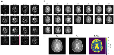 |
123 | Balanced steady-state free precession imaging and associated rapid relaxation time mapping on a point-of-care 46 mT Halbach MRI scanner
Chloé Najac1, Florian Birk2,3, Tom O’Reilly1, Klaus Scheffler2,3, Andrew Webb1, and Rahel Heule2,3,4
1C.J. Gorter MRI Center, Department of Radiology, Leiden University Medical Center, Leiden, Netherlands, 2Department of High-Field Magnetic Resonance, Max Planck Institute for Biological Cybernetics, Tübingen, Germany, 3Department of Biomedical Magnetic Resonance, University of Tübingen, Tübingen, Germany, 4Center for MR Research, University Children's Hospital, Zurich, Switzerland Keywords: Low-Field MRI, Data Acquisition Point-of-care imaging with low-field MRI (<0.1T) is a potential game changer for low-income countries and the intensive care unit. The main challenge is the low SNR. Balanced steady-state free precession (bSSFP) sequences are fast and SNR-efficient. However, bSSFP is very sensitive to B0 inhomogeneities resulting in banding artifacts. We evaluated the feasibility of using bSSFP on our 46 mT MRI scanner. By acquiring 17 bSSFP datasets with linearly increasing frequency offsets (from 0 to 1/TR), we could reconstruct banding-free maximum-intensity images as well as F0 and F-1 SSFP configurations, which we employed for rapid relaxation time mapping. |
|
1588.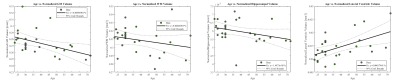 |
124 | On the estimation of age-related volumetric brain changes using ultra-low-field MR images
Peter Hsu1, Daniel K. Sodickson1,2, Hersh Chandarana1, Justin Fogarty1, Patricia Johnson1,2, and Jelle Veraart1,2
1Center for Biomedical Imaging, Department of Radiology, New York University Grossman School of Medicine, New York, NY, United States, 2Center for Advanced Imaging Innovation and Research (CAI2R), Department of Radiology, New York University Grossman School of Medicine, New York, NY, United States Keywords: Low-Field MRI, Brain, ultra-low-field MRI, point-of-care MRI The high cost of clinical MRI has severely limited its accessibility in many regions. Ultra-low-field (ULF) MRI systems have been developed to address this issue, providing diagnostic imaging at a significantly lower cost. However, computational tools that are used for clinical scans are often not applicable to ULF images which suffer from both SNR and resolution loss. Here, we compare two approaches for improving the resolution of ULF images and two techniques for extracting volumetric information from each approach. We show that given effective post-processing, ULF images can capture expected age-related volumetric changes in the brain. |
|
1589. |
125 | Optimized Extremity Coil System for Gradient-Free Low-Field MRI using the Bloch-Siegert Shift for RF Spatial Encoding
Sai Abitha Srinivas1, Christopher E Vaughn1, Jonathan B Martin 1, and William A Grissom1
1Biomedical Engineering, Vanderbilt University, Nashville, TN, United States Keywords: Low-Field MRI, Low-Field MRI Traditional B0 gradients have several drawbacks including high acoustic noise, PNS, bulkiness, and high cost. To address this, we present a coil system for Bloch-Siegert (BS) RF encoding comprising an optimized square root solenoid with a bucking coil for high efficiency encoding and a nested uniform saddle coil for the imaging Tx/Rx coil at 47.5mT (2MHz), a field strength that is especially attractive due to its low SAR and accessibility. The coil designed for in-vivo imaging was evaluated in simulation including SAR measurements and experimentally on a resolution phantom using a 3D BS phase-encoded imaging and optimized ‘U’ shaped pulses. |
|
1590.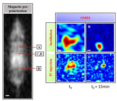 |
126 | Overhauser-enhanced MRI at ultra-low fields: a new system for imaging of free radicals in rodents
Dahmane Boudries1, Philippe Massot1, Elodie Parzy1, Seda Seren1, Philippe Mellet2, Jean-Michel Franconi1, Sylvain Marque3, Florian Fidler4, Stefan Wintzheimer5, Markus Mützel5, and Eric Thiaudiere1
1CNRS, Bordeaux, France, 2INSERM, Bordeaux, France, 3CNRS, Marseille, France, 4Fraunhofer, Würzburg, Germany, 5Pure devices, Rimpar, Germany Keywords: Low-Field MRI, Molecular Imaging, Ultra low field, OMRI, DNP, Magnetic prepolarization A new MRI system at Ultra-low field (206 µT) was designed for Overhauser-enhanced MRI. It allows both conventional MRI with pre-polarization at 20 mT and Dynamic Nuclear Polarization in the 70MHz range in living rats. Anatomical images in 3D allowed a correct visualization of the rat body shape. OMRI with injected or instilled stable and non-toxic nitroxides permitted to detect the free radicals in the lungs, the kidneys and the bladder. The work opens the way of molecular imaging of abnormal proteolysis in the context of a variety of diseases in large animals. |
|
1591. |
127 | Magnetic Resonance Elastography at 0.055 Tesla: A Preliminary Study
Shi Su1,2, Ye Ding1,2, Jiahao Hu1,2, Vick Lau1,2, Yujiao Zhao1,2, Junhao Zhang1,2, Christopher Man1,2, Alex T.L. Leong1,2, and Ed X. Wu1,2
1Laboratory of Biomedical Imaging and Signal Processing, the University of Hong Kong, Hong Kong, China, 2Department of Electrical and Electronic Engineering, the University of Hong Kong, Hong Kong, China Keywords: Low-Field MRI, Elastography Recent development of ultra-low-field (ULF) MRI presents opportunities for low-cost and portable imaging in point-of-care scenarios or/and low- and mid-income countries. Magnetic resonance elastography (MRE) is an essential part of MR abdominal imaging especially for chronic liver diseases. In this study, we explore the MRE at 0.055 Tesla. We demonstrate the feasibility of MRE based on phantom experiments at 0.055 Tesla. |
|
1592.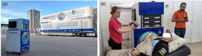 |
128 | Portable MRI for major sporting events – a case study on the MotoGP World Championship
Teresa Guallart-Naval1,2, José Miguel Algarín2,3, Rubén Bosch1, Francisco Juan-Lloris4, Eduardo Pallás2,3, Juan Pablo Rigla1, Pablo Martínez4, José Borreguero1, José María Benlloch2,3, Fernando Galve2,3, and Joseba Alonso2,3
1Tesoro Imaging SL, Valencia, Spain, 2Institute for Instrumentation in Molecular Imaging (i3M), Universitat Politècnica de València, Valencia, Spain, 3Institute for Instrumentation in Molecular Imaging (i3M), CSIC, Valencia, Spain, 4Physio MRI SL, Valencia, Spain Keywords: Low-Field MRI, Low-Field MRI, Portable MRI, Sport event MRI Here we study the potential MR value of a low-field portable system for use in major sporting events, specifically in the Motorcycle Grand Prix held in Valencia (Spain) between November 3rd and 6th, 2022. The system was transported in a small truck, installed in the main surgery room of the circuit medical facilities, and operational around 30 minutes after arrival. Overall, 15 subjects were scanned in four days, with a total of 21 extremity acquisitions. This work demonstrates that portable MRI machines can aid the diagnostic capabilities of medical staff in sporting events and competitions. |
|
1593.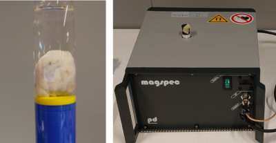 |
129 | Accelerated imaging of resected lymph nodes at high spatial resolution using a portable low-field MRI scanner
Arthur de Lange1, Lejla Alić1, Bennie ten Haken1, and Frank F.J. Simonis1
1TechMed Centre, University of Twente, Enschede, Netherlands Keywords: Low-Field MRI, New Devices After sentinel lymph nodes are detected using SPIONs and excised, their characterization is important to detect possible metastases. In this research a low-field (0.5T) tabletop MRI scanner was tested for this purpose using 4x accelerated high resolution 3D acquisition. Both simulations and experiments on excised pig lymph nodes showed promising results, with the accelerated scans showing similar image quality with respect to fully sampled datasets. This protocol shows lymph nodes can be imaged at 0.25 mm isotropic resolution within reasonable scan times. Clinical usage should be proven by scanning true metastatic lymph nodes. |
|
1594.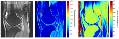 |
130 | Relaxation Times of the Musculoskeletal Tissues at 0.55 T
Iman Khodarahmi1, Mary Bruno1, Ryan Brown1, Jan Fritz1, and Mahesh B Keerthivasan2
1NYU Langone School of Medicine, New York, NY, United States, 2Siemens Medical Solutions USA Inc., Malvern, PA, United States Keywords: Joints, MSK Modern 0.55 T MRI promises to increase worldwide healthcare access through lower costs. Knowledge of the tissue relaxation parameters is essential to achieve the desired image contrast. Here for the first time, we quantify the T1 and T2 relaxation times of the musculoskeletal tissues through a Bloch model-based fitting approach. Our results show that the T1 relaxation time of non-fluid tissues is about 47% of those at 1.5 T. T2 relaxation times are about 70% of that of 1.5 T for synovial fluid, bone marrow and subcutaneous fat, and approximately similar to that of 1.5 T for cartilage and muscle. |
|
1595.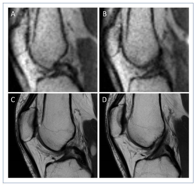 |
131 | Point-of-Care Knee MRI at 0.064T: Expanded Evaluation and Initial Comparison with Clinical Knee MRI at 3T
Jennifer M Watchmaker1, Ding Xia2, Idoia Corcuera-Solano1, Etan Dayan1, Justin Ngeow1, Fang Liu3, Zahi Fayad2, Mingqian Huang1, and Li Feng2
1Diagnostic, Molecular and Interventional Radiology, Icahn School of Medicine at Mount Sinai, New York, NY, United States, 2Biomedical Engineering and Imaging Institute, Icahn School of Medicine at Mount Sinai, New York, NY, United States, 3Athinoula A. Martinos Center for Biomedical Imaging, Harvard Medical School, Boston, MA, United States Keywords: Tendon/Ligament, Low-Field MRI, knee, hardware At last year’s ISMRM, a pioneering work was presented to demonstrate the initial feasibility and performance of point-of-care knee MRI at 0.064T using a Hyperfine Swoop portable MRI scanner. This work extended that study for further evaluation in an expanded cohort of healthy subjects and subjects with knee pathology, and compare low-field imaging with clinical 3 Tesla imaging in a subset of volunteers. |
|
1596. |
132 | Multi-reader evaluation of real-time MRI of the actively moving wrist at 0.55 Tesla
Abhijit J Chaudhari1, Robert D Boutin2, Yongwan Lim3, Sophia Cui4, Robert M Szabo5, and Krishna S Nayak3
1Radiology, University of California Davis, Sacramento, CA, United States, 2Radiology, Stanford University, Palo Alto, CA, United States, 3University of Southern California, Los Angeles, CA, United States, 4Siemens Medical Solutions, Malvern, PA, United States, 5Orthopaedic Surgery, University of California Davis, Sacramento, CA, United States Keywords: Joints, Skeletal, Real-time imaging; Wrist Kinematics; Dynamic imaging. We acquired real-time MRI of the actively moving wrist in humans utilizing a high-performance 0.55T system. Resulting images and associated static wrist scans were assessed by two expert readers blinded to acquisition parameters. Our results show that images acquired at a high temporal resolution of 12.6 ms per frame demonstrate minimal image degradation compared to images acquired at temporal resolution of 100 ms or higher, and enable improved characterization of wrist motion. These benefits support further studies to assess high-performance 0.55T systems for the evaluation of dynamic dysfunction of the wrist. |
|
1597.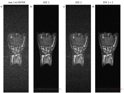 |
133 | High-performance, Shielding-free MSK Imaging at 100mT Enabled by Retrospective Active EMI Cancellation
Mauro Spreiter1, Reina Ayde1,2, Necati Ozcakal1, Tobias Senft1, Najat Salameh1,2, and Mathieu Sarracanie1,2
1Center for Adaptable MRI Technology (AMT Center), Department of Biomedical Engineering, University of Basel, Allschwil, Switzerland, 2AMT Center, Institute of Medical Sciences, School of Medicine, Medical Sciences & Nutrition, University of Aberdeen, Aberdeen, United Kingdom Keywords: Low-Field MRI, Low-Field MRI Electromagnetic interference cancellation method EDITER is implemented in an unshielded low-field MRI scanner at 100mT by addition of only two EMI detection coils to the acquisition chain. The method’s effectiveness is illustrated in high resolution MSK images of healthy volunteer’s hands. Full 3D images with sub-millimeter in-plane or isotropic millimeter voxel dimensions can be obtained in clinically acceptable acquisition times. This approach, in combination with acceleration strategies, opens perspectives in MSK imaging in the field with portable scanners and/or in low resource environments. |
|
1598.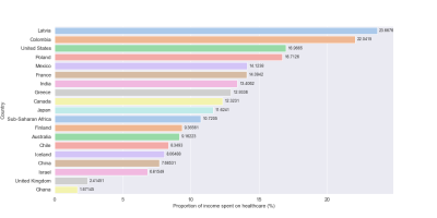 |
134 | Bringing MRI to Low- and Middle- Income Countries: Directions, Challenges and Potential Solutions
Sola Adeleke1, Sanjana Murali2, Hao Ding2, Fope Adedeji3, Cathy Qin4, Johnes Obungoloch5, Tamir Sirkis6, Iris Asllani7, Ntobeko Ntusi8, Regina Mammen9, Udunna Anazodo10, and Thoralf Niendorf11
1School of Biomedical engineering and imaging sciences, King's College London, London, United Kingdom, 2School of Medicine, Imperial College London, London, United Kingdom, 3School of Medicine, University College London, London, United Kingdom, 4Department of Radiology, Imperial College Healthcare Trust, London, United Kingdom, 5Department of Biomedical Engineering, Mbara University of Science and Technology, Mbara, Uganda, 6Royal Berkshire NHS Foundation Trust, Reading, United Kingdom, 7Department of Biomedical Engineering, Rochester Institute of Technology, New York, NY, United States, 8Department of Medicine, University of Cape Town and Groote Schuur Hospital, Cape Town, South Africa, 9Department of Cardiology, Essex Cardiothoracic Centre, Chelmsford, United Kingdom, 10Montreal Neurological Institute, McGill University, Montreal, QC, Canada, 11Berlin Ultrahigh Field Facility, Berlin, Germany Keywords: Low-Field MRI, MR Value, Accessible MRI; Affordable MRI; Sustainable MRI While innovations in MRI technology continue to advance healthcare in the global north, there is a persistent disparity in MRI access and research opportunities in low- and middle-income countries (LMICs). Reasons for this longstanding disparity include technological, economic, geopolitical, and social factors. As awareness of the situation expands, concerted efforts are underway to address it by developing sustainable approaches designed with and for local communities. Here, we provide a framework of such approaches by tackling different aspects of MRI development and access in LMICs. |
|
 |
1599.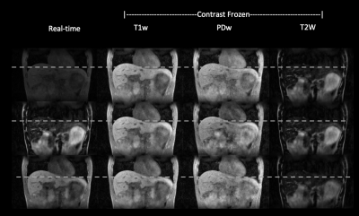 |
135 | Low-field MR Multitasking for an integrated abdominal MRgRT imaging framework
Junzhou Chen1,2, Ye Tian3, Namgyun Lee3, Mingsong Cao4, Wensha Yang5, Sophia Cui6, Krishna Nayak3, Debiao Li2,7, Anthony Christodoulou2,7, and Zhaoyang Fan1,5
1Radiology, University of Southern California, Los Angeles, CA, United States, 2Bioengineering, University of California, Los Angeles, Los Angeles, CA, United States, 3Electrical Engineering, University of Southern California, Los Angeles, CA, United States, 4University of California, Los Angeles, Los Angeles, CA, United States, 5Radiation Oncology, University of Southern California, Los Angeles, CA, United States, 6Siemens Medical Solutions, Los Angeles, CA, United States, 7Cedars-Sinai Medical Center, Los Angeles, CA, United States Keywords: Low-Field MRI, Radiotherapy MR applications in radiation therapy has shown great promise for highly conformal treatment. However, MRgRT in the abdomen remains challenging due to motion and the sheer number of organs. In this work, we have demonstrated at 0.55T an integrated multi-task MR platform for adaptive radiation planning and treatment monitoring in the abdomen. We propose using a hybrid GRE-TSE sequence, from a single "pre-beam" scan we are able to generate a volumetric, multi-contrast and motion resolved images for adaptive treatment planning as well as enabling multi-contrast 3D real-time display during treatment. |
1600.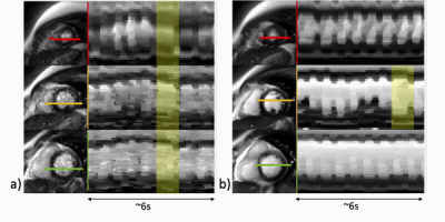 |
136 | Simultaneous multi-slice real-time cardiac MRI at 0.55T
Ecrin Yagiz1, Parveen Garg2, Krishna S. Nayak1, and Ye Tian1
1Ming Hsieh Department of Electrical and Computer Engineering, University of Southern California, Los Angeles, CA, United States, 2Division of Cardiology, Department of Medicine, Keck School of Medicine, University of Southern California, Los Angeles, CA, United States Keywords: Low-Field MRI, Low-Field MRI A standard cine MRI exam typically collects a stack of short-axis slices to cover all left ventricular myocardium and uses electrocardiogram gating and breath-holds. Real-time imaging methods are often used to resolve issues with insufficient gating signal or breath-hold failure. In this work, we demonstrate that real-time SMS cardiac imaging at 0.55T provides sufficient blood-myocardium contrast and regional wall motion evaluation with three-fold acceleration compared to real-time single-band and Cartesian breath-hold ECG-gated cine. We also show an alternative reconstruction approach, clustered locally low rank that can improve image quality. |
|
1601.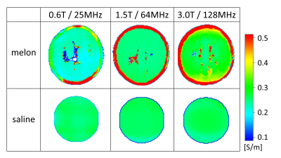 |
137 | On the frequency dependence of electrical conductivity
Ulrich Katscher1 and Peter Vernickel1
1Philips Research Europe, Hamburg, Germany Keywords: Low-Field MRI, Electromagnetic Tissue Properties Electric conductivity depends on the frequency of the probing electric field. The impact of the frequency differs between human tissue types, e.g., it is lower for body fluids than for cellular tissue types. The upcoming mid-field systems suggest to investigate this frequency dependence with MRI. This study investigated two phantoms, with and without cellular structure, to explore the frequency dependence of conductivity using “Electrical Properties Tomography”. The conductivity results obtained at three different Larmor frequencies were used to estimate the conductivity at lower frequencies in the range of kHz as required for, e.g., EEG source localization. |
|
1602.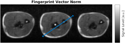 |
138 | Optimized MR Fingerprinting OPTIMUM in vivo in the vicinity of a metallic implant at low field
Gabriel Zihlmann1,2, Mathieu Sarracanie1,2, and Najat Salameh1,2
1Center for Adaptable MRI Technology (AMT center), Department of Biomedical Engineering, University of Basel, Allschwil, Switzerland, 2AMT center, Institute of Medical Sciences, school of Medicine, Medical Sciences & Nutrition, University of Aberdeen, Aberdeen, United Kingdom Keywords: Low-Field MRI, MR Fingerprinting Magnetic susceptibility changes at interfaces induce artefacts, and hence compromises MRI diagnostic value. Low-Field (LF) MRI is inherently less sensitive to susceptibility artifacts making it suitable for imaging near metallic implants. In addition, the dispersion in T1-contrast is enhanced at LF. To evaluate the potential for new contrast mechanisms, quantitative imaging would be key if acquisition times are compatible with clinical constraints. In that context, model-based multi-parametric OPTIMUM was performed in a healthy volunteer’s forearm carrying a titanium plate from plate-osteosynthesis surgery. Performance of two signal models, one oblivious to intravoxel dephasing and the other accounting for it was compared. |
|
1603.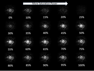 |
139 | Outer Volume Saturation using Adiabatic Inversion Pulses in an Inhomogeneous Transmit Field
Muller De Matos Gomes1, Sourajit Mustafi1, Meredith Sadinski1, Kartiga Selvaganesan2, and Aleksandar Nacev1
1Promaxo, Oakland, CA, United States, 2Yale School of Engineering & Applied Science, Yale, New Haven, CT, United States Keywords: RF Pulse Design & Fields, Low-Field MRI A method for saturating the outer volume in a single sided low field MRI scanner has been developed. By saturating the outer volume using this method, magnetization from regions where the gradient fields are highly nonlinear may be removed from the image, resulting in less image artifacts. |
|
 Back to Meeting Home
 Back to the Program-at-a-Glance
The International Society for Magnetic Resonance in Medicine is accredited by the Accreditation Council for Continuing Medical Education to provide continuing medical education for physicians.
 Back to Meeting Home
 Back to the Program-at-a-Glance
 View Presentation Video