Digital Poster
Phantoms & Repeatability II
ISMRM & ISMRT Annual Meeting & Exhibition • 03-08 June 2023 • Toronto, ON, Canada

| Computer # | |||
|---|---|---|---|
5219.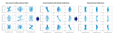 |
121 |
Gradient Non-Linearity Calibration for Improved Spatial Position
Accuracy with a High-Performance Gradient Using a Fiducial
Phantom
Nastaren Abad1,
Eric Fiveland1,
Seung-Kyun Lee1,
Yihe Hua1,
Shengzhen Tao2,
Joshua Trazasko3,
Matt A. Bernstein3,
and Thomas K.F Foo1
1GE Research, Niskayuna, NY, United States, 2Mayo Clinic, Jacksonville, FL, United States, 3Mayo Clinic, Rochester, MN, United States Keywords: System Imperfections: Measurement & Correction, System Imperfections: Measurement & Correction, Gradient Non-Linearity; Spatial Fidelity; High Performance Gradients Spatial encoding in MR-systems is subject to gradient non-linearity. If ignored or inadequately calibrated during system install, non-linear gradient fields manifest as spatial distortions impacting image quality, diminish accuracy for applications requiring MR guided intervention and introduce systematic errors in quantitative imaging such as diffusion MRI. High-performance, high-efficiency gradient systems, such as MAGNUS, require accurate calibration for imaging precision. In this study, a fiducial phantom was utilized to characterize and correct for residual distortions after standard gradient calibrations. Results highlight that distortion due to gradient non-linearity can be successfully reduced by phantom-based calibration for improved accuracy inline with QC metrics. |
|
5220.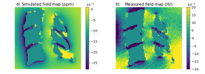 |
122 |
Development of a realistic human spine phantom to mimic static
B0 field inhomogeneity in the cervical spinal cord
Brunnhilde M, A-S Ponsi1,
Laura Beghini1,
Sebastian A.D Sandbu1,
Annelen Dogger Schmidt1,
and S. Johanna Vannesjo1
1Department of Physics, Norwegian University of Science and Technology, Trondheim, Norway Keywords: Phantoms, Spinal Cord, Susceptibility, Field inhomogeneity, High field MRI, Shims, Artifacts, Field simulation Anthropomorphic phantoms could facilitate testing of novel MRI methods. This is especially important for spinal cord imaging at 7T, which is severely affected by local B0 field inhomogeneity due to susceptibility differences between tissues, particularly between bone and soft tissues. Here we built a 3D-printed model of vertebrae C3-C5, which was included in a spherical phantom. Measured field maps of the phantom were compared with simulations, showing similar features in the field distortion. The phantom also exhibited a spatially periodic pattern of signal drop-out around the intervertebral junctions in multi-echo GRE, similar to what is commonly observed in in-vivo data. |
|
5221.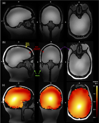 |
123 |
Realistic human head phantoms for 7T sequence development,
parallel transmission & spectroscopy methodology
Sascha Brunheim1,
Yannik Völzke1,
Astrid Dubbins1,
Eberhard D. Pracht1,
and Tony Stöcker1,2
1German Center for Neurodegenerative Diseases (DZNE), Bonn, Germany, 2Physics and Astronomy, University of Bonn, Bonn, Germany Keywords: Phantoms, RF Pulse Design & Fields, Spectroscopy An optimized human head phantom design for 7 Tesla field strength and its standardized fabrication process is presented. Two different phantom versions were produced: The first gel-only version can be set up quickly and provided realistic T1 and T2 values for its brain, muscle, and lipid compartments. The second version, with its liquid brain compartment containing several metabolites, can be utilized for spectroscopy sequence development. With the 3D model and the validated recipes mimicking head and brain tissue, extensions to other applications can be more easily achieved, such as system characterization, diffusion weighted imaging, or realistic flow phantoms. |
|
5222.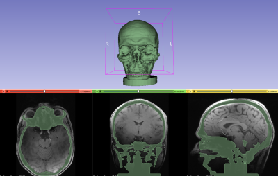 |
124 |
Design and Production of a 3D Printed Anthropomorphic Head
Phantom for MRI – The Story so Far
George Michael John Bruce1,2,
Pauline Hall-Barrientos1,2,
and David Brennan1,2
1MRI Physics, Department of Clinical Physics and Bioengineering, NHS Greater Glasgow and Clyde, Glasgow, United Kingdom, 2School of Medical, Veterinary and Life Sciences, University of Glasgow, Glasgow, United Kingdom Keywords: Phantoms, Phantoms, 3D Printing Commercial phantom objects for use in MR can be expensive and poorly representative of human anatomy. 3D printing provides the potential to produce cheaper, novel and reproducible phantoms . This project investigates the potential use of 3D printed materials in the construction of an anthropomorphic head phantom for MRI. It can be effectively split into two different, smaller projects: investigation into MR properties of 3D printed materials and design of an anthropomorphic head phantom for MRI. |
|
5223.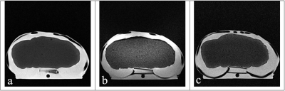 |
125 |
Design and Validation of a Multi-Modality Lung Phantom
Donald L Bowen1,
Tyler J Touchet1,
Duncan J Maitland1,
and Mary P McDougall2
1Biomedical Engineering, Texas A&M University, College Station, TX, United States, 2Biomedical Engineering and Electrical Engineering, Texas A&M University, College Station, TX, United States Keywords: Phantoms, Multimodal Phantoms often mimic only one specific property of the body, and without relevant anatomical structure. We report the design and construction of an anatomic phantom that accurately mimics the in vivo properties of the lungs for both ultrasound and magnetic resonance imaging. The phantom demonstrated qualitative likeness to the lungs under ultrasound and quantitatively similar tissue relaxation characteristics using magnetic resonance imaging. Future work will determine if the phantom can be used to validate early-stage medical devices that operate within the lungs. |
|
5224. |
126 |
A Pre-Prepared, Low-Cost, Mass-Produced Phantom Regulated by the
Food Standards Agency for Assessing Imaging Characteristics
within MRI
Aaron Axford1,
Jordan McGing1,
Catarina Rua2,
Mark Symms3,
Damian J Tyler1,4,
and James T Grist1,4,5,6,7
1Oxford Centre for Clinical Magnetic Resonance Research, University of Oxford, Oxford, United Kingdom, 2Invicro LLC, London, United Kingdom, 3GE Healthcare, London, United Kingdom, 4Department of Physiology, Anatomy, and Genetics, University of Oxford, Oxford, United Kingdom, 5Department of Radiology, Oxford University Hospitals, Oxford, United Kingdom, 6Institute of Cancer and Genomic Sciences, University of Birmingham, Birmingham, United Kingdom, 7University of Bologna, Bologna, Italy Keywords: Phantoms, Relaxometry A potential low-cost quantitative MR image assessment phantom was investigated by calculating relaxation times and the magnetization transfer ratio over multiple scans. An imaging protocol comprised of an inversion recovery sequence with varying inversion times, a spin echo with varying echo times, and an Enhanced Fast Gradient Echo 3D sequence with and without a magnetization transfer pulse, was developed and used to acquire images of the phantom, a block of store-bought jelly. The imaging data was used to calculate a T1 time of 109±1ms, a T2 time of 12.8±0.2ms, and an MTR value of 17.4±0.2%. |
|
5225.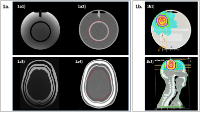 |
127 |
Do anthropomorphic phantoms enhance compliance with the
professional bodies' quality assurance guidelines for MRI in
radiotherapy
Meshal Alzahrani1,2,
David Broadbent3,
Irvin Teh1,
Bashar Al-Qaisieh3,
Adrian Walker4,
Rachel Lamb4,
and Richard Speight3
1Leeds Institute of Cardiovascular and Metabolic Medicine, University of Leeds, Leeds, United Kingdom, 2Department of Diagnostic Radiology, Faculty of Applied Medical Sciences, King Abdulaziz University, Jeddah, Saudi Arabia, 3Department of Medical Physics and Engineering, Leeds Teaching Hospitals NHS Trust, Leeds, United Kingdom, 4Leeds Test Objects, Boroughbridge, United Kingdom Keywords: Phantoms, Radiotherapy With the increasing use of magnetic resonance imaging (MRI) in radiotherapy (RT), guidelines for the use of MRI in RT have recently been published, including recommendations for quality assurance (QA) tests. This project investigates whether anthropomorphic phantoms can be more beneficial to comply with these guidelines compared to other phantoms. The performance of the anthropomorphic phantom has been compared to other phantoms. The anthropomorphic phantom has been shown to be useful in QA for MRI-CT registration and end-to-end test that cannot be performed by other phantoms. |
|
5226.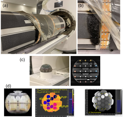 |
128 |
Assessing Scanner Changes after Major Repairs using Traceable
Phantoms from the NIST/NIBIB Lending Library
Stephen E Russek1,
Kathryn E Keenan1,
Karl F Stupic1,
Cassandra M Stoffer1,
Teryn S Wilkes2,
and Lena Sherbakov2
1NIST, Boulder, CO, United States, 2Intermountain Neuroimaging Consortium, University of Colorado, Boulder, CO, United States Keywords: System Imperfections: Measurement & Correction, Quantitative Imaging We demonstrate the utility of periodic use of standard phantoms to assess changes in MRI scanner performance after unexpected major repairs. We present characterization of a 3T scanner before and after an unexpected gradient failure and replacement using a traceable system phantom and an isotropic diffusion phantom from the NIST/NIBIB Medical Phantom Lending Library. No major changes were observed in gradient calibration, geometric distortion, imaging uniformity, and resolution. Post-repair improvements were noted in SNR, relaxation time, water diffusivity measurements, although both pre and post-repair data were acceptable. |
|
5227.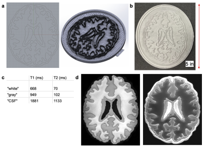 |
129 |
UV-Curable Hydrogels for Quantitative T1/T2 in Slice Phantoms
Szu Ting Tung1,
Jolene Huey1,
Michael Lustig 1,
Ana Claudia Arias1,
and Anita Flynn1
1Department of Electrical Engineering and Computer Science, University of California, Berkeley, Berkeley, CA, United States Keywords: Phantoms, Multi-Contrast We developed a high-volume, low error UV-curable hydrogel formation process that can target specific T1 and T2 parameters. Our process relies on the polymerization of water-soluble photoinitiator TPO-Lithium in acrylamide (monomer), polyethylene-glycol-diacrylate (crosslinker), de-ionized (DI) water, and paramagnetic ions. We characterize the linearizing effect of varying paramagnetic ion concentrations in determining T1 and T2 values, and present an application to construct a quantitative anatomy-mimicking brain slice phantom containing UV gels. |
|
5228.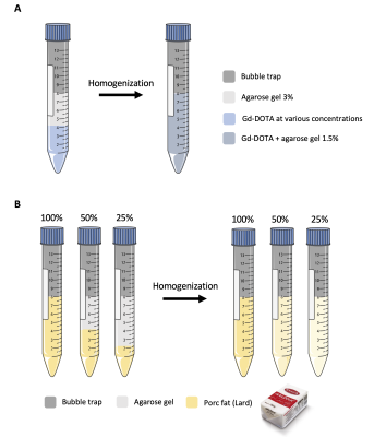 |
130 |
Development of an easy-manufacturable and cheap phantom for
quality control of T1, T2 and fat fraction mapping.
Laurence Dallet1,
Sylvain Miraux1,
François Maingault1,
and Emeline J Ribot1
1Centre de Résonance Magnétiques des Systèmes biologiques UMR5536, CNRS/Université de Bordeaux, Bordeaux, France Keywords: Phantoms, Quantitative Imaging, quality control A quality control phantom was developed through a simple and easy protocol for T1, T2 and Fat fraction mapping. The phantom is constituted of 8 tubes containing Gd-DOTA or commercially available animal fat, each at various concentrations and stabilized in agarose gels. A 3D printed bubble trap was placed on top of the gels to limit the contrast agent dilution over time. The solutions are stable for at least 6 months and the manufacturing protocol is highly reproducible. |
|
5229.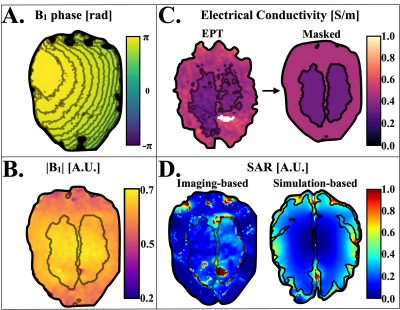 |
131 |
Imaging-Based Subject-Specific SAR Maps using B1-mapping and
Electrical Properties Tomography in a heterogenous Brain Phantom
Jessica A. Martinez1,
Adriano Troia1,
Alessandro Arduino1,
Kevin Moulin2,3,
Umberto Zanovello1,
Oriano Bottauscio1,
and Luca Zilberti1
1Advanced Materials Metrology and Life Science, Istituto Nazionale di Ricerca Metrologica, Torino, Italy, 2CREATIS Laboratory, University of Lyon, Lyon, France, 3Department of Radiology, University Hospital Saint-Etienne, Saint-Etienne, France Keywords: Safety, Safety, Specific Absorption Rate, SAR In this work a heterogeneous realistic brain phantom, with electric and magnetic properties in the range of healthy gray-matter and white-matter, was constructed. The phantom was scanned at 3 T where T1 maps, T2 maps, magnitude, and phase information of the B1 field were acquired. The electrical conductivity was derived using a convection reaction EPT approach and the E field was obtained by solving Ampere’s law from the B1 field. From this information, imaging-based subject-specific SAR maps were acquired and compared to simulation-based SAR maps. The analysis time (acquisition and SAR calculation) was under 6 minutes. |
|
| 5230. | WITHDRAWN | ||
5231.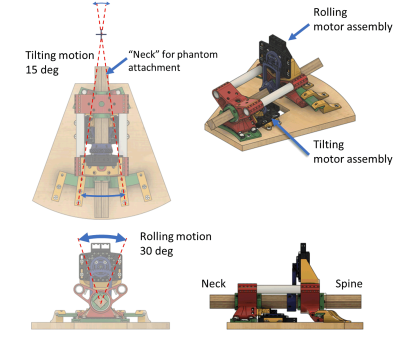 |
132 |
Computer-controlled non-metallic phantom platform to simulate
head motion with two degrees of freedom
Jacob Horne1,
Reggie Taylor2,
and Tong Xu3
1Department of Mechanical & Aerospace Engineering, Carleton University, Ottawa, ON, Canada, 2Institute of Mental Health Research, The Royal Ottawa Mental Health Centre, Ottawa, ON, Canada, 3Department of Physics, Carleton University, Ottawa, ON, Canada Keywords: Phantoms, Motion Correction, Dynamic phantom A computer-controlled non-metallic head phantom platform was designed and constructed from 3D-printed parts. Driven by pneumatic stepper motors, the platform can simulate head motion with two degrees of freedom: tilting and rolling, with angular step resolution of 0.0833 degree and 0.227 degree, respectivily. A "neck/spine" design allows different static head phantoms to be mounted to the platform while still be able to fit inside standard head coils. The platform was tested on a PET/MRI scanner and successfully produced motion artifact with simulated motion. |
|
5232.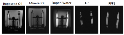 |
133 |
What should I use to fill my Ultra-high Field MRI phantom?
Felix Horger1,2,3,
Jyoti Mangal1,2,3,
Raphael Tomi-Tricot1,2,3,4,
David Carmichael1,2,3,
Joseph Hajnal1,2,3,
and Shaihan Malik1,2,3
1Biomedical Engineering Department, School of Biomedical Engineering & Imaging Sciences, King's College London, London, United Kingdom, 2Centre for the Developing Brain, School of Biomedical Engineering & Imaging Sciences, King's College London, London, United Kingdom, 3London Collaborative Ultra high field System (LoCUS), London, United Kingdom, 4MR Research Collaborations, Siemens Healthcare Limited, Frimley, United Kingdom Keywords: High-Field MRI, Phantoms We explored commonly used substances for phantoms, air, water, oil and Perfluoropolyether, for their suitability for imaging at ultra-high field (7T). We illustrate the impact of susceptibility matching and how to prevent transmit field doming by selecting a substance with low dielectric constant. This work provides insights into the variety and expected magnitude of effects and can serve as guidance when designing a phantom for a specific application. |
|
5233.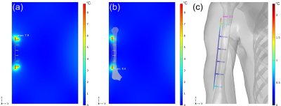 |
134 |
Is the ASTM F2182 Gel Phantom Adequate for Determining
Worst-Case RF-Induced Heating of Implanted Medical Devices?
Grant Monroe Baker1,
Eric David Anttila1,
Alan Ross Leewood1,
and David Carl Gross1
1MED Institute, West Lafayette, IN, United States Keywords: Safety, Safety, Heating, RF-Induced Heating, MRI Safety Evaluations, Simulation ASTM F2182 was developed to evaluate RF-induced heating near passive implanted medical devices by subjecting an implant within an average tissue-mimicking (i.e., electrical and thermal properties) phantom to controlled RF exposure. The purpose of this study was to determine if the ASTM F2182 phantom is adequate for determining worst-case RF-induced heating of implanted medical devices. Results from this study display that device temperature rises in the ASTM F2182 are significantly higher than in-vivo temperature rises, regardless of tissue-specific properties and local blood perfusion incorporation to the phantom, indicating that the ASTM gel phantom is adequate for RF-induced heating evaluations. |
|
5234.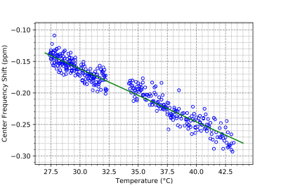 |
135 |
Hydroxyethylcellulose as a tissue mimicking phantom gel:
Characterizing the temperature response at 0.5T
Diego F Martinez1,
Curtis N. Wiens2,
Chad T. Harris2,
Will B. Handler1,
and Blaine A. Chronik1,3
1The xMR Labs, Physics and Astronomy, Western University, London, ON, Canada, 2Research and Development, Synaptive Medical, Toronto, ON, Canada, 3Medical BioPhysics, Western University, London, ON, Canada Keywords: Phantoms, Thermometry Temperature Mapping at 0.5T offers an accessible method for non-invasively tracking thermal procedures, relying on tissue temperature response of properties such as Proton Resonant Frequency, Apparent Diffusion Coefficient, or T1. To evaluate performance of temperature mapping using these properties, Hydroxyethyl Cellulose – a tissue mimicking gel – was formulated using the ISO 10974:2018 standard and doped with CuSO4 to match representative tissue T1 values. Temperature response of each of these parameters was: PRF parameter α= (-8.74 ± 0.12) x 10-3 ppm/°C, T1 parameter (12.02 ± 0.07) ms/°C, and ADC parameter (5.34 ± 0.063) x 10-5 mm2/s/°C, all in acceptable range. |
|
5235.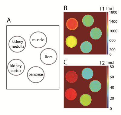 |
136 |
Relaxometry phantoms: Are there alternatives to paramagnetic
additives?
Victor Fritz1 and
Fritz Schick1
1Section on Experimental Radiology, Department of Diagnostic and Interventional Radiology, University of Tübingen, Tübingen, Germany Keywords: Phantoms, Relaxometry Phantoms used for relaxometry usually consist of a gelling agent doped with paramagnetic salt. However, since the use of paramagnetic additives can be disadvantageous for certain purposes, there is a great interest in finding other suitable substances. In this work, soy-lecithin-agar gels are presented and evaluated as an alternative phantom material for the construction of relaxometry phantoms with tissue-like relaxation times. They were found to work quite well. Soy-lecithin-agar gels are easy to prepare and allow independent adjustment of T1 and T2. Relaxation times of different tissues (muscle, liver, pancreas, kidney) could be successfully mimicked. |
|
5236.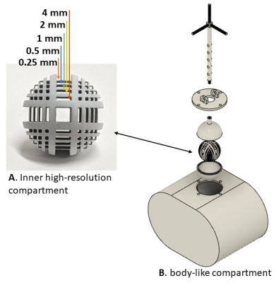 |
137 |
An open-source platform for simulating internal organ motion:
Preliminary results
Eddy Solomon1 and
Leeor Alon2
1Radiology, Weill Cornell Medical College, New York, NY, United States, 2Center for Advanced Imaging Innovation and Research, New York University School of Medicine, New York, NY, United States Keywords: Phantoms, Motion Correction Motion-correction techniques require an objective quantitative assessment under controlled conditions. In this work, we present an open-source platform for simulating internal ‘organ’ motion under realistic scan conditions. The phantom was composed of two partitions which include a high-resolution inner compartment translated in space using a Lorentz force, within a larger body-like second compartment. The motion phantom was controlled using custom-made software and was tested with a radial MR sequence, demonstrating motion-resolved capabilities. |
|
5237.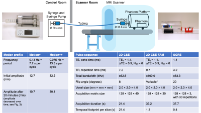 |
138 |
Development of an MR-Compatible Motion Phantom to Evaluate
Motion-Robust Quantitative MRI
Jiayi Tang1,2,
James Rice2,3,
Jack Gwertzman3,
Scott B. Reeder1,2,4,5,6,
Alejandro Roldán-Alzate2,3,
and Diego Hernando1,2
1Medical Physics, University of Wisconsin-Madison, Madison, WI, United States, 2Radiology, University of Wisconsin-Madison, Madison, WI, United States, 3Mechanical Engineering, University of Wisconsin-Madison, Madison, WI, United States, 4Biomedical Engineering, University of Wisconsin-Madison, Madison, WI, United States, 5Medicine, University of Wisconsin-Madison, Madison, WI, United States, 6Emergency Medicine, University of Wisconsin-Madison, Madison, WI, United States Keywords: Phantoms, Motion Correction Chemical-shift encoded (CSE) MRI can quantify PDFF and R2* as biomarkers for liver fat and iron deposition, respectively, but conventional methods (3D-CSE) are not motion-robust. Recent methods (2D-CSE-FAM) have been proposed which demonstrate motion-robustness; the evaluation of these methods urges the development of a MR-compatible motion phantom. In this work, we constructed such a phantom, validated it via video tracking, and used it to show motion artifacts and quantification biases in 3D-CSE, identify a potential source of bias in axial R2* 2D-CSE-FAM acquisitions, and confirm the motion-robustness of coronal 2D-CSE-FAM. |
|
5238.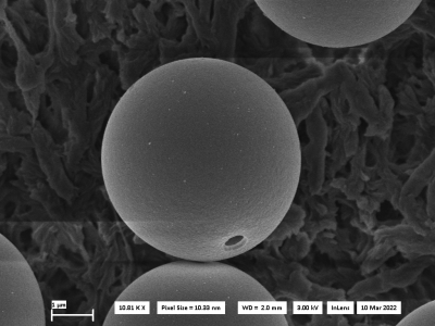 |
139 |
Colloidal Cell Mimics as Reference System for Diffusion MRI
Experiments
Henrik zu Jeddeloh1,2,
Dominik Ludwig1,
Julian Rauch1,2,
Frederik Laun3,
Mark Ladd1,2,4,
Karel Klika5,
and Tristan Anselm Kuder1,2
1Division of Medical Physics in Radiology, German Cancer Research Center (DKFZ), Heidelberg, Germany, 2Faculty of Physics and Astronomy, Heidelberg University, Heidelberg, Germany, 3Institute of Radiology, University Hospital Erlangen, Friedrich-Alexander-Universität Erlangen-Nürnberg (FAU), Erlangen, Germany, 4Faculty of Medicine, Heidelberg University, Heidelberg, Germany, 5Molecular Structure Analysis, German Cancer Research Center (DKFZ), Heidelberg, Germany Keywords: Phantoms, Diffusion/other diffusion imaging techniques, Cell Mimics, Polymerized Spheres In order to verify modern diffusion MRI sequences, a well-defined reference system consisting of polymerized, hollow microspheres with a hole in the surface is synthesized. This reference system can model the diffusion properties of cells with varying size1,3 and membrane permeability2. To produce these cell mimics3, tripropyleneglycol-monomethylether monomer was mixed with an ammonia solution with polystyrene particles to initiate a nucleation process. The droplets were inflated in an NaOH solution and then hardened under UV exposure. Lastly, the polystyrene particles were dissolved. Scanning Electron Microscope images and DWI measurements were acquired to verify the quality of the cell mimics. |
|
 Back to Meeting Home
 Back to the Program-at-a-Glance
The International Society for Magnetic Resonance in Medicine is accredited by the Accreditation Council for Continuing Medical Education to provide continuing medical education for physicians.
 Back to Meeting Home
 Back to the Program-at-a-Glance
 View Presentation Video