Digital Poster
Low-Field & Point-of-Care/Portable MRI II
ISMRM & ISMRT Annual Meeting & Exhibition • 03-08 June 2023 • Toronto, ON, Canada

Digital Poster
Low-Field & Point-of-Care/Portable MRI II
| Computer # | |||
|---|---|---|---|
1760.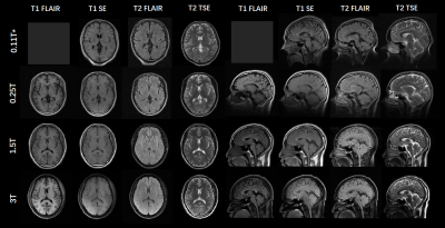 |
121 | A SNR and contrast comparison study at 0.11T, 0.25T, 1.5T and 3T MRI systems
Yueqi Qiu1, Haoran Bai1, Hao Chen1, and Zhiyong Zhang1
1School of Biomedical Engineering, Shanghai Jiao Tong University, Shanghai Jiao Tong University, Shanghai, China Keywords: Low-Field MRI, Low-Field MRI Low-field MRI systems have seen a renaissance recently due to improvements in technology and its low costs. However, properties like SNR, T1 and T2 values of low fields have caused a lot of concern because they always change with field strengths. This study conducted the SNR comparison based on phantom and fitted the quantitative mapping of human brains at 0.11T, 0.25T, 1.5T and 3T to design a set of protocols enabling similar contrasts of the typical clinical images at different fields. The impacts of lowered magnetic field strengths on imaging quality were shown in this study. |
|
1761.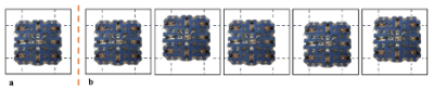 |
122 | Stop-Motion RF for Improving Spatial Resolution in Non-Linear Gradient-Free Low-Field MRI
Anja Samardzija1, Kartiga Selvaganesan1, Younghyun Ha2, Zhehong Zhang1, Chenhao Sun2, Heng Sun1, Gigi Galiana2, and Todd Constable2
1Biomedical Engineering, Yale University, New Haven, CT, United States, 2Department of Radiology and Biomedical Imaging, Yale School of Medicine, New Haven, CT, United States Keywords: Low-Field MRI, RF Arrays & Systems We developed a stop-motion imaging technique in which a 3x3 radiofrequency coil that transmits Bloch-Siegert encodings is placed in a different position within the field-of-view for each MR signal acquisition. This study is performed using a non-linear gradient-free low-field MRI system. High spatial resolution is achieved through stop-motion imaging. We show that stop-motion imaging done with a total of 200 encodings (20 imaging positions and 10 encodings per position) outperformed stationary imaging performed with 200 encodings (1 imaging position and 200 encodings per position). |
|
1762.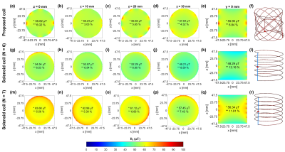 |
123 | Widened-bandwidth RF volume coil using crossed elliptical winding for Halbach-array-based portable MRI knee scanner
MEENA RAJENDRAN1 and SHAOYING HUANG1
1Engineering Product Developement, Singapore University of Technology and Design, Singapore, Singapore Keywords: Low-Field MRI, Low-Field MRI, RF coil A widened-bandwidth RF volume coil using crossed elliptical winding pattern (symmetric in azimuthal direction) is proposed for Halbach-array-based portable MRI knee scanner. The fractional bandwidth (FBW) is 0.51% centered at 2.84 MHz (B0 = 67mT) with a high B1 field strength (57µT) and a high homogeneity (94%) within a cylindrical volume of diameter and length of both 95mm. Compared to solenoid coils of comparable dimensions, it is an increase of 18% in FBW, an improvement in homogeneity by 6.1%, and only a compromise of 1.9% in the field strength, which is the best combination. |
|
1763.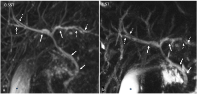 |
124 | Abdominal MRI at 0.55 T: Initial evaluation and optimization in healthy subjects
Anupama Ramachandran1, Hero Hussain1, Mishal Mendiratta Lala2, Jacob Richardson3, Nancy Dudek2, Joel Morehouse2, Katherine Wright4, Vikas Gulani3, and Nicole Seiberlich3
1Radiology, University of Michigan, Ann Arbor, MI, United States, 2University of michigan, Ann arbor, MI, United States, 3Radiology, University of michigan, Ann arbor, MI, United States, 4University of Michigan, Ann arbor, MI, United States Keywords: Low-Field MRI, Body, Abdomen Abdominal MRI including MRCP was performed in 15 healthy subjects on both a 0.55T and a 1.5T MR system. Image quality (IQ) was rated by two radiologists (22 and 17 yrs experience). All sequences were rated acceptable at 0.55T. In comparison to 1.5T, IQ scores at 0.55T were higher for DWI and 3D MRCP, and lower for the other sequences. Acquisition times were longer at 0.55T for all sequences except 3D MRCP. Thus, acceptable quality abdominal images can be obtained on a commercial 0.55T system with slightly lower IQ ratings of some of the sequences compared to 1.5T. |
|
1764.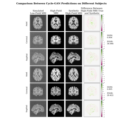 |
125 | Denoising Simulated Low-Field MRI (70mT) using Denoising AutoEncoders (DAE) and Cycle-Consistent Generative Adversarial Network (Cycle-GAN)
Fernando Vega1,2,3, Abdoljalil Addeh1,2,3, and M. Ethan MacDonald1,2,3,4
1Biomedical Engineering, University of Calgary, Calgary, AB, Canada, 2Electrical & Software Engineering, University of Calgary, Calgary, AB, Canada, 3Hotchkiss Brain Institute, University of Calgary, Calgary, AB, Canada, 4Department of Radiology, University of Calgary, Calgary, AB, Canada Keywords: Low-Field MRI, Machine Learning/Artificial Intelligence, MRI, Unpaired Image Translation In this work, a denoising Cycle-GAN is implemented to yield high-field, high resolution, high signal-to-noise ratio MRI images from simulated low-field, low resolution, low signal-to-noise MRI images. Resampling and additive Rician noise were used to simulate low-field MRI. Images were utilized to train a DAE and Cycle-GAN, with paired and unpaired cases, respectively. Both networks were evaluated using SSIM and PSNR image quality metrics. This work demonstrates the use of advanced machine learning to improve low-field MRI images that can outperform classical denoising autoencoders and does not require image pairs. |
|
1765. |
126 | Muscle volumetry from low-field scans and its relation to performance in physical activities
José Miguel Algarín1,2, Teresa Guallart-Naval2,3, Ana Ferri-Caruana4, Rubén Bosch3, Francisco Juan-Lloris5, Eduardo Pallás1,2, Juan Pablo Rigla3, Pablo Martínez5, José Borreguero3, José María Benlloch1,2, Fernando Galve1,2, and Joseba Alonso1,2
1Institute for Instrumentation in Molecular Imaging (i3M), CSIC, Valencia, Spain, 2Institute for Instrumentation in Molecular Imaging (i3M), Universitat Politècnica de València, Valencia, Spain, 3Tesoro Imaging SL, Valencia, Spain, 4Educación Física y Deportiva, Universitàt de València, Valencia, Spain, 5Physio MRI SL, Valencia, Spain Keywords: Low-Field MRI, Low-Field MRI, Sport MRI Here we show the results of the first systematic study performed in our low-field 72 mT MRI scanner. Calf images were acquired from 35 volunteers whose performance was measured during different physical activities before the scan. We segmented the images to determine the cross-sectional area and volume of the gastrocnemius muscles and correlated the results with the muscular activity measurements of the volunteers. We found that the medial gastrocnemius muscle volume correlates significantly with participants weight and unilateral jump capacity. |
|
1766. |
127 | Feasibility of integrating a wearable accelerometer in very low-field MRI to detect motion
Keerthi Sravan Ravi1,2, Kunal Aggarwal3, John Thomas Vaughan Jr.2, Yun Soung Kim4, and Sairam Geethanath5
1Biomedical Engineering, Columbia University, New York, NY, United States, 2Columbia Magnetic Resonance Research Center, Columbia University, New York, NY, United States, 3Accessible MR Lab, BMEII, Diagnostic, molecular and interventional radiology, Icahn School of Medicine at Mt. Sinai, New York City, NY, United States, 4Biomedical Engineering and Imaging Institute, Dept. of Diagnostic, Molecular and Interventional Radiology,, Mt.Sinai, New York, NY, United States, 5Accessible MR Laboratory, Biomedical Engineering and Imaging Institute, Dept. of Diagnostic, Molecular and Interventional Radiology,, Mt. Sinai, New York, NY, United States Keywords: Low-Field MRI, Low-Field MRI These artifacts degrade image quality, often causing misdiagnosis. A 6-axis motion tracking sensor (ICM-20649, TDK-InvenSense) with a full-scale range of +-4000 degrees per second for the gyroscope and +-30g for the accelerometer was integrated with a 50mT scanner. The sensor’s readings can successfully be processed to detect motion. However, it resulted in zipper artifacts and degraded image quality in a phantom experiment. Still, the sensor’s placement on the forehead or the temples or the chin might not significantly impact brain data when coupled with slab-wise shimming. |
|
1767.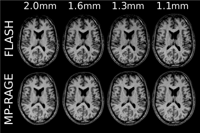 |
128 | Optimization of the magnetization-prepared rapid gradient echo (MP-RAGE) sequence for 0.55 T
Jessica Schäper1,2 and Oliver Bieri1,2
1Department of Biomedical Engineering, University of Basel, Basel, Switzerland, 2Division of Radiological Physics, Department of Radiology, University Hospital Basel, Basel, Switzerland Keywords: Low-Field MRI, Brain, MP-RAGE, low-field, white matter, gray matter The MP-RAGE is the most commonly used sequence for structural brain imaging in clinical routine. Consequently, the optimization of imaging parameters for maximum contrast and signal is an important task. In this work, we implemented a previously developed computation and calculated optimal parameters for 0.55T. Additionally, the obtained images were compared with a gray-white matter contrast-optimized FLASH. Computing the contrast-to-noise ratio revealed a clear benefit for MP-RAGE over FLASH at mid to low resolutions, while a trend towards similar or even higher contrast-to-noise ratios for FLASH as compared to MP-RAGE at high resolutions was observed. |
|
1768.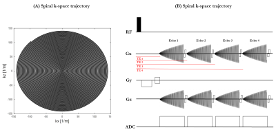 |
129 | An initial exploration of spiral imaging for quantitative fetal T2* mapping on a low-cost low-field MRI scanner
Jordina Aviles Verdera1,2, David Leitao1,2, Daniel West1,2, Joseph V Hajnal 1,2, Shaihan Malik1,2, Raphael Tomi-Tricot1,2,3, and Jana Hutter1,2
1Center for the Developing Brain, School of Biomedical Engineering & Imaging Sciences, King's College London, London, United Kingdom, 2Biomedical Engineering Department, School of Biomedical Engineering & Imaging Sciences, King's College London, London, United Kingdom, 3MR Research Collaborations, Siemens Healthcare Limited, Frimley, United Kingdom Keywords: Low-Field MRI, Data Acquisition Fetal MRI is an important complementary technique to ultrasound to monitor fetal health. Low-field MRI benefits from reduced distortion, increased B1 homogeneity, wider bore sizes for maintained field homogeneity and longer T2*. The latter is particularly beneficial for single-shot techniques as it allows more flexible and longer read-out strategies. This abstract explores, for the first time, spiral read-out for fetal brain T2* relaxometry and offers preliminary quantitative fetal brain T2* results. Next steps include further exploration of flexible read-out strategies such as undersampled interleaved spirals to work towards higher temporal resolution and motion robustness. |
|
1769.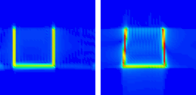 |
130 | General implementation of frequency–space reconstruction
Marko Havu1, Koos Zevenhoven1, Antti Mäkinen1, and Risto J Ilmoniemi1
1Department of Neuroscience and Biomedical Engineering, Aalto University, Espoo, Finland Keywords: Low-Field MRI, Image Reconstruction In low-field and ultra-low-field MRI, concomitant fields can cause severe blurring and distortion in traditional Fourier reconstruction. We have developed an improved image reconstruction framework based on our previously published method that uses a frequency–space formulation. It provides more accurate reconstruction results with the cost of increased computation time. Known inhomogeneity and other field nonidealities can be incorporated into the model to improve the quality of the reconstruction. The framework will be published as open source. |
|
1770. |
131 | Characterization of Concomitant Gradient Fields and their effects on a 46 mT Halbach-Based point-of-care MRI system
Bart de Vos1, Rob Remis2, and Andrew Webb1,2
1C.J. Gorter MRI Center, Leiden University Medical Center, Leiden, Netherlands, 2Circuits and Systems, Delft University of Technology, Delft, Netherlands Keywords: Low-Field MRI, Artifacts, Concomitant fields Low field Halbach systems are susceptible to concomitant gradient effects. The associated fields are significantly different from clinical systems and are presented here for the first time. The corresponding equations and associated effects are verified using a spin echo sequence on a 46 mT Halbach system where a gradient strength of 10 mT/m on a phantom with maximum dimension of 200 mm is observed to create distortions. These are most evident in the transverse plane, where the phase encoding gradient causes blurring and overlapping the other transverse gradient lobe results in an additional warping effect. |
|
1771.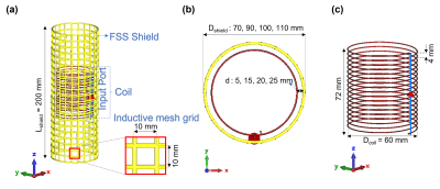 |
132 | Study of Frequency selective surface (FSS) to shield solenoids for low-field portable MRI
MEENA RAJENDRAN1 and SHAOYING HUANG1
1Engineering Product Developement, Singapore University of Technology and Design, Singapore, Singapore Keywords: Low-Field MRI, Low-Field MRI The lightweight frequency selective surface(FSS) using high-pass inductive mesh grids is proposed as shielding of RF solenoids at 2.84MHz for an MRI system using a Halbach array(average B0=67mT). Its effects on the coil B1-sensitivity and the shielding effectiveness are examined. A solenoid(diameter=60mm) was used as an example and the shielding of 5-25mm away was examined. The 5mm-away-FSS-shielded coil has 50.1% (in simulation) and 91.3% (in measurements) higher B1-sensitivity and comparable shielding effectiveness within a defined FoV compared to the copper-shielded coil of the same dimensions. The measured data agree with the simulated ones. |
|
1772.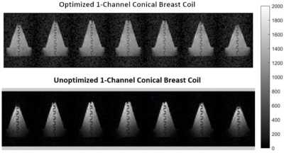 |
133 | Breast and Chest RF Design and Optimization for Ultra-low Field MRI
Torben P.P Hornung1,2,3, Neha Koonjoo1,2, Susu Yan3,4, Matthew S Rosen1,2,5, and Thomas R Bortfeld3,4
1Department of Radiology, A.A Martinos Center for Biomedical Imaging / MGH, Charlestown, MA, United States, 2Harvard Medical School, Boston, MA, United States, 3Department of Physics, ETH Zürich, Zürich, Switzerland, 4Department of Radiation Oncology, MGH, Boston, MA, United States, 5Department of Physics, Harvard University, Cambridge, MA, United States Keywords: Low-Field MRI, Breast, Hybrid & Novel System Technology MRI guidance in Proton Therapy can improve breast cancer targeting accuracy during treatment planning. This study focuses on RF coil optimization for breast imaging at ultra-low field where proton beam deflection is minimal. Our previous optimization algorithm was further improved to adapt to any breast-shaped coil and any anatomical region. It was also developed for organs-at-risk imaging with a deep view into the chest. This new optimization method was tested for a single-channel conical-shaped coil and validated with imaging. The optimized coil showed a more homogenous B-field compared to its unoptimized evenly spaced wound coil in agreement with the simulations. |
|
1773.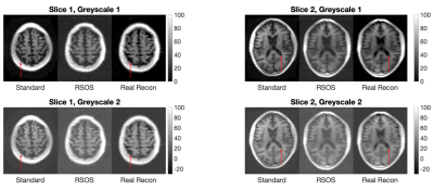 |
134 | Improved T1w Imaging at Ultra-Low Field by use of phase-sensitive reconstruction
Samson Lecurieux Lafayette1, Francesco Padormo2, Paul Cawley1, Emil Ljunberg3,4, Rui Teixeira2, David Edwards1, Mary Rutherford1, Tomoki Arichi1, and Joseph Hajnal1
1Perinatal Imaging & Health, King's College, London, United Kingdom, 2Hyperfine, London, United Kingdom, 3Department of Neuroimaging, King's College, London, United Kingdom, 4Department of Medical Radiation Physics, Lund University, Lund, Sweden Keywords: Low-Field MRI, Brain A phase-sensitive reconstruction is used on adult and neonatal T1w acquisitions on a ultra-low field point-of-care MRI system. We show improved image contrast and reduction in confusing intensities associated with rectification of negative signals. |
|
1774.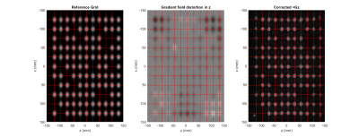 |
135 | Gradient non-linearity estimation in an Open MRI scanner
Olivier E Mougin1, Paul Glover1, Richard Bowtell1, and Penny A Gowland1
1SPMIC, University of Nottingham, Nottingham, United Kingdom Keywords: Low-Field MRI, Gradients Open magnet provide advantageous facility for imaging the human body in a natural way. However field inhomogeneities can reduce the utilisable imaging field-of-view. We present a simple way to map the static field inhomogeneity and the non-linear gradient inhomogeneity in order to correct them during the image reconstruction. |
|
1775. |
136 | Demonstrating the Scalability of Spokes-and-Hub Magnet Designs
Liliana Edmonds1, Irene Kuang1, Elfar Adalsteinsson1,2, and Jacob White1
1Electrical Engineering and Computer Science, Massachusetts Institute of Technology, Cambridge, MA, United States, 2Institute for Medical Engineering and Science, Massachusetts Institute of Technology, Cambridge, MA, United States Keywords: Low-Field MRI, Low-Field MRI The easily constructed Aubert ring-inspired “spokes-and-hub” magnets show promise as a topology for handheld MR imaging, particularly for educational purposes. In this paper we show the scalability of the spokes-and-hub design, and associated tools and RF signal processing, by scaling up the magnet. Constructing a 2x larger magnet yields a volume large enough to potentially image a finger (with which we hope to inspire students). We used our simulator to design and optimize the magnet topology, then constructed it from off-the-shelf small bar magnets, and verified its fields by direct Hall-effect measurements as well as indirectly through spin echoes. |
|
1776.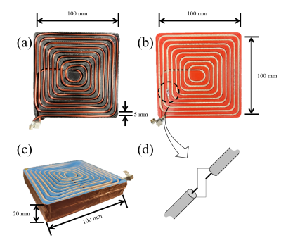 |
137 | EM shielded spiral RF coil design for array coil decoupling at ultra-low field MRI
Yonghyun Ha1, Kartiga Selvaganesan2, Chenhao Sun1, Zhehong Zhang2, Anja Samardzija2, Heng Sun2, Gigi Galiana1, and Todd Constable1
1Radiology and Biomedical Imaging, Yale University, New Haven, CT, United States, 2Biomedical Engineering, Yale University, New Haven, CT, United States Keywords: RF Arrays & Systems, RF Arrays & Systems We introduced a new RF coil made with Moebius cable and RF shield bowl that can be tuned with relatively low capacitance and has good isolation between the coils at low field. B1 amplitude, isolation between the coils, and SNR of the NMR signal of the proposed coil were compared with reference coils (made with copper wire and Moebius cable without an RF shield). Both bench measurements and MR experiments have demonstrated that this coil performs well as part of a low field coil array. |
|
1777. |
138 | Optimization and Construction of a Low-Field MRI System Using Permanent Magnets
Mikah Mellors1 and Rebecca Feldman2
1University of Toronto, Toronto, ON, Canada, 2University of British Columbia, Kelowna, BC, Canada Keywords: Low-Field MRI, Magnets (B0) A low-field tabletop spoke-and-hub permanent magnet array was constructed and assessed for homogeneity as a B0 field. Spatial encoding in the x and y directions was achieved by tilting the hubs relative to one other to generate a linearly increasing field in the tilt direction. A non-array RF coil was designed and optimized for low field strengths, where the signal-to-noise ratio is limited by the weak signal from the sample. The optimized coil design was tuned and matched to the resonant frequency of the spoke-and-hub magnet. |
|
 Back to Meeting Home
 Back to the Program-at-a-Glance
The International Society for Magnetic Resonance in Medicine is accredited by the Accreditation Council for Continuing Medical Education to provide continuing medical education for physicians.
 Back to Meeting Home
 Back to the Program-at-a-Glance
 View Presentation Video