Digital Poster
Progress & Challenges in RF Coils II
ISMRM & ISMRT Annual Meeting & Exhibition • 03-08 June 2023 • Toronto, ON, Canada

Digital Poster
Progress & Challenges in RF Coils II
| Computer # | |||
|---|---|---|---|
4580.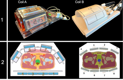 |
161 |
Prostate Imaging at 7 Tesla: Comparison of Two 8-channel
Transmit/Receive RF Local Body Arrays
Markus W. May1,2,
Oliver Kraff1,
Jenni Schulz3,
Stephan Orzada4,
Sebastian Schmitter5,
Thomas Meurs6,
Mark van Uden6,
Tom Scheenen3,
and Harald H. Quick1,2
1Erwin L. Hahn Institute for MRI, University Duisburg-Essen, Essen, Germany, 2High-Field and Hybrid MR Imaging, University Hospital Essen, Essen, Germany, 3Department of Medical Imaging, Radboud University Medical Center, Nijmegen, Netherlands, 4Medical Physics in Radiology (E020), German Cancer Research Center (DKFZ), Heidelberg, Germany, 5DZHK (German Center for Cardiovascular Research), DZHK (German Center for Cardiovascular Research), Partner Site Berlin, Berlin, Germany, 6Tesla Dynamic Coils, Zaltbommel, Netherlands Keywords: RF Arrays & Systems, Body The purpose of this study is to compare two different coil array designs for prostate imaging. We compared an in-house developed 8chpTx/Rx micro stripline element coil array with meanders to a recently introduced, commercially available 8chpTx/Rx coil array based on fractionated dipoles for 1H prostate imaging at 7T. Both coil arrays showed comparable results in the phantom and in vivo examinations. The commercially available coil array performs slightly better in the region of the prostate and is well suited for future prostate imaging at 7T. |
|
4581.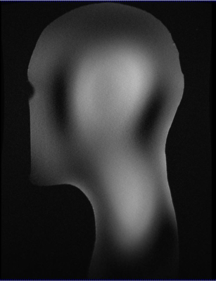 |
162 |
Eight-channel transceiver array for combined head and neck
imaging at 7 Tesla
Divya Baskaran1,
Paul McElhinney1,
Sydney Williams1,
Sarah Allwood Spiers2,
David Porter1,
and Shajan Gunamony1
1Imaging Centre of Excellence, University of Glasgow, Glasgow, Scotland, 2MRI Physics, NHS Greater Glasgow and Clyde, Glasgow, Scotland Keywords: High-Field MRI, RF Arrays & Systems, Neurovascular imaging Combined full brain and neck MRI has been proven inevitable in diagnosis of variety of neurodegenerative diseases such as multiple sclerosis, and amyotrophic lateral sclerosis. High spatial resolution and signal-to-noise ratio provided by the ultra-high field MRI systems (>7T) could be valuable in identification of such subtle morphologies which are not noticeable at standard MRI systems. Current state-of-the-art requires change of coil for imaging either brain or C-spine or equipped with 16-channel transmission capability. This work presents the design and validation of novel neurovascular transceiver array with 8 channels (industry standard) for combined head and neck imaging at 7T. |
|
4582.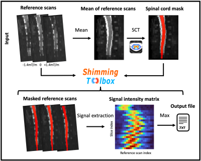 |
163 |
Slice-wise z-shimming for spinal cord fMRI with Shimming Toolbox
Alexandre D'Astous1,
Merve Kaptan2,
Daniel Papp1,
Alice Dabbagh2,
Jürgen Finsterbusch3,
Falk Eippert2,
Eva Alonso-Ortiz1,
and Julien Cohen-Adad1,4,5,6
1Neuropoly Lab, Institute of Biomedical Engineering, Polytechnique Montreal, Montreal, QC, Canada, 2Max Planck Institute for Human Cognitive and Brain Sciences, Leipzig, Germany, 3Department of Systems Neuroscience, University Medical Center Hamburg-Eppendorf, Hamburg, Germany, 4Mila - Quebec AI Institute, Montreal, QC, Canada, 5Functional Neuroimaging Unit, Centre de recherche de l'Institut universitaire de gériatrie de Montréal, Montreal, QC, Canada, 6Centre de recherche du CHU Sainte-Justine, Université de Montréal, Montreal, QC, Canada Keywords: Shims, Spinal Cord Spinal cord fMRI is challenging, in part due to B0 inhomogeneities. An EPI, z-shimming signal intensity based approach was implemented to automatically and dynamically shim the spinal cord. The technique was implemented in the open-source Shimming Toolbox. Z-shimming was tested on a healthy participant and showed a 14% increase in signal intensity as well as 19% increase in tSNR. The technique was generalized to allow for the use of any shim coil geometry as well as non-EPI data. |
|
4583.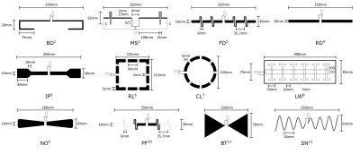 |
164 |
Simulation and Comparison of Transmit Elements for 7T
Head-Imaging with a Large Diameter Transmit Coil
Max Joris Hubmann1,2,
Robert Kowal 2,3,
Stephan Orzada4,
Piet Wagner5,
Frank Seifert5,
Oliver Speck2,3,
and Holger Maune2
1Siemens Healthineers GmbH, Magdeburg, Germany, 2Otto-von-Guericke University, Magdeburg, Germany, 3Research Campus STIMULATE, Magdeburg, Germany, 4Deutsches Krebsforschungszentrum Heidelberg, Heidelberg, Germany, 5Physikalisch-Technische Bundesanstalt (PTB), Berlin, Germany Keywords: Non-Array RF Coils, Antennas & Waveguides, Non-Array RF Coils, Antennas & Waveguides 12 transmission coil designs for 7T head imaging are compared using EM simulations for a large diameter transmit coil. The highest power and SAR efficiency are achieved by loop coils, a passively-fed dipole shows the highest intrinsic decoupling and an IMARS the lowest load dependence. The results provide insight to each element's performance. To make a final decision on a coil element, the array performance must also be evaluated in the futur, as this may differ from the performance of the single element performance. Thus, the decision on a particular transmit element depends on the coil's application and configuration. |
|
4584.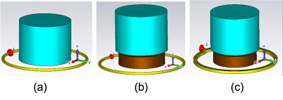 |
165 |
Denoising Effect of Double-Loop Spiral Coil with ultra-High
Dielectric Constant Materials for MRI Applications
Parisa Lotfi poshtgol1,
Navid P. Gandji1,
Michael Lanagan2,
Soo-Han Soon3,
Wei Chen3,
and Qing X Yang1
1Center for NMR Research, Departments of Neurosurgery and Radiology, College of Medicine, Pennsylvania State University, Hershey, PA, United States, 2Engineering Science and Mechanics, Pennsylvania State University, state college, PA, United States, 3Center for Magnetic Resonance Research, Department of Radiology, University of Minnesota, Minneapolis, MN, United States Keywords: Non-Array RF Coils, Antennas & Waveguides, High-Field MRI, High dielectric material We investigated the denoising effect of employing double-loop spiral coil (DLSC) versus the conventional single loop surface coil in the presence of ultrahigh dielectric constant materials (uHDCM). The DLSC confines the conservative E-field distribution between the loops which as a result reduces the E-field in the sample, therefore reduces the noise level. Reduction of conservative E-Field makes the non-conservative E-field more dominant in the sample, which synergistically with the noise reduction allows increasing transmit efficiency and receive sensitivity for MRI application. |
|
4585.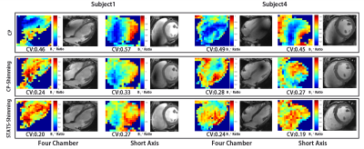 |
166 |
Parallel transmission of cardiac imaging using statistical
initial phase optimization at 5T
Jiaxu Li1,2,
Liqiang Zhou3,
Bin Liu3,
Nan Li1,4,
Zhenhua Shen3,
Xiaoliang Zhang5,
and Ye Li1,4
1Paul C. Lauterbur Imaging Research Center, Shenzhen Institutes of Advanced Technology, Chinese Academy of Sciences, Shenzhen, China, 2School of Pharmacy and Bioengineering, Chongqing University of Technology, Chongqing, China, 3Shanghai United Imaging Healthcare, Shanghai, China, 4Key Laboratory for Magnetic Resonance and Multimodality Imaging of Guangdong Province, Shenzhen, China, 5Department of Biomedical Engineering, State University of New York at Buffalo, Buffalo, NY, United States Keywords: Parallel Transmit & Multiband, Shims High-field magnetic resonance imaging is one of the main development directions of magnetic resonance cardiac imaging because of its high signal-to-noise ratio and more anatomical details. However, with the increase of field strength, the homogeneity of B1 field is a serious challenge, and parallel transmission is one of the important methods to solve this problem.In this work, we proposed a clinically applicable radiofrequency shimming method to improve the imaging quality of cardiac imaging at 5T MRI.Statistical methods were employed to find out a better initial phase of an eight-channel coil and enhance the stability of cardiac imaging. |
|
4586.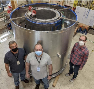 |
167 |
Compact 7T: Progress in Construction and Assembly of a
Low-Cryogen, High-Performance, Head-only High-Field MRI System
Thomas Foo1,
Mark Vermilyea1,
Gene Conte1,
Chris Van Epps1,
Justin Ricci1,
Minfeng Xu1,
Anbo Wu1,
Shike Huang1,
Seung-Kyun Lee1,
Wolfgang Stautner1,
Bai Ye1,
Doug Kelley2,
John Huston III3,
Matt A Bernstein3,
Yunhong Shu3,
Christopher Hess4,
and Duan Xu4
1GE Research, Niskayuna, NY, United States, 2GE Healthcare, Waukesh, WI, United States, 3Mayo Clinic, Rochester, MN, United States, 4University of California-San Francisco, San Francisco, CA, United States Keywords: High-Field MRI, Magnets (B0), Cryogenics; Low-Cryogen; Compact Steps in the construction of an 8 metric ton, 7.0 T MRI scanner for imaging the brain are described. The system nearing completion uses only 12 liters of liquid helium and has outside dimensions comparable to a clinical whole-body 3.0 T scanner, with a similar footprint. As such, the new Compact 7.0 T system fits into any whole-body 3.0 T room, simplifying installation. The system is in the final stages of assembly. |
|
4587.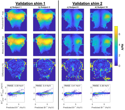 |
168 |
Improved multi-channel pTx B1+ mapping in the head and neck at
7T by combining RF shims and transmit voltages
Matthijs H.S. de Buck1,
Peter Jezzard1,
and Aaron T. Hess1
1Wellcome Centre for Integrative Neuroimaging, FMRIB, Nuffield Department of Clinical Neurosciences, University of Oxford, Oxford, United Kingdom Keywords: Parallel Transmit & Multiband, High-Field MRI, B1+ mapping Acquiring robust multi-channel B1+ maps in both the head and neck using pTx head coils at 7T can be challenging due to the combination of low RF penetration into the neck, the limited dynamic range of multi-channel B1+ mapping techniques, and B0 sensitivity. We present a pipeline for large dynamic range pTx field mapping along with a publicly available 10-subject database of head and neck field maps for a commonly used multi-channel head transmit coil. The pipeline is evaluated by comparing the predicted B1+ to the measured B1+ for random transmit shim configurations in the head and neck. |
|
4588.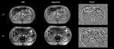 |
169 |
Susceptibility-weighted imaging (SWI) of the kidney at 5T –
initial results
Liyun Zheng1,2,3,
Chun Yang1,2,
Yongming Dai4,
and Mengsu Zeng1,2
1Shanghai Institute of Medical Imaging, Shanghai, China, 2Department of Radiology, Zhongshan Hospital, Fudan University, Shanghai, China, 3Shenzhen United Imaging Research Institute of Innovative Medical Equipment, Shenzhen, China, 4MR Collaboration, Central Research Institute, United Imaging Healthcare, Shanghai, China Keywords: High-Field MRI, Kidney At high magnetic field strengths (B0 ≥ 3 T), susceptibility-weighted imaging (SWI) is an appealing approach for increased signal-to-noise ratio (SNR). To our knowledge, implementation of SWI on the kidney at higher field MRI (> 3T) has not been attempted yet. This initial study proved that it is possible to obtain SWI at 5T for renal with sufficient image quality. For 5T SWI, SNR of cortex and medulla and CNR of cortex/medulla were significantly higher than at 3T, leading to improved vessel conspicuity and corticomedullary discrimination. |
|
4589.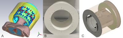 |
170 |
16-Channel 11.7T Transmit Array with Integrated Field Probes
Son Chu1,
Vincent Gras2,
Nicolas Boulant2,
and Shajan Gunamony1
1University of Glasgow, Glasgow, United Kingdom, 2University of Paris-Saclay, CEA, CNRS, BAOBAB, NeuroSpin, Gif sur Yvette, France Keywords: RF Arrays & Systems, RF Arrays & Systems Transmit arrays are essential to mitigate B1+ inhomogeneity at extremely high static magnetic field strengths. In addition, the transmit elements must be arranged in multiple rows to achieve whole-brain coverage at 11.7T. Furthermore, concurrent field monitoring probes offer the possibility to improve MR image quality by correcting subject-specific k-space trajectories and monitoring real-time dynamic field fluctuations. The placement of these probes must be carefully controlled to achieve proper probe conditioning as well as to preserve the RF performance of the transmit array. This work presents the numerical simulation and optimization of a 16-channel-transmit array with integrated field probes. |
|
4590.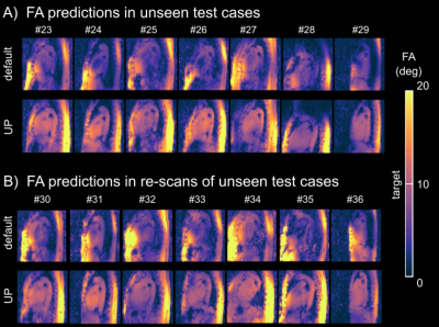 |
171 |
Reproducibility and variability of tailored and universal
nonselective excitation pulses at 7T for human body imaging:
Re-scans after one year
Christoph Stefan Aigner1,
Jean Pierre Bassenge1,2,
Sebastian Dietrich1,
Max Lutz1,
Felix Krüger1,
and Sebastian Schmitter1,3,4
1Physikalisch-Technische Bundesanstalt (PTB), Berlin and Braunschweig, Germany, 2Working Group on Cardiovascular Magnetic Resonance, Experimental and Clinical Research Center, a joint cooperation between the Charité Medical Faculty and the Max-Delbrück Center for Molecular Medicine, Berlin, Germany, 3Medical Physics in Radiology, German Cancer Research Center (DKFZ), Heidelberg, Germany, 4University of Minnesota, Center for Magnetic Resonance Research, Minneapolis, MN, United States Keywords: RF Pulse Design & Fields, Parallel Transmit & Multiband 3D ultrahigh field (UHF) imaging of the body, particularly the heart, is highly challenging due to the inhomogeneous transmit field (B1+) leading to spatially varying flip-angle (FA) patterns. This study demonstrates that pre-computed universal pulses (UPs) allow for calibration-free 3D cardiac FA homogenization at 7T, despite inter-subject variations because of sex, age, BMI, and coil-placement differences. Furthermore, the performance is robust across multiple MRI operators or re-scans after one year. UPs are, therefore, suited to significantly simplify UHF cardiac imaging by removing the need for lengthy calibration prescans. |
|
4591.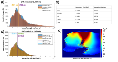 |
172 |
A Comparison Between Shielded and Unshielded RF Antennas for 7T
Body MRI
Tobey D Haluptzok1,
Simon Schmidt1,
Russell L Lagore1,
and Gregory J Metzger1
1Center for Magnetic Resonance Research, University of Minnesota, Minneapolis, MN, United States Keywords: RF Arrays & Systems, High-Field MRI To more fully realize the benefit of UHF MRI, coil designs must be optimized to for pTx and intrinsic SNR performance. In this abstract we compare 5 RF coil array block systems. The first comparison ran is pTx performance, the second comparison is of intrinsic array SNR, and the third tests robustness to top loading. The shielded and unshielded three-loop dipole elements performed better than the single-loop dipole and the shieled helped with top load insensitivity. From these results we conclude that the three-loop dipole coil blocks have superior performance and that the shield doesn’t hurt performance. |
|
4592.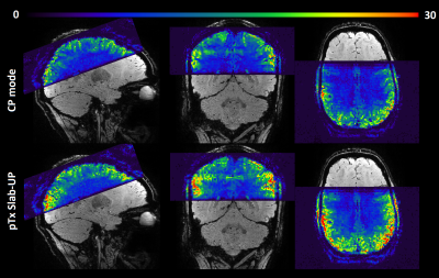 |
173 |
PASTeUR package extension to slab-selective excitations and
3D-EPI for plug-and-play parallel transmit at 7 Tesla
Redouane Jamil1,
Aurélien Massire2,
Franck Mauconduit1,
Vincent Gras1,
Mathieu Naudin3,4,5,
Rémy Guillevin3,4,5,
Eberhard Pracht6,
Tony Stöcker6,7,
Nicolas Boulant1,
and Rüdiger Stirnberg6
1Paris-Saclay University, CEA, CNRS, BAOBAB, NeuroSpin, Gif-sur-Yvette, France, 2Siemens Healthcare SAS, Saint-Denis, France, 3CHU Poitiers, Poitiers, France, 4LRCOM I3M, DACTIM LMA CNRS 7348, University of Poitiers, Poitiers, France, 5Laboratory of Applied Mathematics, UMR CNRS 7348, University of Poitiers, Poitiers, France, 6German Center for Neurodegenerative Diseases (DZNE), Bonn, Germany, 7Department of Physics and Astronomy, University of Bonn, Bonn, Germany Keywords: Parallel Transmit & Multiband, Parallel Transmit & Multiband, High-Field MRI; fMRI Keywords: Parallel Transmit & Multiband; High-Field MRI; RF Pulse Design & Fields; fMRI PASTeUR is a package of sequences using non-selective parallel transmission Universal Pulses (UP) as a plug-and-play pTX solution alleviating B1+ inhomogeneities. Here, we extend the UP solution to slab-selective excitations while preserving a plug-and-play philosophy. A slab of any thickness/orientation/rotation can be acquired without subject-specific calibrations. In vivo experiments performed with an fMRI protocol on a 3D-EPI show that the improved B1+ homogeneity can result in higher SNR. Moreover, the slab-selective UP coupled with parallel imaging and segmented k-space acquisitions pushed further the quality of 3D-EPI. |
|
4593.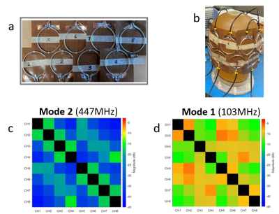 |
174 |
Design and Test of a Flexible Two-row CTL Array and Its
Detunable Resonant Elements for 10.5T MR Imaging
Xiaoliang Zhang1,
Matt Waks2,
Lance DelaBarre2,
Komlan Payne1,
Kamil Ugurbil2,
and Gregor Adriany2
1Department of Biomedical Engineering, State University of New York at Buffalo, Buffalo, NY, United States, 2Radiology, University of Minnesota, Minneapolis, MN, United States Keywords: New Devices, High-Field MRI A flexible patch array using small-sized 5-cm coaxial transmission line (CTL) resonators is designed, constructed and tested, aiming for human head imaging at the ultrahigh field of 10.5T. Bench tests on decoupling performance among resonant elements are shown. For full utility, a detuning circuit is successfully developed, showing the ecellent detuning capability for 5-cm small CTL loops at 447 MHz. This study suggests that it is possible to build high-density receive-only coil arrays using small-sized CTL resonators at 10.5T. |
|
4594.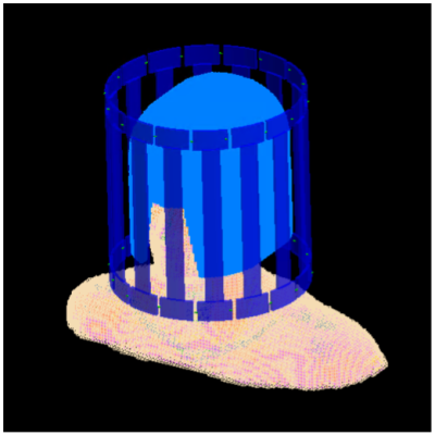 |
175 |
Impact of a High-Permittivity Helmet on Transmit Efficiency
Across a Wide Range of Field Strengths and Relative
Permittivities
Giuseppe Carluccio1,2 and
Christopher Michael Collins1,2
1Radiology, NYU Grossman School of Medicine, New York, NY, United States, 2Center for Advanced Imaging Innovation and Research (CAI2R), New York University, New York, NY, United States Keywords: Non-Array RF Coils, Antennas & Waveguides, Non-Array RF Coils, Antennas & Waveguides, High-Permittivity Materials High-Permittivity Materials are used in MRI to manipulate the B1 field distribution for example to provide a stronger transmit efficiency. For a given coil, the transmit efficiency depends on the frequency. Here we examine the effects of a high-permittivity helmet on the transmit efficiency in exciting the brain with a birdcage coil over a range of frequencies. The optimal frequency providing the maximum transmit efficiency is not significantly impacted by the presence of the helmet, while the maximum efficiency can increase by about 100% compared to when no helmet is used. |
|
4595.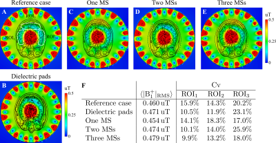 |
176 |
Metasurface for passive RF shimming to improve brain imaging at
7T
Vladislav Koloskov1,
Alena Shchelokova1,
and Andrew Webb2
1School of Physics and Engineering, ITMO University, St. Petersburg, Russian Federation, 2Department of Radiology, C.J. Gorter Center for High Field MRI, Leiden University Medical Center, Leiden, Netherlands Keywords: Hybrid & Novel Systems Technology, Hybrid & Novel Systems Technology, metasurface, passive shimming We propose a new passive radiofrequency (RF) shimming approach using a flexible, compact, low-cost, and long-term stability metasurface to improve brain imaging at 7T. The metasurface can be manufactured as a periodic structure based on a set of metal crosses connected by the capacitors, thus having a thickness of less than 1 mm. Numerical studies with the human voxel model showed up to 6% homogeneity improvement at the regions of interest (ROI) in the presence of the metasurface in comparison with the conventional case, which also outperforms the effect of the dielectric pad’s placement. |
|
 Back to Meeting Home
 Back to the Program-at-a-Glance
The International Society for Magnetic Resonance in Medicine is accredited by the Accreditation Council for Continuing Medical Education to provide continuing medical education for physicians.
 Back to Meeting Home
 Back to the Program-at-a-Glance
 View Presentation Video