Digital Poster
ML/AI for Acquisition & Reconstruction II
ISMRM & ISMRT Annual Meeting & Exhibition • 03-08 June 2023 • Toronto, ON, Canada

Digital Poster
ML/AI for Acquisition & Reconstruction II
| Computer # | |||
|---|---|---|---|
3875.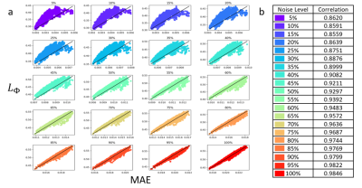 |
121 |
Estimating Uncertainty of Deep Learning for Tomographic Image
Reconstruction Through Local Lipschitz
Danyal Bhutto1,2,
Bo Zhu2,
Jeremiah Zhe Liu3,4,
Neha Koonjoo2,5,
Bruce R Rosen2,5,
and Matthew S Rosen2,5,6
1Biomedical Engineering, Boston University, Boston, MA, United States, 2Athinoula A. Martinos Center for Biomedical Imaging, Charlestown, MA, United States, 3Google, Mountain View, CA, United States, 4Biostatistics, Harvard University, Cambridge, MA, United States, 5Harvard Medical School, Boston, MA, United States, 6Physics, Harvard University, Boston, MA, United States Keywords: Machine Learning/Artificial Intelligence, Image Reconstruction As deep learning approaches for image reconstruction become increasingly used in the radiological space, strategies to estimate reconstruction uncertainties become critically important to ensure images remain diagnostic. We estimate reconstruction uncertainty through calculation of the Local Lipschitz value, demonstrate a monotonic relationship between the Local Lipschitz and Mean Absolute Error, and show how a threshold can determine whether the deep learning technique was accurate or if an alternative technique should be employed. We also show how our technique can be used to identify out-of-distribution test images and outperforms baseline metrics, i.e. deep ensemble and Monte-Carlo dropout. |
|
3876.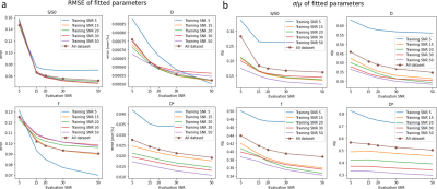 |
122 |
Fast Uncertainty Estimation of IVIM parameters using Bayesian
Neural Networks
Zehuan Zhang1,
Matej Genči1,
Hongxiang Fan1,
Wayne Luk1,
and Andreas Wetscherek2
1Department of Computing, Imperial College London, London, United Kingdom, 2Joint Department of Physics, The Institute of Cancer Research, London, United Kingdom Keywords: Machine Learning/Artificial Intelligence, Diffusion/other diffusion imaging techniques, Uncertainty estimation We transformed the state-of-the art IVIM-NET for IVIM parameter fitting into a Bayesian Neural Network (BNN). BNNs can estimate uncertainty for quantitative MRI parameters, which is relevant for clinical decision making. We found that training on data with the highest SNR outperformed IVIM-BNNET models trained on matching SNR regarding parameter errors and uncertainties. A region with artificially increased noise could be identified from IVIM-BNNET's uncertainty output. Compared with traditional fitting, IVIM-BNNET achieved comparable accuracy, while being 21 times faster and providing less correlated parameter estimates. Monte-Carlo dropout rate 0.4 provided the best trade-off between low errors and low uncertainty. |
|
3877.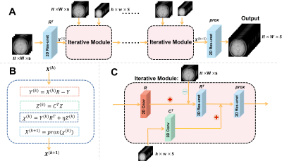 |
123 |
Super-Resolution Reconstruction of CEST images for human brain
at 3T based on deep learning
Sirui Wu1,
WenXuan Chen1,
Yibing Chen2,
Zhongsen Li1,
and Xiaolei Song1
1Center for Biomedical Imaging Research, Department of Biomedical Engineering, Tsinghua University, Beijing 100084, China, Beijing, China, 2Xi’an Key Lab of Radiomics and Intelligent Perception, School of Information Sciences and Technology, Northwest University, Xi’an, Shaanxi 710069, China, Xi'an, China Keywords: Machine Learning/Artificial Intelligence, CEST & MT, Super-resolution, Unrolled network, quantitation CEST MRI suffers from the low resolution, leading to the disability to image small structures. We proposed a new optimization process adapting the CEST data form. Based on deep learning, our optimization flow is unfolded into an unrolled network for super resolution, termed as SC-Net. Besides, we introduced ROI-based normalization loss to guarantee quantitation accuracy. Results showed that SC-Net could reconstruct high-resolution image series and quantitative maps (PSNR = 44.27dB, CNR APTw = 41.79dB), when down sampling rate = 8. And the ROI-based normalization loss could calibrate errors. In conclusion, SC-Net has the potential to image small regions and acceleration. |
|
3878.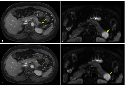 |
124 |
Investigation of deep learning reconstruction in MR imaging of
pancreatic space occupying lesions
Meng Zhang1,
Zheng Ye1,
Bo Zhang2,
Miaoqi Zhang2,
and Zhenlin Li1
1Department of Radiology, West China Hospital, Sichuan University, Chengdu, China, 2MR Research, GE Healthcare, Beijing, China Keywords: Machine Learning/Artificial Intelligence, Image Reconstruction At present, the resolution of magnetic resonance image is still limited to the detection of pancreatic space occupying lesions. In this work, quantitative and qualitative analysis of pancreatic images of T1WI and DWI with built-in DL Recon was carried out. The results showed that the images obtained by the sequence with built-in DL Recon were better than the original images in terms of SNR, CNR and subjective scores. It is confirmed that deep learning reconstruction can improve image resolution and has potential in the detection of space occupying lesions in pancreas. |
|
3879.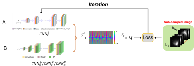 |
125 |
Accurate Estimation of Background Phase in Virtual Conjugate
Coil Expansion Combined with Wave Encoding
Congcong Liu1,
Zhuoxu Cui1,
Sen Jia1,
Zhilang Qiu2,
Xin Liu1,
Hairong Zheng1,
Dong Liang1,
and Haifeng Wang1
1Shenzhen Institutes of Advanced Technology, Chinese Academy of Sciences, Shenzhen, China, 2Case Western Reserve University, Cleveland, OH, United States Keywords: Machine Learning/Artificial Intelligence, Image Reconstruction The wave encoding model with virtual conjugate coil (Wave-VCC) extension can provide more powerful MRI-accelerated imaging performance. However, estimating the background phase covering the full frequency range is un-tractable by only acquiring the middle auto-calibration signals (ACS) line during reconstruction. Here, combining a neural network without training, a new method to generate more accurate background phase in Wave-VCC is proposed. Including ablation experiments and comparison, experiments were carried out to verify the feasibility and performance of the proposed methods, respectively. |
|
3880.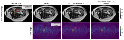 |
126 |
Deep Learning Reconstruction of Dynamic Free-breathing Fetal
Heart MRI to Improve Clinical Pipeline
Denis Prokopenko1,
Daniel Rueckert2,3,
and Joseph V. Hajnal1
1Biomedical Engineering Department, School of Biomedical Engineering and Imaging Sciences, King's College London, London, United Kingdom, 2Department of Informatics, Technical University of Munich, Munich, Germany, 3Department of Computing, Imperial College London, London, United Kingdom Keywords: Machine Learning/Artificial Intelligence, Image Reconstruction Dynamic free-breathing fetal heart MRI requires high spatial and temporal resolution, which could be reconstructed by kt-SENSE from undersampled data guided by priors of the same anatomy. Doubled acquisition time and uncontrolled fetal motion between the 2 acquisitions affects the data quality for reconstruction. We explored an alternative deep learning approach using a 3D U-Net based model with time-averaged skip connection and data consistency. Assessment of the model set a baseline for prior preconstruction and underlines important pitfalls that will drive further improvements to achieve optimal reconstruction quality. |
|
3881.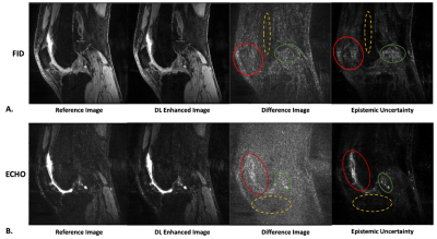 |
127 |
Uncertainty Estimation for Deep Learning-Based Enhancement of
Undersampled Dual-Echo Steady-State Knee MRI
Ashmita Deb1,2,
Shu-Fu Shih1,2,
Zhaohuan Zhang1,2,
and Holden H Wu1,2
1Department of Radiological Sciences, David Geffen School of Medicine, University of California Los Angeles, Los Angeles, CA, United States, 2Department of Bioengineering, University of California Los Angeles, Los Angeles, CA, United States Keywords: Machine Learning/Artificial Intelligence, Data Analysis, Image Enhancement Deep learning (DL) based image enhancement of undersampled 3D dual-echo steady-state knee MRI can achieve faster computation times compared to compressed sensing reconstruction. However, it is hard to interpret how DL models work. This introduces the risk of DL-enhanced images containing inaccuracies without the user’s knowledge and thus confounding diagnosis. This work aimed to calculate pixel-wise uncertainty maps for DL-enhanced images by incorporating Monte Carlo Dropout into a 2D UNET to estimate epistemic uncertainty. Analysis showed that the DL-enhanced images achieved good image quality and the spatial uncertainty maps reflected errors, compared to reference images. |
|
3882.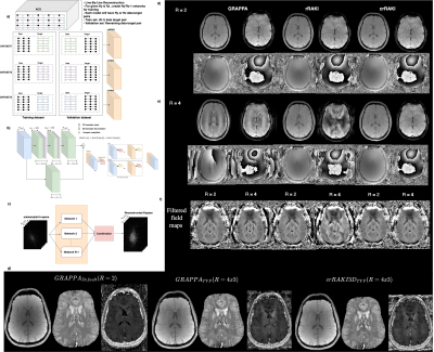 |
128 |
Accelerating Phase and Quantitative susceptibility mapping with
Scan-Specific Complex Convolutional Neural Networks
Swetali Nimje1,2,
Ludovic de Rochefort1,
and Thierry Artières2
1Aix Marseille University, CNRS, CRMBM, Marseille, France, 2Aix Marseille University, Ecole Centrale de Marseille, CNRS, LIS, Marseille, France Keywords: Machine Learning/Artificial Intelligence, Quantitative Susceptibility mapping MRI data is inherently complex-valued, the vast majority of deep learning frameworks do not yet support complex-valued data. Most reconstruction networks separate real and imaginary components into two separate real-valued channels, which may not be the most efficient way to represent complex numbers. Phase is essential for many MRI applications, including phase contrast velocity mapping and Quantitative Susceptibility Mapping (QSM) etc. We propose a new crRAKI, a scan-specific complex-valued residual convolutional neural network for 2D/3D MRI data for accelerating phase mapping and QSM. A comparison is made with GRAPPA and rRAKI for the accelerated reconstruction of MRI images. |
|
3883.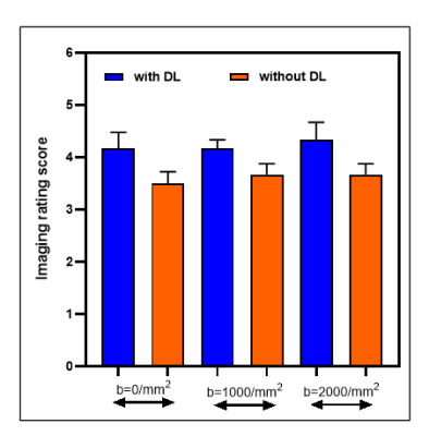 |
129 |
Evaluation of deep learning-based reconstruction for qualitative
and quantitative DW-MRI in head and neck cancers
Ramesh Paudyal1,
Akash Deelip Shah2,
Amaresha Shridhar Konar1,
Jaemin Shin3,
Eve LoCastro1,
Nisha Bagchi4,
Maggie Fung3,
Suchandrima Banerjee5,
Nancy Lee6,
and Amita Shukla-Dave1,2
1Medical Physics, Memorial Sloan Kettering Cancer Center, New York, NY, United States, 2Radiology, Memorial Sloan Kettering Cancer Center, New York, NY, United States, 3GE Health Care, New York, NY, United States, 4Kimmel Medical College, Thomas Jefferson University, Philadelphia, PA, United States, 5GE Health Care, Menlo Park, CA, United States, 6Radiation Oncology, Memorial Sloan Kettering Cancer Center, New York, NY, United States Keywords: Machine Learning/Artificial Intelligence, Tumor The head and neck (HN) region have complex anatomical structures that affect the image quality of diffusion-weighted MRI. Therefore deep learning (DL)-based Reconstruction (Recon) for DW-MRI could be a promising method that can help improve image sharpness and signal-to-noise ratio (SNR) without increasing signal averaging. The present study aimed to evaluate the performance of qualitative and quantitative multiple b-value DW-MRI powered by DL-based Recon for tumors in the HN region. The DL-based recon method improved the DW image quality and SNR compared to those without DL recon. |
|
3884.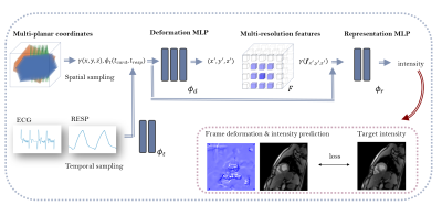 |
130 |
Implicit CINE: a deep-learning super-resolution model for
multi-planar real-time MRI
Nora Vogt1,
Karyna Isaieva1,
Jean-Sébastien Louis1,
Christian Weihsbach2,
Mattias Paul Heinrich2,
Freddy Odille1,3,
Pierre-André Vuissoz1,3,
Jacques Felblinger1,3,
and Julien Oster1,3
1IADI, Université de Lorraine, INSERM U1254, Nancy, France, 2Institut für Medizinische Informatik, Universität zu Lübeck, Lübeck, Germany, 3CIC-IT, INSERM 1433, Université de Lorraine and CHRU Nancy, Nancy, France Keywords: Machine Learning/Artificial Intelligence, Cardiovascular Cardiac CINE MRI plays an essential role in cardiac diagnosis, but not all patients are eligible for 3D imaging, which is associated with long acquisition times. We propose a deep-learning super-resolution model to generate 3D CINE from multi-planar 2D real-time MRI using external signals for cardiac and respiratory motion estimation. The proposed neural field model is trained on a single subject and performs non-rigid motion compensation and implicit representation learning in an end-to-end manner. A preliminary study with three healthy volunteers demonstrates promising reconstruction performance and computation times compared to traditional registration-based approaches. |
|
3885.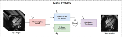 |
131 |
Accelerated MRI using Dual-domain Transformer-Based
Reconstruction and Learning-based Undersampling
Guan Qiu Hong1,2,3,
William Morley2,
Matthew Wan2,
Yuan Tao Wei1,2,3,
Yang Su2,
and Hai-Ling Margaret Cheng1,2,3
1Institute of Biomedical Engineering, University of Toronto, Toronto, ON, Canada, 2The Edward S. Rogers Sr. Department of Electrical and Computer Engineering, University of Toronto, Toronto, ON, Canada, 3Translational Biology & Engineering Program, Ted Rogers Centre for Heart Research, Toronto, ON, Canada Keywords: Machine Learning/Artificial Intelligence, Image Reconstruction Deep learning architectures such as Convolutional Neural Networks (CNNs) are widely used alongside fixed k-space undersampling for reconstructing highly undersampled MRI k-space data. However, CNN-based architectures may perform sub-optimally due to their limited ability to capture long range dependencies, and fixed undersampling patterns may not be amenable to optimal reconstruction. We propose dual-domain (image and k-space), transformer-based reconstruction architectures paired with learning-based undersampling to reconstruct undersampled k-space data. Experimental results demonstrate improved reconstruction quality compared to CNN-based (UNet) architecture across 5x to 100x acceleration factors; dual domain outperformed single domain reconstructions. |
|
3886.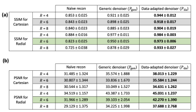 |
132 |
Data-adapted Neural Network Denoisers as a Regularization Engine
for Low-latency Image Reconstruction in Accelerated Cardiac
Perfusion MRI
Dilek Mirgun Yalcinkaya1,2,
Hazar Benan Unal1,
Subha Raman3,4,
Abolfazl Hashemi2,
Rohan Dharmakumar3,4,
and Behzad Sharif1,3,4
1Laboratory for Translational Imaging of Microcirculation, Indiana University (IU) School of Medicine, Indianapolis, IN, United States, 2Elmore Family School of Electrical and Computer Engineering, Purdue University, West Lafayette, IN, United States, 3Weldon School of Biomedical Engineering, Purdue University, West Lafayette, IN, United States, 4Krannert Cardiovascular Research Center, IU School of Medicine/IU Health Cardiovascular Institute, Indianapolis, IN, United States Keywords: Machine Learning/Artificial Intelligence, Image Reconstruction, Plug-and-play, denoiser In this work, we demonstrated that a deep-learning based denoiser trained on a limited dataset of first-pass myocardial perfusion MRI studies enables low-latency image reconstruction using the plug-and-play iterative reconstruction framework. Our proof-of-concept results suggest that the data-adapted denoiser resulted in superior performance versus a generic denoiser especially if there are constraints on the number of iterations (total computation time) which is the case in certain clinical settings specifically interventional MRI. Our findings also imply that radial sampling may be a more desirable data acquisition strategy for PnP-based image reconstruction in first-pass MP MRI studies. |
|
3887.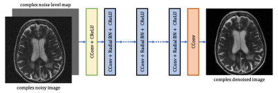 |
133 |
MRI Denoising with a Non-Blind Deep Complex-Valued Convolutional
Neural Network
Quan Dou1,
Zhixing Wang1,
Xue Feng1,
and Craig Meyer1
1Biomedical Engineering, University of Virginia, Charlottesville, VA, United States Keywords: Machine Learning/Artificial Intelligence, Machine Learning/Artificial Intelligence Signal-to-noise ratio is crucial for MR image analysis. In this work, we proposed a non-blind complex-valued convolutional neural network for MRI denoising. Compared to existing denoising methods, the proposed method shows better performance on handling noise at different levels and on spatially variant noise. |
|
3888. |
134 |
Deep Learning Reconstruction for Magnetic Resonance Image
Quality Improvement in Lumbar Endplate Inflammation
Xinyang Lv1,
Zheng Ye1,
Miaoqi Zhang2,
Bo Zhang2,
and Zhenlin Li1
1radiology department, West China Hospital,Sichuan University, Chengdu, China, 2MR Research, GE Healthcare, Beijing, China Keywords: Machine Learning/Artificial Intelligence, Image Reconstruction Magnetic resonance imaging (MRI) is a useful tool to diagnose lumbar endplate inflammation. It is thus important to improve diagnostic accuracy by improving image quality. In this study, we compared signal-to-noise ratio (SNR), contrast noise ratio (CNR) and subjective scores between original images and deep learning reconstruction (DL Recon) images in 31 patients diagnosed with lumbar endplate inflammation. It was observed that the deep learning reconstructed images outperformed conventional images in terms of both subjective scores and objective values. |
|
3889. |
135 |
Reconstruction of accelerated MR cholangiopancreatography using
supervised and self-supervised 3D Variational Networks
Jonas Kleineisel1,
Bernhard Petritsch1,
Thorsten A. Bley1,
Herbert Köstler1,
and Tobias Wech1
1Department of Diagnostic and Interventional Radiology, University Hospital of Würzburg, Würzburg, Germany Keywords: Machine Learning/Artificial Intelligence, Image Reconstruction Magnetic resonance cholangiopancreatography suffers from long examination times and artifacts originating from residual motion. To shorten the acquisition protocol, we acquired data at 12-fold undersampling and investigated a 3D Variational Network (VN) architecture for reconstruction. We compared a self-supervised training scheme and a supervised network trained on synthetic data. We find that the self-supervised method is only able to provide competitive reconstructions if the network is initialized with pre-trained weights, and even then does not offer superior performance over the supervised approach. For the presented exemplary data, the supervised VN showed comparable image quality as a reference Compressed Sensing model. |
|
3890. |
136 |
Deep Learning Reconstruction in MRI: Comparison of Image Quality
in Patients With Hepatic Malignancy
Yuqi Tan1,
Zheng Ye1,
Miaoqi Zhang2,
Bo Zhang2,
and Zhenlin Li1
1West China Hospital of Sichuan University, Chengdu, China, 2GE Healthcare, MR Research, Beijing, China Keywords: Machine Learning/Artificial Intelligence, Image Reconstruction Improving image resolution by denoising is an important research goal in MRI reconstruction. An emerging technique, deep learning reconstruction (DLR), has shown great potential in MRI denoising. In this study, we included 37 patients with pathologically diagnosed hepatic malignancy, and compared the image quality of DLR and original reconstruction regarding dual echo T1 weighted sequence, diffusion weighted imaging (DWI) and fat-suppressed T1 weighted gadolinium-enhancement. It was shown that DLR significantly improved the image quality by reducing background noise, thus making hepatic malignancy more conspicuous. |
|
3891.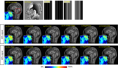 |
137 |
Overview of Complex-valued Image Reconstruction for CS-MRI Using
Real-valued CNN with Symmetrical Signal Under-Sampling
Shohei Ouchi1,
Itona Fukatsu1,
Kazuki Yamato1,
and Satoshi Ito1
1Utsunomiya University, Utsunomiya, Japan Keywords: Machine Learning/Artificial Intelligence, Machine Learning/Artificial Intelligence Complex-valued CNN based image reconstruction methods have been proposed to correspond to MR images with a spatial phase variation. However, using those CNN may lead to over-fitting because CNN layers for complex numbers are requires large number of parameters than real-valued CNN. We previously proposed a reconstruction method for complex-valued image using a real-valued DnCNN by introducing a symmetrical k-space under-sampling. In this study, we introduced this method to U-Net and ADMM-CSNet. Reconstruction experiments showed that a real-valued CNN has the possibility to have the same or better performance as a complex-valued CNN without perform complex calculations. |
|
3892.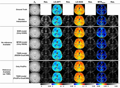 |
138 |
A Two-Stage Super-Resolution (TSSR) CEST Model Using
Deep-Learning Reconstruction
Wenxuan Chen1,
Sirui Wu1,
and Xiaolei Song1
1Center for Biomedical Imaging Research, Department of Biomedical Engineering, Tsinghua University, Beijing, China Keywords: Machine Learning/Artificial Intelligence, Machine Learning/Artificial Intelligence, Super-resolution CEST is an important source of new contrast in MRI. However, it is time-consuming to obtain high-resolution CEST images. We proposed a deep learning based Two-Stage Super-Resolution (TSSR) model for CEST images. Compared with conventional SISR or MFSR models, TSSR model can effectively utilize the correlations among slices and those among saturation offsets for CEST images. We acquired brain CEST images on 14 volunteers using a 3T clinical MR scanner. Initial results suggested that the proposed TSSR model outperformed other methods for all the evaluation indicators. Our work showed the potential in reconstruction of high-resolution CEST images from low-resolution ones. |
|
3893.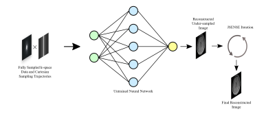 |
139 |
Joint Estimation of Coil Sensitivity and Image by Using
Untrained Neural Network without External Training Data
Gulfam Ahmed Saju1,
Zhiqiang Li2,
Reza Abiri3,
Tianming Liu4,
and Yuchou Chang1
1Computer and Information Science, University of Massachusetts Dartmouth, North Dartmouth, MA, United States, 2Neuroradiology, Barrow Neurological Institute, Phoenix, AZ, United States, 3Electrical, Computer and Biomedical Engineering, University of Rhode Island, Kingston, RI, United States, 4Computer Science, University of Georgia, Athens, GA, United States Keywords: Machine Learning/Artificial Intelligence, Parallel Imaging Training data for MRI reconstruction are difficult to be acquired in clinical practice. In addition, machine learning or deep learning-based MRI reconstruction suffers the distribution shift problem between training data and testing data. Generalization error always exists, so reconstructed images are unstable. We proposed a joint estimation of coil sensitivity and image using the prior of an untrained neural network (UNN). Coil sensitivity map improvement gradually enhances the UNN prior and the image to be reconstructed in an iterative optimization process. The method outperforms other MRI reconstruction methods by suppressing noise and aliasing artifacts. |
|
3894.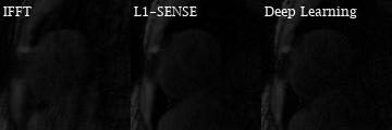 |
140 |
Deep learning-based Motion-corrected Rapid Image Reconstruction
for High-resolution Cartesian First-pass Myocardial Perfusion
Imaging at 3 T
Junyu Wang1 and
Michael Salerno1
1Cardiovascular Medicine, Stanford University, Stanford, CA, United States Keywords: Machine Learning/Artificial Intelligence, Image Reconstruction Keywords: artificial intelligence, image reconstruction, perfusion
Cardiac magnetic resonance first-pass contrast-enhanced myocardial perfusion imaging is valuable for evaluating coronary artery disease1. 2D Cartesian perfusion imaging using compressed sensing (CS)-based reconstructions such as L1-SENSE2 enables fast and high-resolution imaging, but whole-heart coverage cannot be achieved without simultaneous multi-slice (SMS) acquisitions and the CS-based iterative reconstruction takes ~30 minutes per slice. To address this, we have developed a deep learning-based motion-corrected rapid image reconstruction for high-resolution Cartesian perfusion imaging at 3 Tesla, for both 2D and SMS MB=2 acquisitions, which provides fast and high-quality motion-corrected reconstruction and makes rapid online reconstruction feasible. |
|
 Back to Meeting Home
 Back to the Program-at-a-Glance
The International Society for Magnetic Resonance in Medicine is accredited by the Accreditation Council for Continuing Medical Education to provide continuing medical education for physicians.
 Back to Meeting Home
 Back to the Program-at-a-Glance
 View Presentation Video