Digital Poster
ML/AI for Recon & Super Resolution I
ISMRM & ISMRT Annual Meeting & Exhibition • 03-08 June 2023 • Toronto, ON, Canada

Digital Poster
ML/AI for Recon & Super Resolution I
| Computer # | |||
|---|---|---|---|
4031.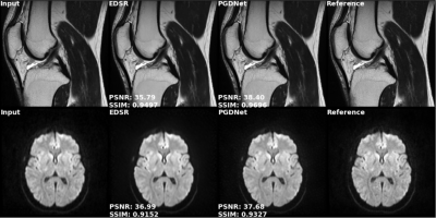 |
101 |
Adaptive Deep Learning MR Image Enhancement with Property
Constrained Unrolled Network
Zechen Zhou1,
Ryan Chamberlain1,
Praveen Gulaka1,
Enhao Gong1,
Greg Zaharchuk1,
and Ajit Shankaranarayanan1
1Subtle Medical Inc, Menlo Park, CA, United States Keywords: Machine Learning/Artificial Intelligence, Machine Learning/Artificial Intelligence An unrolled network with explicit MR image degradation modeling and physical property constraints, termed PGDNet, is proposed for adaptive image denoising and deblurring. Preliminary evaluation demonstrated that PGDNet can achieve similar/superior image quality enhancement compared to the conventional task-specific networks, and can outperform others in joint denoising and deblurring tasks. PGDNet provides a promising solution for adaptive MR image denoising and deblurring to restore the image quality of accelerated clinical MR scans. |
|
4032. |
102 |
Performance Evaluation of Deep Learning-based Image
Reconstruction for Head and Neck Imaging Protocol
Amaresha Shridhar Konar1,
Jaemin Shin2,
Ramesh Paudyal1,
Abhay Dave3,
Maggie Fung2,
Suchandrima Banerjee4,
Vaios Hatzoglou5,
and Amita Shukla-Dave1,5
1Medical Physics, Memorial Sloan Kettering Cancer Center, New York City, NY, United States, 2GE Healthcare, New York City, NY, United States, 3Touro College of Osteopathic Medicine, New York, NY, United States, 4GE Healthcare, Menlo Park, CA, United States, 5Radiology, Memorial Sloan Kettering Cancer Center, New York City, NY, United States Keywords: Machine Learning/Artificial Intelligence, Quantitative Imaging MRI has excellent extracranial soft-tissue contrast to detect tumors in the head and neck (HN) region. Technical challenges arise due to MRI related artifacts. In routine radiological practice, HN MR imaging protocols are optimized specifically to the subsites. We aimed to evaluate the performance of the HN imaging protocol that include qualitative T1w, T2w, and quantitative diffusion MRI powered by a novel deep learning (DL) based reconstruction (recon) using the ACR and QIBA diffusion phantoms. This phantom study showed that qualitative T1w and T2w images and multiple b-value DWI data powered with DL recon substantially improves the image quality. |
|
4033.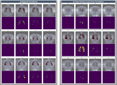 |
103 |
Image sequence partitioning in CRNN-based reconstruction
improves ventilation defect detection of 3D-PREFUL with
undersampled data
Maximilian Zubke1,2,
Filip Klimeš1,2,
Andreas Voskrebenzev1,2,
Marius Wernz1,2,
Till F Kaireit1,2,
Agilo L Kern1,2,
Marcel Gutberlet1,2,
Robin A Müller1,2,
Frank Wacker1,2,
and Jens Vogel-Claussen1,2
1Institute of Diagnostic and Interventional Radiology, Hannover Medical School, Hannover, Germany, 2Biomedical Research in Endstage and Obstructive Lung Disease Hannover (BREATH), German Center for Lung Research (DZL), Hannover, Germany Keywords: Machine Learning/Artificial Intelligence, Lung 3D-phase-resolved-functional-lung (3D-PREFUL) MRI enables a non-contrast-enhanced detection of lung ventilation defects. Convolutional, recurrent neural networks (CRNN) can accelerate data acquisition by reconstructing dynamic lung images from undersampled data. However, a remaining bias in reconstruction led to incorrect detection of ventilation defects. Reducing the number of respiratory phases per reconstruction step improved the accuracy of the defect detection. This improvement is demonstrated by comparison of 2 complementary ventilation defect metrics derived from the original data and the 2x, 4x and 6x undersampled data of Asthma, COPD and post-COVID-19 patients reconstructed from a CRNN in partitions of 30,20 and 10 respiratory phases. |
|
4034.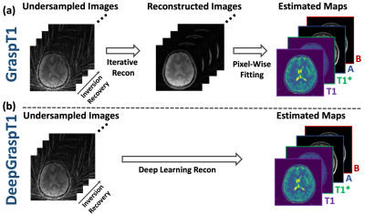 |
104 |
DeepGraspT1: Deep Learning-Enabled GRASP T1 Mapping
Haoyang Pei1,2,
Ding Xia1,
Fang Liu3,
and Li Feng1
1Biomedical Engineering and Imaging Institute and Department of Radiology, Icahn School of Medicine at Mount Sinai, New York, NY, United States, 2Department of Electrical and Computer Engineering, NYU Tandon School of Engineering, New York, NY, United States, 3Athinoula A. Martinos Center for Biomedical Imaging, Massachusetts General Hospital, Harvard Medical School, Boston, MA, United States Keywords: Machine Learning/Artificial Intelligence, Machine Learning/Artificial Intelligence Golden-angle RAdial Sparse Parallel (GRASP) MRI has recently been extended for rapid, accurate and robust T1 mapping (GraspT1) that can be performed during free breathing. However, GraspT1 implements a conventional T1 mapping framework that reconstructs an image series from undersampled dynamic k-space in the first step and then performs pixel-wise parameter fitting in the second step. This leads to a slow and cumbersome pipeline to obtain T1 maps. In this work, we developed deep learning-based GraspT1 (DeepGraspT1), which directly estimates T1 maps from undersampled k-space and enables additional acceleration that outperforms conventional iterative reconstruction. |
|
4035.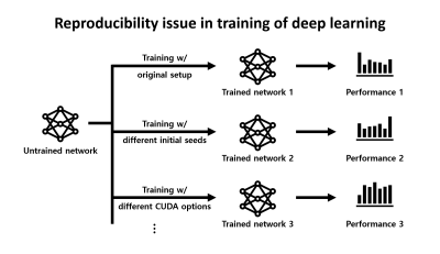 |
105 |
Exploring reproducibility in deep learning-based parallel
imaging reconstruction
Chungseok Oh1,
Hongjun An1,
and Jongho Lee1
1Seoul National University, Seoul, Korea, Republic of Keywords: Machine Learning/Artificial Intelligence, Machine Learning/Artificial Intelligence The performance of a deep neural network can be affected by software and hardware setups when training the network and, therefore, can vary from training to training. This issue of reproducibility, which can be referred to as an “intrinsic” reproducibility of deep learning, can be critical for academic research because reproducibility is a key requirement for journal papers. In this study, we explore this intrinsic reproducibility issue for deep learning-powered parallel imaging reconstruction by using a popular end-to-end variational network. This study may provide minimal requirements for reproducible research in network training. |
|
4036.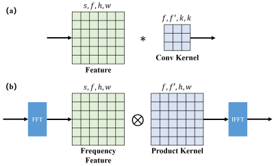 |
106 |
Fouier Convolution Nerual Network for MRI reconstruction
Haozhong Sun1,
Yuze Li1,
Runyu Yang1,
Zhongsen Li1,
and Huijun Chen1
1Center for Biomedical Imaging Research, Tsinghua University, Beijing, China Keywords: Machine Learning/Artificial Intelligence, Machine Learning/Artificial Intelligence Various image reconstruction methods have been proposed to reduce Magnetic resonance (MR) image acquisition time. One of recent trends is convolution neuron network (CNN) based deep learning model. However, most of these CNN models keep architecture of stacking small filters (e.g. 1×1 or 3×3) and the effective receptive field of these networks is limited, which is undesired for reconstruction because the random undersampling pattern causes global artifact. We proposed Fourier convolution block (FCB) to replace regular convolution filters. FCB can achieve both global receptive field and high computing efficiency by multiplication in frequency domain. |
|
4037.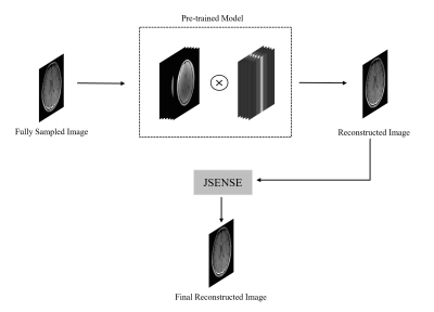 |
107 |
Improving JSENSE Using an Initial Reconstruction with an
Unrolled Deep Network Prior
Gulfam Ahmed Saju1,
Zhiqiang Li2,
Reza Abiri3,
Tianming Liu4,
and Yuchou Chang1
1Computer and Information Science, University of Massachusetts Dartmouth, North Dartmouth, MA, United States, 2Neuroradiology, Barrow Neurological Institute, Phoenix, AZ, United States, 3Electrical, Computer and Biomedical Engineering, University of Rhode Island, Kingston, RI, United States, 4Computer Science, University of Georgia, Athens, GA, United States Keywords: Machine Learning/Artificial Intelligence, Parallel Imaging JSENSE iteratively optimizes sensitivity maps and the image, so the sensitivity profile can be gradually improved during the reconstruction process. The initially reconstructed image in the first iteration is obtained by the initially estimated coil sensitivity maps. The initial coil sensitivity profiles may be inaccurate and therefore degrade the quality of the subsequent image quality and coil sensitivity map estimation in the iterative optimization process. We propose to use unrolled deep network prior to replace the initial reconstruction in the conventional JSENSE for improving the image reconstruction quality. Experimental results show that the proposed method outperforms CG-SENSE and JSENSE. |
|
4038.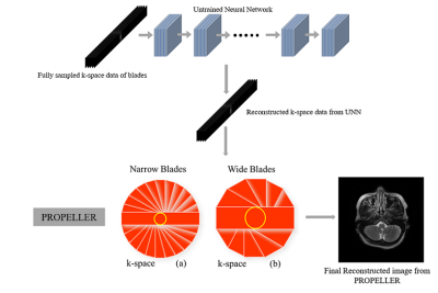 |
108 |
Incorporating Untrained Neural Network Prior in PROPELLER
Imaging
Gulfam Ahmed Saju1,
Zhiqiang Li2,
Reza Abiri3,
Tianming Liu4,
and Yuchou Chang1
1Computer and Information Science, University of Massachusetts Dartmouth, North Dartmouth, MA, United States, 2Neuroradiology, Barrow Neurological Institute, Phoenix, AZ, United States, 3Electrical, Computer and Biomedical Engineering, University of Rhode Island, Kingston, RI, United States, 4Computer Science, University of Georgia, Athens, GA, United States Keywords: Machine Learning/Artificial Intelligence, Motion Correction Periodically Rotated Overlapping ParallEL Lines with Enhanced Reconstruction (PROPELLER) MRI technique enables the correction of motion artifacts resulted from patient motions in a scanner. Undersampling the blades can increase data acquisition speed and reduce potential motions caused by pains in a short time but may degrade image quality. Deep neural networks may support the blade reconstruction with undersampled data but motion patterns are difficult to be acquired for building a training dataset. To avoid the acquisition of training data, this abstract proposes an untrained neural network-based PROPELLER reconstruction technique to enhance image quality with undersampled blades. |
|
4039.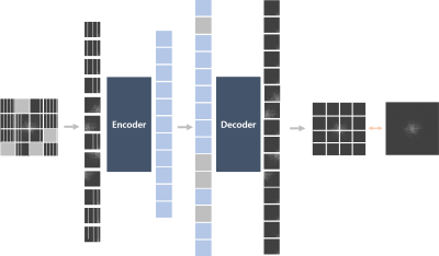 |
109 |
MR reconstruction in k-space using Vision Transformer boosted
with Masked Image Modeling
Jaa-Yeon Lee1 and
Sung-Hong Park2
1Korea Advanced Institute of Science & Technology, Daejeon, Korea, Republic of, 2Korea Advanced Institute of Science & Technology, Daejoen, Korea, Republic of Keywords: Machine Learning/Artificial Intelligence, Machine Learning/Artificial Intelligence, Vision Transformer, Masked Image Modeling Masked image modeling (MIM) skim has recently been shown to pre-train vision transform (ViT) and works as an effective data augmentation. In this study, we proposed MR reconstruction algorithm using ViT with MIM in k-space. The proposed method showed better performance than the original 1D and 2D ViT. Our study showed that MIM can be used to enhance the reconstruction quality to help learn the data distribution and that combining the loss in both k-space and spatial domain reconstructs images with better perceptuality. |
|
4040.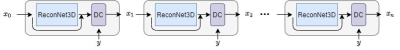 |
110 |
Deep learning acceleration and reconstruction for single-channel
signals
Yongquan Ye1,
Zhongqi Zhang1,
Eric Z Chen2,
Xiao Chen2,
Shanhui Sun2,
and Jian Xu1
1United Imaging, Houston, TX, United States, 2United Imaging Intelligence, Cambridge, MA, United States Keywords: Machine Learning/Artificial Intelligence, Image Reconstruction The capacity of the deep learning ReconNet3D model for single-channel image reconstruction with highly under-sampled k-spaces is demonstrated. Without the need for coil sensitivity information, the proposed method can achieve an acceleration factor of 4 on a dual-channel VTC coil. Supporting high-factor acceleration with limited coil channels can be very beneficial for imaging with surface coils or preclinical animal scans. |
|
4041.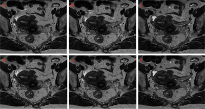 |
111 |
Deep Learning Based Reconstruction Improved Image Quality for
rectum T2-weighted imaging
Weiming Feng1,
Lan Zhu1,
Yihan Xia1,
Kangning Wang1,
Yong Zhang2,
Jiankun Dai3,
Guifeng Fu3,
and Huan Zhang1
1Ruijin Hospital, Shanghai Jiao Tong University School of Medicine, Shanghai, China, 2GE Healthcare, Shanghai, China, 3GE Healthcare, Beijing, China Keywords: Machine Learning/Artificial Intelligence, Image Reconstruction, rectum; magnetic resonance imaging; T2-weighted imaging High-resolution MRI is of much significance in preoperatively staging rectal cancer. However, the motion artifact from intestinal peristalsis inevitably affects image quality then the accuracy of staging. Deep learning reconstruction (DLRecon) that uses artificial neural networks to extract patterns and makes predictions from large data sets, has been verified in related studies for improving image quality and reducing scanning time. In this study, rectum T2-weighted imaging (T2WI) reconstructed with DLRecon and conventional reconstruction were evaluated, and the results indicate that DLRecon could be employed for better image quality without extra scanning time in clinical practice. |
|
4042.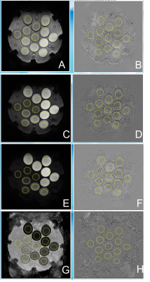 |
112 |
Quantitative Evaluation of Deep Learning Reconstruction of
Diffusion-weighted MRI using a DWI Phantom
Xiangchuang Kong1,
Shengzhen Tao1,
Eric H. Middlebrooks1,
Thomas Benkert2,
Xiangzhi Zhou1,
and Chen Lin1
1Department of Radiology, Mayo Clinic, Jacksonville, FL, United States, 2Siemens Medical Solutions USA, Inc., Jacksonville, FL, United States, JACKSONVILLE, FL, United States Keywords: Machine Learning/Artificial Intelligence, Diffusion/other diffusion imaging techniques, deep learning reconstruction; DWI phantom In a quantitative phantom study, deep learning (DL) reconstruction is shown to improve the SNR ratio of DWI images while preserving measured ADC values. The average SNR gain is similar to that achieved with better gradient performance between Siemens Prisma and Vida. Such benefits of DL recon should allow better quality and/or shorter scanning of DWI in clinical applications. |
|
4043.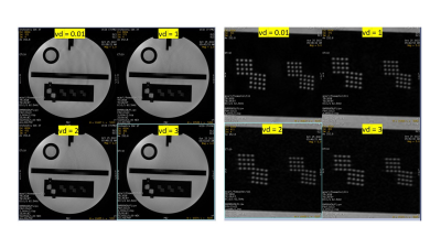 |
113 |
Deep Learning Reconstruction for 4-fold Accelerated 2DFSE
Imaging: optimization of variable density undersampling
Michael Carl1,
Rafi Brada2,
Nir Mazor2,
Daniel V Litwiller3,
and Maggie Fung4
1GE Healthcare, San Diego, CA, United States, 2GE Research, Herzliya, Israel, 3GE Healthcare, Denver, CO, United States, 4GE Healthcare, New York, NY, United States Keywords: Machine Learning/Artificial Intelligence, Machine Learning/Artificial Intelligence In this work we use variable-density prospective undersampling of the phase-encode k-space lines (ky) in 2D fast spin-echo (2DFSE) followed by deep learning (DL) reconstruction. We were able to achieve an acceleration of R=4 while maintaining high image quality. |
|
4044.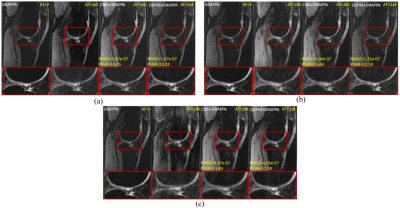 |
114 |
Highly Accelerated Multi-channel 3D Knee Imaging Using Denoising
Diffusion Probabilistic Model (DDPM) and GRAPPA
Ruiying Liu1,
Jee Hun Kim2,
Hongyu Li1,
Peizhou Huang1,
Xiaojuan Li2,
and Leslie Ying1
1Department of Biomedical Engineering, Department of Electrical Engineering, University at Buffalo, Buffalo, NY, United States, 2Program of Advanced Musculoskeletal Imaging (PAMI), Cleveland Clinic, Cleveland, OH, United States Keywords: Image Reconstruction, Machine Learning/Artificial Intelligence Deep learning methods have achieved superior reconstruction results in MRI reconstruction. Recently, denoising diffusion probabilistic models (DDPM) have demonstrated great potential in image-processing tasks. In this study, we combine denoising diffusion probabilistic models (DDPM) and GRAPPA for highly accelerated 3D imaging. The method sequentially performs DDPM and GRAPPA with specially designed sampling masks such that the benefits of the diffusion model and the availability of multi-channel data can be utilized jointly. Our results demonstrate that the proposed method can achieve an acceleration factor of up to 16 which is the product of the factors achieved by DDPM and GRAPPA alone. |
|
4045.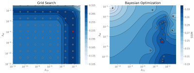 |
115 |
Finding Optimal Regularization Parameter for Undersampled
Reconstruction using Bayesian Optimization
Alberto Di Biase1,2,
Claudia Prieto1,2,
and Rene Botnar1,2
1Department of Electrical Engineering, Pontificia Universidad Católica de Chile, Santiago, Chile, 2Millennium Institute for Intelligent Healthcare Engineering (iHEALTH), Santiago, Chile Keywords: Image Reconstruction, Machine Learning/Artificial Intelligence, Compressed sensing We present a Bayesian optimization approach to find the optimal regularization parameters for undersampled MRI reconstructions such as Compressed Sensing (CS). Bayesian optimization can find optimal points for expensive to evaluate functions by efficient sampling the next points to try by maximizing the expected improvement. Additionally, the use of pruning can speed up optimization by stopping early unpromising trials. We demonstrate the effectiveness of this optimization technique by finding optimal parameters for undersampled MRI using Total Variation and CS-Wavelet regularization. The parameters found with the proposed approach are comparable to does found by grid search but requiring shorter computational times. |
|
4046. |
116 |
Quantitative evaluation of denoising algorithms without
noise-free ground-truth data
Laura Pfaff1,2,
Fabian Wagner1,
Julian Hossbach1,2,
Elisabeth Preuhs1,
Dominik Nickel2,
Tobias Wuerfl2,
and Andreas Maier1
1Pattern Recognition Lab, Friedrich-Alexander-Universität Erlangen-Nürnberg, Erlangen, Germany, 2Magnetic Resonance, Siemens Healthcare GmbH, Erlangen, Germany Keywords: Image Reconstruction, Machine Learning/Artificial Intelligence In MRI, the quantitative evaluation of denoising methods is often limited due to the lack of noise-free ground-truth data. We show how to still approximate the quality metrics mean squared error (MSE) and peak signal-to-noise-ratio (PSNR) without access to ground-truth data by using Stein’s unbiased risk estimator (SURE). The proposed method can be employed to evaluate learning- and non-learning-based denoising approaches, assuming an additive Gaussian noise model with known distribution. Our experiments further reveal that the accuracy of our evaluation method increases with the number of test samples available. |
|
4047.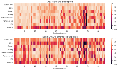 |
117 |
Impact of Deep Learning-Based MR Image Reconstruction Algorithms
on Abdominal MRI Radiomic Features
Hailong Li1,
Vinicius Vieira Alves1,
Amol Pednekar1,
Mary Kate Manhard1,
Joshua Greer1,
Andrew T. Trout1,
Lili He1,
and Jonathan R. Dillman1
1Cincinnati Children's Hospital Medical Center, Cincinnati, OH, United States Keywords: Image Reconstruction, Machine Learning/Artificial Intelligence, deep learning reconstruction Deep learning (DL)-based techniques are increasingly being applied to assist with or improve image reconstruction. However, the impact of DL-based algorithms on radiomics is not well understood. This study aims to evaluate the impact of two commercially available DL-based reconstruction pipelines: (1) SmartSpeed (Philips Healthcare, U.S. FDA-cleared); and (2) SmartSpeed with Super Resolution (SmartSpeed+SuperRes, not U.S. FDA-cleared to date) on MRI radiomic features. Our analysis showed that compared to conventional image reconstruction technique, 42 out of 86 investigated radiomic features from SmartSpeed images were highly correlated whereas only 13 features from SmartSpeed+SuperRes images had high correlations with conventional image features. |
|
4048.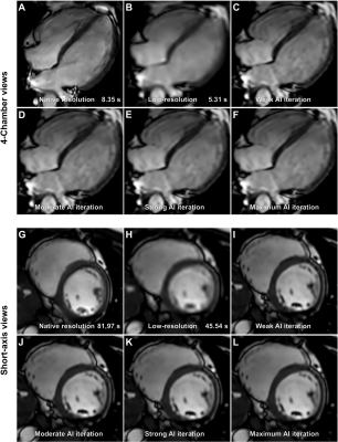 |
118 |
AI assisted upscaling of low-resolution cardiac MRI reduces
acquisition time and yields qualitatively comparable images to
standard techniques
Dmitrij Kravchenko1,2,
Alexander Isaak1,2,
Narine Mesropyan1,2,
Claus Christian Pieper1,
Daniel Kuetting1,2,
Leon M. Bischoff1,2,
Shuo Zhang3,
Christoph Katemann3,
Johannes M. Peeters4,
Oliver Weber3,
Ulrike Attenberger1,
and Julian Luetkens1,2
1Diagnostic and interventional radiology, University Hospital Bonn, Bonn, Germany, 2Quantitative Imaging Laboratory Bonn, Bonn, Germany, 3Philips GmbH Market DACH, Hamburg, Germany, 4Philips MR Clinical Science, Best, Netherlands Keywords: Machine Learning/Artificial Intelligence, Cardiovascular AI assisted upscaling of low-resolution cine bSSFP images yields comparable image quality to conventional images with no clinically significant difference in volumetric data at a reduction of acquisition time by a factor of 1.5 to 2. Keywords: Artificial intelligence, acceleration, Superresolution, cardiac MRI |
|
4049. |
119 |
Self-Supervised Denoising for Longitudinal MRI
Matt Hemsley1,2,
Liam S.P Lawrence1,2,
and Angus Z Lau1,2
1Medical Biophysics, University of Toronto, Toronto, ON, Canada, 2Physical Sciences Platform, Sunnybrook Research Institute, Toronto, ON, Canada Keywords: Machine Learning/Artificial Intelligence, Cancer, Denoising, Self-Supervised In MR guided radiation therapy, images of patients are acquired daily. However, scan times are long to acquire images with an acceptable SNR for treatment planning and adaptation. In this study we test a self-supervised machine learning based approach for denoising data that can utilize previous scans of the same patient to improve quality of the denoised image. Results on a numerical phantom and clinical images are presented and compared to a popular non-machine learning denoising algorithm. |
|
4050. |
120 |
Safeguarded Deep Unfolding Network for Parallel MR Imaging
Zhuo-Xu Cui1,
Sen Jia2,
Jing Cheng2,
Qingyong Zhu1,
and Dong Liang1,3
1Medical AI Research Center, Shenzhen Institutes of Advanced Technology, Chinese Academy of Science, Shenzhen, China, 2Paul C. Lauterbur Research Center for Biomedical Imaging, Shenzhen Institutes of Advanced Technology, Chinese Academy of Science, Shenzhen, China, 3Pazhou Lab, Guangzhou, China Keywords: Machine Learning/Artificial Intelligence, Image Reconstruction This study proposes a safeguarded methodology for network unrolling. Specifically, focusing on parallel MR imaging, we unroll a zeroth-order algorithm, of which the network module represents a regularizer itself so that the network output can still be covered by a regularization model. Furthermore, inspired by the idea of deep equilibrium models, before backpropagation, we carry out the unrolled network to converge to a fixed point and then prove that it can tightly approximate the real MR image. In a case where the measurement data contain noise, we prove that the proposed network is robust against noisy interferences. |
|
 Back to Meeting Home
 Back to the Program-at-a-Glance
The International Society for Magnetic Resonance in Medicine is accredited by the Accreditation Council for Continuing Medical Education to provide continuing medical education for physicians.
 Back to Meeting Home
 Back to the Program-at-a-Glance
 View Presentation Video