Digital Poster
ML/AI for Disease Detection, Diagnosis & Prediction
ISMRM & ISMRT Annual Meeting & Exhibition • 03-08 June 2023 • Toronto, ON, Canada

Digital Poster
ML/AI for Disease Detection, Diagnosis & Prediction
| Computer # | |||
|---|---|---|---|
1878.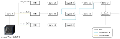 |
61 | Deep Learning Assisted Diagnosis of Prostate Cancer: Using a Multi-scale Neural Network Based on Points of Interest
Weiting Huang1, GuoRui Hou 2, Chen Wang3, and Kai Ai4
1Department of Magnetic Resonance, Lanzhou University Second Hospital, Lanzhou, China, 2Department of Magnetic Resonance, Xijing Hospital, Xi'an, China, 3Department of Radiology, Xijing Hospital, Xi'an, China, 4Philips Healthcare, Xi’an, China Keywords: Prostate, Machine Learning/Artificial Intelligence, Spatial Transformer Network, Transfer learning In this study, we propose a method to predict clinically significant state cancer based on MRI points of interest (POI) and classification network with multi-mode and multi-scale. Instead of the traditional method of manual delineation region-of-interest (ROI) to assist prediction, our method utilizes multi-scale input combined with Spatial Transformer Network (STN) to automatically adjust the adjust the scale of interest. This work also explored the possibility of predicting the grade of prostate cancer in a small amount of data using the method of transfer learning. Experiments show that this method has high prediction performance. |
|
1879.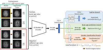 |
62 | Age and gender prediction from minimally processed 3D structural brain MRI through multi-task contrastive learning
Vick Lau1,2, Christopher Man1,2, Shi Su1,2, Ye Ding1,2, Jiahao Hu1,2, Junhao Zhang1,2, Yujiao Zhao1,2, Alex T. L. Leong1,2, and Ed X. Wu1,2
1Laboratory of Biomedical Imaging and Signal Processing, The University of Hong Kong, Hong Kong SAR, China, 2Department of Electrical and Electronic Engineering, The University of Hong Kong, Hong Kong SAR, China Keywords: Machine Learning/Artificial Intelligence, Machine Learning/Artificial Intelligence Predicting brain age from structural MRI (sMRI) is potentially valuable as the deviation of predicted age from chronological age can be a biomarker for characterising brain health conditions. Currently, extensive pre-processing of sMRI data is required for most deep learning methods. This study presents a multi-task contrastive learning framework for simultaneous brain age prediction and gender classification from minimally processed, noisy 3D T1-weighted images. By including gender classification task and supervised contrastive learning, we demonstrate that leveraging gender information in training and better representation learning can boost age prediction accuracy for both in-domain and out-of-domain datasets. |
|
1880.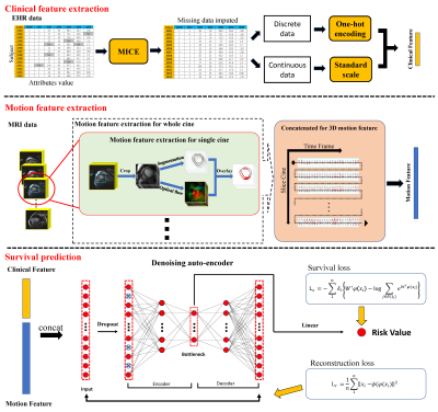 |
63 | Deep learning-based prognostic model using non-enhanced cardiac cine MRI in patients with heart failure.
Yifeng Gao1, Zhen Zhou1, Bing Zhang2, Saidi Guo2, Kairui Bo1, Shuang Li1, Nan Zhang1, Hui Wang1, Yang Guang3, Heye Zhang2, Tong Liu4, Jianxiu Lian5, and Lei Xu1
1Department of Radiology, Beijing Anzhen Hospital, Beijing, China, 2School of Biomedical Engineering, Sun Yat-Sen University, Guangzhou, China, 3National Heart and Lung Institute, Imperial College London, London, United Kingdom, 4Department of Cardiology, Beijing Anzhen Hospital, Beijing, China, 5Philips Healthcare, Beijing, China Keywords: Machine Learning/Artificial Intelligence, Heart, Deep Learning; Heart Failure We proposed a multi-source deep-learning model including traditional functional parameters and myocardial strain derived from cardiovascular magnetic resonance, as well as clinical features such as laboratory tests, electrocardiograms, and echocardiography. Meanwhile, we innovatively integrated cardiac motion characteristics through deep learning algorithms and fast neural convolution networks to construct deep learning heart failure prediction model. The results showed that compared with the traditional cox model, the deep learning model had higher efficacy for prognosis evaluation in patients with heart failure and could provide risk stratification in patients with heart failure, which may further guide clinical decision-making. |
|
1881.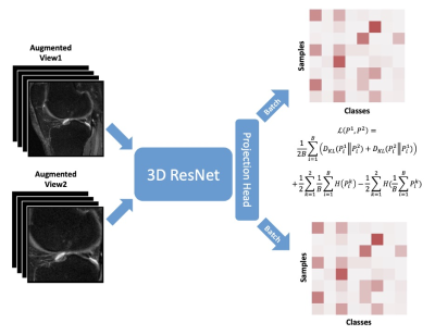 |
64 | Total Knee Replacement Prediction using Twin Class Distribution Estimation
Chaojie Zhang1, Shengjia Chen1, Haoxu Huang2, Haresh Rengaraj Rajamohan3, Jungkyu Park1, Noah Kasmanoff1, Kyunghyun Cho3, Gregory Chang1, Richard Kijowski1, and Cem M. Deniz1
1Department of Radiology, New York University Langone Health, New York, NY, United States, 2Courant Institute of Mathematical Sciences, New York University, New York, NY, United States, 3Center for Data Science, New York University, New York, NY, United States Keywords: Machine Learning/Artificial Intelligence, Machine Learning/Artificial Intelligence Our study implemented the self-supervised learning method, Twin Class Distribution Estimation, with unlabeled knee MR images. The self-supervised pretraining improves the downstream analysis in predicting total knee replacement within 9 years using labeled knee MR images. The self-supervised features are shown to be efficient classifiers in TKR prediction. |
|
1882.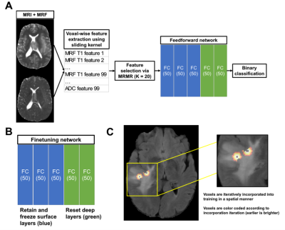 |
65 | Semi-Supervised Learning with Spatial Pseudo Labeling for Peritumoral Infiltration Prediction in Glioblastoma using MR Fingerprinting
Walter Zhao1, Xiaofeng Wang2, Charit Tippareddy3, Hamed Akbari4,5, Anahita Fathi Kazerooni4,5, Christos Davatzikos4,5, Marta Couce6, Andrew E. Sloan6,7,8,9, Chaitra Badve3, and Dan Ma1
1Department of Biomedical Engineering, Case Western Reserve University, Cleveland, OH, United States, 2Department of Quantitative Health Sciences, Cleveland Clinic, Cleveland, OH, United States, 3Department of Radiology, Case Western Reserve University and University Hospitals Cleveland Medical Center, Cleveland, OH, United States, 4Center for Biomedical Image Computing and Analytics, Perelman School of Medicine, University of Pennsylvania, Philadelphia, PA, United States, 5Department of Radiology, Perelman School of Medicine, University of Pennsylvania, Philadelphia, PA, United States, 6Department of Pathology, Case Western Reserve University and University Hospitals Cleveland Medical Center, Cleveland, OH, United States, 7Department of Neurosurgery, Case Western Reserve University and University Hospitals Cleveland Medical Center, Cleveland, OH, United States, 8Seidman Cancer Center and Case Comprehensive Cancer Center, Cleveland, OH, United States, 9Piedmont Health, Atlanta, GA, United States Keywords: Machine Learning/Artificial Intelligence, Cancer Existing glioblastoma (GB) infiltration models are often limited by lack of true infiltration labels and employ the assumption that edema closer to tumor has higher infiltrative potential relative to distant edema. Here, we propose a semi-supervised learning scheme that incorporates pretraining on the near-far heuristic and spatial pseudo labeling using true infiltration labels for voxel-wise tumor infiltration prediction. Our results show improved classification performance following finetuning on labeled infiltration data compared to training on the near-far heuristic alone and indicate the potential in employing MR fingerprinting-based models to guide GB diagnosis and treatment. |
|
1883.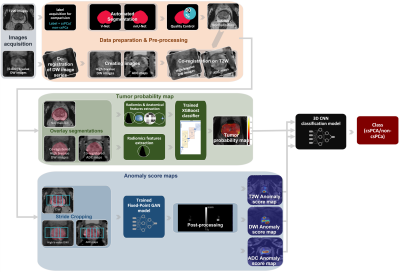 |
66 | Biparametric MRI classification model for prostate cancer detection using a combination of prediction maps
Mohammed R. S. Sunoqrot1,2, Rebecca Segre1,3, Gabriel A. Nketiah1,2, Alexandros Patsanis1, Tone F. Bathen1,2, and Mattijs Elschot1,2
1Department of Circulation and Medical Imaging, Norwegian University of Science and Technology (NTNU), Trondheim, Norway, 2Department of Radiology and Nuclear Medicine, St. Olavs Hospital, Trondheim University Hospital, Trondheim, Norway, 3Department of Mechanical and Aerospace Engineering, Politecnico di Torino, Torino, Italy Keywords: Machine Learning/Artificial Intelligence, Prostate Biparametric MRI (bpMRI) is a valuable tool for the diagnosis of prostate cancer (PCa). Computer-aided detection and diagnosis (CAD) systems have the potential to improve the robustness and efficiency of PCa detection) compared with conventional radiological reading. This work explores the combination of the results of two PCa CAD systems as input to a new classification model. We show that this is promising approach that can potentially improve the final detection of PCa. |
|
1884.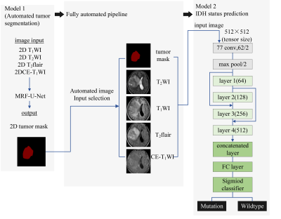 |
67 | Prediction of IDH Mutation Status of Gliomas Using Pre-operative MR Images: a Fully Automatic Convolutional Neural Networks Based Approach.
Xiaohua Chen1, Zhiqiang Chen2, Zhuo Wang1, Shaoru Zhang1, Yunshu Zhou1, Shili Liu1, Ruodi Zhang1, Yuhui Xiong3, and Aijun Wang4
1Clinical medicine school of Ningxia Medical University, Yinchuan, China, 2Department of Radiology ,the First Hospital Affiliated to Hainan Medical College, Haikou, China, 3GE Healthcare MR Research, Beijing, China, 4Department of Radiology, General Hospital of Ningxia Medical University, Yinchuan, China Keywords: Machine Learning/Artificial Intelligence, Brain Using an automated method of convolutional neural networks (CNNs), we aimed to predict the IDH mutation status of gliomas from conventional preoperative MRI in this study. We conclude that the Markov Random Field-U-Net network can accurately segment the tumor region. Using a modified 34-layer Resnet network, we were subsequently able to predict IDH mutation status effectively. Consequently, the model has the potential to be utilized more broadly as a practical tool with reproducibility, this model has the potential to be a practical tool for the non-invasive characterization of gliomas to help the individualized treatment planning. |
|
1885.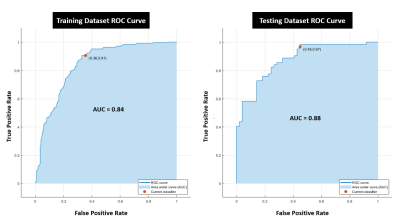 |
68 | Prediction of active Multiple Sclerosis lesions through use of logistic regression classifier and first-order features
Vivian S. Nguyen1,2, Adam J. Hasse3, Emily Tao1, Jihye Jang4, Adil Javed5, Timothy J. Carroll3, and Keigo Kawaji1,2
1Biomedical Engineering, Illinois Institute of Technology, Chicago, IL, United States, 2Medicine - Cardiology, University of Chicago Medical Center, Chicago, IL, United States, 3Radiology, University of Chicago Medical Center, Chicago, IL, United States, 4Philips Healthcare, Gainesville, FL, United States, 5Neurology, University of Chicago Medical Center, Chicago, IL, United States Keywords: Machine Learning/Artificial Intelligence, Multiple Sclerosis Multiple Sclerosis is a neuroinflammatory disease in which the immune system attacks nerve fibers and myelin sheaths, leading to the formation of lesions through white matter. Gadolinium-enhanced MRI is used to diagnose and track the progression of MS. Active MS lesions enhance with gadolinium, but there is an interest in prediction of lesion enhancement based on lesion features. In this study, we examined first-order features derived from T1w pre-contrast MS lesions acquired on multiple 3T imagers at a single center to train a logistic regression classifier to classify lesions as active or inactive. |
|
1886.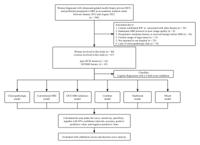 |
69 | Prediction of ductal carcinoma in situ with microinvasion postoperatively in women with biopsy-confirmed ductal carcinoma in situ
Zhou Huang1, Xue Chen2, Nan Jiang1, Su Hu1, and Chunhong Hu1
1Department of Radiology, The First Affiliated Hospital of Soochow University, Suzhou, China, 2Department of Radiology, The Affiliated Suzhou Hospital of Nanjing Medical University, Suzhou, China Keywords: Machine Learning/Artificial Intelligence, Breast To predict ductal carcinoma in situ with microinvasion (DCISMI) based on clinicopathologic, conventional breast magnetic resonance imaging (MRI), and dynamic contrast enhanced MRI (DCE-MRI) radiomics signatures in women with biopsy-confirmed ductal carcinoma in situ (DCIS) to choose high-risk women who may benefit from sentinel lymph node biopsy at initial surgery. The mixed model showed better AUC values than both clinicopathological and DCE-MRI radiomics models in both training/test sets with heterogeneous enhancement and radiomics scores as significant independent predict factors. The mixed model showed the greatest overall net benefit for upstaging and the second was the combine model. |
|
1887.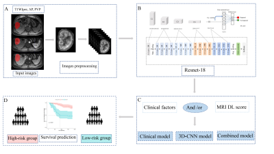 |
70 | Development and validation of a deep learning model trained on MRI for the prediction of hepatocellular carcinoma survival
Lidi Ma1, Congrui Li2, Haixia Li3, Kan Deng3, Cheng Zhang1, Weijing Zhang1, and Chuanmiao Xie1
1Department of Radiology, Sun Yat-sen University Cancer Center, Guangzhou, China, 2Department of Diagnostic Radiology, Hunan Cancer Hospital, Central South University, Changsha, China, 3Philips Healthcare, Guangzhou, China Keywords: Machine Learning/Artificial Intelligence, Liver In this study, we developed a deep learning model based on GD-DTPA-enhanced MRI data to predict the overall survival (OS) of patients with HCC. Our results showed that 3D-CNN model based on GD-DTPA-enhanced MRI can non-invasively predict the OS of patients with HCC. The combined model integrating the deep learning score and clinical factors showed a higher predictive value than the clinical and 3D-CNN models and may be more useful in guiding clinical treatment decisions to improve the prognosis of patients with HCC. |
|
1888.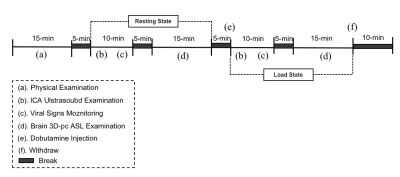 |
71 | Machine learning methods for cerebral perfusion status prediction
Linkun Cai1, Haijun Niu1, Erwei Zhao2, Yawen Liu1, Tingting Zhang1, Dong Liu3, Penggang Qiao4, Pengling Ren4, Wei Zheng2, and Zhenchang Wang4
1School of Biological Science and Medical Engineering,Beihang University, Beijing, China, 2National Space Science Center,Chinese Academy of Sciences, Beijing, China, 3Department of Ultrasound, Beijing Friendship Hospital, Beijing, China, 4Department of Radiology, Beijing Friendship Hospital, Beijing, China Keywords: Machine Learning/Artificial Intelligence, Machine Learning/Artificial Intelligence The diagnosis and evaluation of cerebral perfusion status are crucial for the management of brain diseases. However, the detection method of cerebral perfusion status is complicated. Considering that CBF is mainly supplied by the internal carotid artery (ICA), this paper proposes a novel cerebral perfusion status prediction model, which can automatically quantify the cerebral perfusion level of patients by modeling the association between ICA blood flow and cerebral perfusion. The experimental results on a real-world dataset using machine learning methods can achieve satisfactory performance. Thus, it can be used as an effective adjuvant tool for determining the cerebral perfusion status. |
|
1889.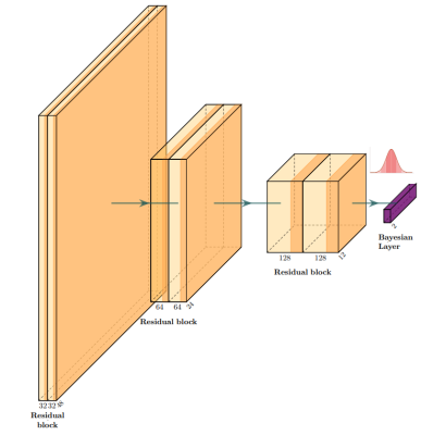 |
72 | BayesNetCNN: incorporating uncertainty in neural networks for image-based classification tasks
Matteo Ferrante1, Tommaso Boccato2, Marianna Inglese2, and Nicola Toschi3,4
1Biomedicine and prevention, University of Rome Tor Vergata, Roma, Italy, 2Biomedicine and prevention, University of Rome Tor Vergata, Rome, Italy, 3BioMedicine and prevention, University of Rome Tor Vergata, Rome, Italy, 4Department of Radiology,, Athinoula A. Martinos Center for Biomedical Imaging and Harvard Medical school, Boston, MA, USA, Boston, MA, United States Keywords: Machine Learning/Artificial Intelligence, Alzheimer's Disease The willingness to trust predictions formulated by automatic algorithms is key in a vast number of domains. We propose converting a standard neural network into a Bayesian neural network and estimating the variability of predictions by sampling different networks at inference time. We use a rejection-based approach to increase classification accuracy from 0.86 to 0.95 while retaining 75% of the test set for Alzheimer disease classification from MRI morphometry images. Estimating uncertainty of a prediction and modulating the behavior of the network to a desirable degree of confidence, represents a crucial step in the direction of responsible and trustworthy AI. |
|
1890.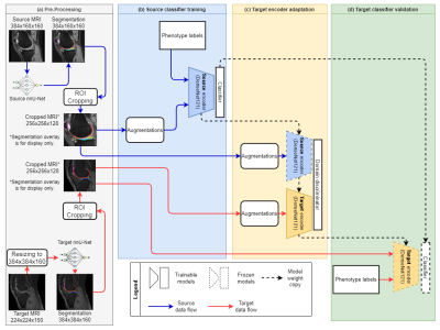 |
73 | Unsupervised Domain Adaptation for Automated Knee Osteoarthritis Phenotype Classification
Junru Zhong1, Yongcheng Yao1, Dόnal G. Cahill2, Fan Xiao3, Siyue Li1, Jack Lee4, Kevin Ki-Wai Ho5, Michael Tim-Yun Ong5, James F. Griffith2, and Weitian Chen1
1CU Lab for AI in Radiology (CLAIR), Department of Imaging and Interventional Radiology, The Chinese University of Hong Kong, Sha Tin, NT, Hong Kong, 2Department of Imaging and Interventional Radiology, The Chinese University of Hong Kong, Sha Tin, NT, Hong Kong, 3Department of Radiology, Shanghai Sixth People's Hospital Affiliated to Shanghai Jiao Tong University School of Medicine, Shanghai, China, 4Centre for Clinical Research and Biostatistics, The Chinese University of Hong Kong, Sha Tin, NT, Hong Kong, 5Department of Orthopaedics & Traumatology, The Chinese University of Hong Kong, Sha Tin, NT, Hong Kong Keywords: Osteoarthritis, Osteoarthritis We propose a knee osteoarthritis phenotype classification system using unsupervised domain adaptation (UDA). A convolutional neural network was initially trained on a large source dataset (Osteoarthritis Initiative, n=3116), then adapted to a small target dataset (n=50). We observed a significant performance improvement compared to the classifiers trained solely on the target dataset. We demonstrated the feasibility of applying UDA for medical image analysis in a small label-free dataset. |
|
1891.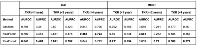 |
74 | RiskForm: A novel risk formulation to improve progressive disease outcome prediction
Haresh Rengaraj Rajamohan1, Kyunghyun Cho1, Richard Kijowski2, and Cem M. Deniz2
1New York University, NEW YORK, NY, United States, 2NYU Langone Health, NEW YORK, NY, United States Keywords: Osteoarthritis, Machine Learning/Artificial Intelligence, Deep Learning We propose a novel risk constraint to improve the performance of deep learning models on progressive disorders. On the Osteoarthritis Initiative (OAI) dataset, the proposed approach outperforms a baseline model trained with the standard cross-entropy loss on predicting total knee replacement (TKR) within 3 different time horizons- 1 year, 2 years and 4 years of the MRI date. It further generalizes better to the external Multicenter Osteoarthritis Study dataset. |
|
 |
1892.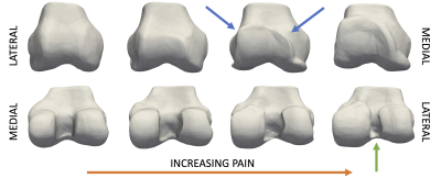 |
75 | Neural Shape Models Predict Knee Pain Better than Conventional Statistical Shape Models: Data from the Osteoarthritis Initiative
Anthony A Gatti1, Dave Van Veen2, Garry E Gold1, Scott L Delp3, and Akshay S Chaudhari1
1Radiology, Stanford University, Stanford, CA, United States, 2Electrical Engineering, Stanford University, Stanford, CA, United States, 3Bioengineering, Stanford University, Stanford, CA, United States Keywords: Osteoarthritis, Machine Learning/Artificial Intelligence MRI-based statistical shape models can predict future disease and distinguish between patient groups. However, these models require thousands of matching points between bones which may introduce biases and their strictly linearly orthogonal features is a limitation. This study built continuous 3D shape representations of the femur using neural implicit representations and used the learned latent space to predict knee pain. The neural shape model can generate arbitrarily high resolution surfaces and predict pain with area under the receiver operating characteristic curve of 0.7 and sensitivity of 0.89, metrics comparable to deep learning methods trained on orders of magnitude more data. |
1893.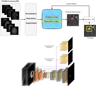 |
76 | Multi-Modal Detection and Localization of Intracranial Aneurysms using 3D nnDetection Deep Learning Model
Maysam Orouskhani1, Shaojun Xia2, Mahmud Mossa-Basha3, and Chengcheng Zhu3
1University of Washington, SEATTLE, WA, United States, 2Peking University Cancer Hospitals & Institution, Beijing, China, 3Department of Radiology, University of Washington, Seattle, WA, United States Keywords: Machine Learning/Artificial Intelligence, Machine Learning/Artificial Intelligence, Aneurysm detection, nnDetection, Aneurysm Localization Intracranial aneurysms are relatively common life-threatening diseases with a prevalence of 3.2% in the general population. Therefore, detection is a vital task in aneurysm management. Lesion detection refers to simultaneously localizing and categorizing the lesions in medical images. In this study, we employed nnDetection framework, a self-configuring framework for 3D medical object detection, to detect and localize the 3D coordination of aneurysms. To capture and extract diverse features of aneurysms, two modalities including TOF-MRA, and structural MRI from ADAM dataset have been used. The performance of the proposed deep learning model was evaluated by free-response receiver operative characteristics |
|
1894.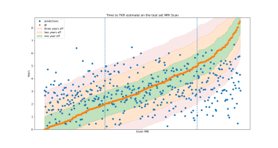 |
77 | Estimating time-to-total knee replacement surgery using deep learning
Eisa Hedayati1, Haresh Rajamohan2, Lily Zhou3, Kyunghyun Cho2, Gregory Chang1, Richard Kijowski1, and Cem M Deniz1
1Radiology, New York University Langone health, New York, NY, United States, 2Center for data Science, New York University, New York, NY, United States, 3Radiology & diagnostic Imaging, University of Alberta, Edmonton, AB, Canada Keywords: Machine Learning/Artificial Intelligence, Osteoarthritis "When do I need my knee replaced? " is a common question of patients with progressed osteoarthritis conditions. However, answering that question is not trivial, especially for cases that do not result in an immediate surgery. To help doctors addressing this question we employed neural networks to recommend an estimated subject-specific date for the total knee replacement (TKR). |
|
1895.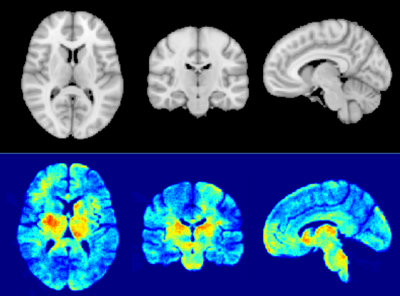 |
78 | GAMER-MRI and modified Layer-wise Relevance Propagation identify on quantitative MRI regions sensitive to clinical disability in MS patients
Po-Jui Lu1,2,3, Benjamin Odry4, Muhamed Barakovic1,2,3, Matthias Weigel1,2,3,5, Robin Sandkühler6, Reza Rahmanzadeh7, Xinjie Chen1,2,3, Mario Ocampo-Pineda1,2,3, Jens Kuhle2,3, Ludwig Kappos2,3, Philippe Cattin6, and Cristina Granziera1,2,3
1Translational Imaging in Neurology (ThINk) Basel, Department of Medicine and Biomedical Engineering, University Hospital Basel and University of Basel, Basel, Switzerland, 2Department of Neurology, University Hospital Basel, Basel, Switzerland, 3Research Center for Clinical Neuroimmunology and Neuroscience Basel (RC2NB), University Hospital Basel and University of Basel, Basel, Switzerland, 4AI for Clinical Analytics, Covera Health, New York, NY, United States, 5Division of Radiological Physics, Department of Radiology, University Hospital Basel, Basel, Switzerland, 6Center for medical Image Analysis & Navigation, Department of Biomedical Engineering, University of Basel, Allschwil, Switzerland, 7Neuroradiology Department, Inselspital, Bern, Switzerland Keywords: Machine Learning/Artificial Intelligence, Multiple Sclerosis The decision process of artificial intelligence is elusive. We proposed a new method that by combining an attention-based convolutional neural network (GAMER-MRI) with the modified Layer-wise Relevance Propagation could reveal relevant regions on quantitative imaging maps in differentiating multiple sclerosis patients with mild-moderate and severe disabilities. The assessment of the relevant regions included the impact of inverting values within the regions and the heatmap on the MNI152 template. Our results show good network performance and identify brain regions relevant to the corticospinal tract. The proposed method might be useful to further explore patterns of brain microstructural alterations associated with disability. |
|
1896.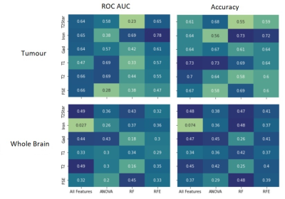 |
79 | Pre-Clinical MRI radiomics and machine learning to predict survival after immunotherapy treatment
Vlora Riberdy1, Alessandro Guida2,3, James Rioux1,2,3, and Kimberly Brewer1,2,3,4,5
1Physics and Atmospheric Science, Dalhousie University, Halifax, NS, Canada, 2Diagnostic Radiology, Dalhousie University, Halifax, NS, Canada, 3Biomedical MRI Research Laboratory, Nova Scotia Health Authority, Halifax, NS, Canada, 4School of Biomedical Engineering, Dalhousie University, Halifax, NS, Canada, 5Microbiology & Immunology, Dalhousie University, Halifax, NS, Canada Keywords: Molecular Imaging, Radiomics Molecular MRI allows for immunotherapy treatment monitoring of glioblastoma, but analysis of multi-parametric data is complex. Machine learning algorithms can be applied to quantitative maps, to identify correlations between radiomic features and treatment outcomes. Feature selection is key when dealing with longitudinal preclinical data with multiple contrasts and small group numbers. We evaluated three feature selection methods in terms of their ability to produce predictive models of survival. The best performance was seen using recursive feature elimination applied to features from iron concentration maps of the tumor, which yielded an ROC AUC of 0.78 and an accuracy of 0.72. |
|
1897.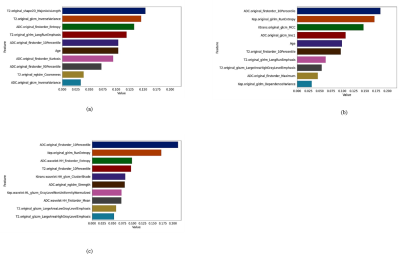 |
80 | Towards an optimal breast lesions predictive model by assessing different MRI protocols’ combinations: Radiomics analysis
Gelareh Valizadeh1, Fereshteh Khodadadi Shoushtari2, Soheila Koopaee1, Hanieh Mobarak Salari1, Mohammad Hossein Golezar3, Masomeh Gity4, and Hamidreza Saligheh Rad1,5
1Quantitative MR Imaging and Spectroscopy Group, Tehran University of Medical Sciences, Tehran, Iran (Islamic Republic of), 2Shiraz University, Shiraz, Iran (Islamic Republic of), 3Shahed University, Tehran, Iran (Islamic Republic of), 4Tehran university, Tehran, Iran (Islamic Republic of), 5Department of Medical Physics and Biomedical Engineering, Tehran University of Medical Sciences, Tehran, Iran (Islamic Republic of) Keywords: Multimodal, Breast This study aimed to assess added value of various combinations of different MRI protocols and artificial intelligence techniques in differentiation capability between malignant and benign breast lesions. 61 benign and 69 malignant lesions were recruited. Radiomics features were extracted from three proposed scenarios, including original images of ADC and T2W (scenario-I), original images of ADC, T2W, and DCE (scenario-II), and the joint of original and the pre-filtered images (using wavelet) from ADC, T2W and DCE (scenario-III). Ten most relevant features were utilized for training 11 machine learning algorithms. Finally, decision tree achieved the highest results of accuracy using scenario-III. |
|
 Back to Meeting Home
 Back to the Program-at-a-Glance
The International Society for Magnetic Resonance in Medicine is accredited by the Accreditation Council for Continuing Medical Education to provide continuing medical education for physicians.
 Back to Meeting Home
 Back to the Program-at-a-Glance
 View Presentation Video