Digital Poster
ML/AI for Recon & Super Resolution II
ISMRM & ISMRT Annual Meeting & Exhibition • 03-08 June 2023 • Toronto, ON, Canada

Digital Poster
ML/AI for Recon & Super Resolution II
| Computer # | |||
|---|---|---|---|
4187.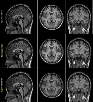 |
101 |
Improving Brain Volume Measurement Workflow using combination of
CS-MRI and Deep Learning based Super-Resolution
Atita Suwannasak1,
Uten Yarach1,
and Prapatsorn Sangpin2
1Chiang Mai university, Chiang Mai, Thailand, 2Philips Healthcare (Thailand), Bangkok, Thailand Keywords: Machine Learning/Artificial Intelligence, Brain For brain volume measurement (BVM), High-resolution (HR) MR images have shown to provide accurate results at small subcortical areas. However, prolonged scan time remains a classical challenge for 3D MRI. We implemented a combined technique, deep learning based super-resolution (DL-SR) and low-resolution Compressed Sensing (CS) 3D-TFE-T1W with acceleration factor 4 to generate Super-resolution (SR) images under one minute scan time. The results show that DL-SR model is able to improve image resolution, in which no significant differences (p>0.01) in quantitative volumetric measurement between reference and DL-SR at subcortical regions, except for caudate region. |
|
4188.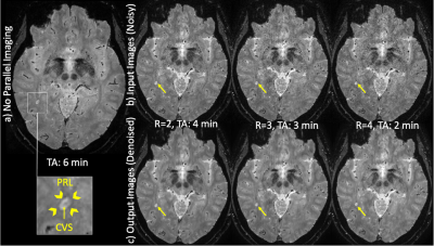 |
102 |
Denoising highly accelerated T2*-weighted brain MRI using a deep
learning convolutional neural network
Bryan Quah1,
Sreekanth Madhusoodhanan Nair1,
Jin Jin2,
Fei Han3,
Brian Renner1,
Elaina Gombos1,
Ke Cheng Liu3,
Sunil Patil3,
John A. Derbyshire4,
Ken Sakaie5,
Emmanuel Obusez5,
Jonathan Lee5,
Mark Elliot6,
Russell T. Shinohara7,
Matthew K. Schindler8,
Jae W. Song6,
Michel Bilello6,
Marwa Kaisey1,
Nader Binesh9,
Marcel Maya9,
Javier Galvan9,
Hui Han10,
Debiao Li10,
Andrew Solomon11,
Daniel S. Reich12,
Nancy L. Sicotte1,
Mark Lowe5,
Daniel Ontaneda13,
Omar Al-Louzi1,
and Pascal Sati1,10
1Department of Neurology, Cedars-Sinai Medical Center, Los Angeles, CA, United States, 2Siemens Healthcare Pty Ltd, Brisbane, Australia, 3Siemens Medical Solutions, PA, United States, 4Functional MRI Facility, National Institute of Mental Health, National Institutes of Health, Bethesda, MD, United States, 5Imaging Institute, Cleveland Clinic, Cleveland, OH, United States, 6Department of Radiology, University of Pennsylvania, Philadelphia, PA, United States, 7Department of Biostatistics, Epidemiology, and Informatics, Penn Statistics in Imaging and Visualization Center, University of Pennsylvania Perelman School of Medicine, Philadelphia, PA, United States, 8Department of Neurology, University of Pennsylvania, Philadelphia, PA, United States, 9Department of Imaging, Cedars-Sinai Medical Center, Los Angeles, CA, United States, 10Biomedical Imaging Research Institute, Cedars-Sinai Medical Center, Los Angeles, CA, United States, 11Larner College of Medicine, The University of Vermont, Burlington, VT, United States, 12Translational Neuroradiology Section, National Institute of Neurological Disorders and Stroke, National Institutes of Health, Bethesda, MD, United States, 13Mellen Center, Department of Neurology, Neurological Institute, Cleveland Clinic, Cleveland, OH, United States Keywords: Machine Learning/Artificial Intelligence, Data Processing, Denoising, Neuroimaging The scan time of high-resolution T2*-weighted brain imaging using 3D echo-planar imaging (3D-EPI) can be significantly reduced by applying Controlled Aliasing In Parallel Imaging Results In Higher Acceleration (CAIPIRINHA). However, this comes at the expense of a significant reduction in image quality. In this study, we evaluated the feasibility of using a deep learning-based approach (DnCNN) to denoise highly accelerated 3D-EPI scans acquired at 3T. Our results show that DnCNN was able to efficiently denoise highly accelerated T2*-weighted brain scans while preserving anatomical and pathological details. |
|
4189.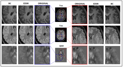 |
103 |
Enhanced-Deep-Super-Resolution Neural Network on Multiple MR
Brain Images
Cristiana Fiscone1,
Nico Curti2,
Matti Ceccarelli3,
David Neil Manners1,
Gastone Castellani2,
Caterina Tonon1,4,
Daniel Remondini5,6,
Raffaele Lodi1,4,
and Claudia Testa4,5
1Department of Biomedical and Neuromotor Sciences, University of Bologna, Bologna, Italy, 2Department of Experimental, Diagnostic and Specialty Medicine, University of Bologna, Bologna, Italy, 3Department of Agricultural and Food Sciences, University of Bologna, Bologna, Italy, 4Functional and Molecular Neuroimaging Unit, IRCCS Istituto delle Scienze Neurologiche di Bologna, Bologna, Italy, 5Department of Physics and Astronomy, University of Bologna, Bologna, Italy, 6INFN Bologna, Bologna, Italy Keywords: Machine Learning/Artificial Intelligence, Visualization, Super Resolution, Generalization Enhanced Deep Super Resolution (EDSR) is a machine learning model aimed to improve image spatial resolution. It was previously trained with general purpose figures and, in this work, directly tested on different MR images: T1w, T2w and Quantitative Susceptibility Mapping (QSM), a quantitative imaging technique. The studied cohort included 28 healthy subjects. Without needing fine-tuning, EDSR shows excellent ability of generalization over new kind of data, improving imaging visualization and outperforming the traditional bicubic upsampling. In future applications, images of patients will be considered to test EDSR reconstruction when there is pathological tissue. |
|
4190.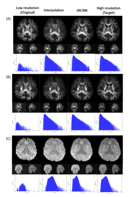 |
104 |
CNN-based three-dimensional superresolution technology in Brain
MRI with generalized q-sampling imaging
Chun-Yuan Shin1,
Yi-Ping Chao2,
Li-Wei Kuo3,4,
Yi-Peng Eve Chang5,
and Jun-Cheng Weng1,6,7
1Department of Medical Imaging and Radiological Sciences, and Department of Artificial Intelligence, Chang Gung University, Taoyuan, Taiwan, 2Department of Computer Science and Information Engineering, and Graduate Institute of Artificial Intelligence, Chang Gung University, Taoyuan, Taiwan, 3Institute of Biomedical Engineering and Nanomedicine, National Health Research Institutes, Miaoli, Taiwan, 4Institute of Medical Device and Imaging, National Taiwan University College of Medicine, Taipei, Taiwan, 5Department of Counseling and Clinical Psychology, Columbia University, New York City, NY, United States, 6Medical Imaging Research Center, Institute for Radiological Research, Chang Gung University and Chang Gung Memorial Hospital at Linkou, Taoyuan, Taiwan, 7Department of Psychiatry, Chang Gung Memorial Hospital, Chiayi, Taiwan Keywords: Machine Learning/Artificial Intelligence, Diffusion/other diffusion imaging techniques Understanding neural connections helps scientists conduct cognitive behavioral research. There are many nerve fiber intersections in the brain that need to be observed, and the size is between 30-50 nanometers. Improving image resolution has become an important issue. Generalized q-sampling imaging (GQI) was used to reveal the fiber geometry of straight and crossing. However, it is difficult to accurately describe fiber bending, fanning, and diverging with low-resolution imaging. In this work, we tried to achieve superresolution with a deep learning method on diffusion magnetic resonance imaging (MRI) images that has the potential to assess crossing, curving, and splaying fiber structures. |
|
4191.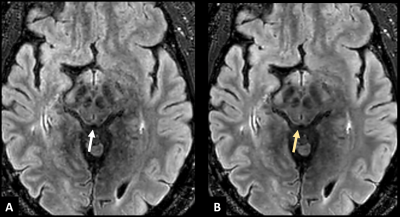 |
105 |
Artificial intelligence-based denoising for clinical magnetic
resonance imaging: from head to toe
Dallas Turley1,2,
Pattana Wangaryattawanich2,
Jalal Andre2,
Majid Chailan2,
Johannes M. Peeters3,
Kim Van de Ven3,
and Orpheus Kolokythas2
1Philips Healthcare, Seattle, WA, United States, 2Department of Radiology, University of Washington, Seattle, WA, United States, 3Philips Healthcare, Best, Netherlands Keywords: Machine Learning/Artificial Intelligence, Machine Learning/Artificial Intelligence Artificial intelligence in MR is a diverse and growing field. In this work, we apply convolutional neural networks (CNN) to accelerated routine clinical protocols to investigate potential image quality improvements. For moderate increase in reconstruction time, CNNs were judged by experienced radiologists to significantly improve image quality by reducing noise and artifact. |
|
4192.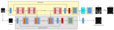 |
106 |
3D MRI super-resolution using convolutional generative
adversarial network with gradient guidance
Wei Xu1,
Jing Cheng1,
and Dong Liang1
1Shenzhen Institutes of Advanced Technology, Chinese Academy of Sciences, ShenZhen, China Keywords: Machine Learning/Artificial Intelligence, Brain The application of MRI has been limited due to the restriction of imaging time and spatial resolution. Super-resolution is an important strategy in clinics to speed up MR imaging. In this work, we propose a novel GAN-based super-resolution method which incorporates gradient features to improve the recovery of local structures of the super-resolution images. Experiments on 3D MR Vessel Wall imaging demonstrate the superior performance of the proposed method. |
|
4193.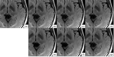 |
107 |
Convolutional-neural-network-based denoising with estimated
noise-based normalization to effectively reduce noise for
various noise levels
Atsuro Suzuki1,
Tomoki Amemiya1,
Yukio Kaneko1,
Suguru Yokosawa1,
and Toru Shirai1
1Imaging Technology Center, FUJIFILM Corporation, Tokyo, Japan Keywords: Machine Learning/Artificial Intelligence, Machine Learning/Artificial Intelligence, Noise reduction To develop a convolutional neural network (CNN)-based denoiser for various noise levels, we propose the use of estimated noise-based normalization in denoising. When the CNN-based denoiser with estimated noise-based normalization was applied to brain FLAIR images with various noise levels, it resulted in values closer to the normalized root mean square error (NRMSE) between the denoised and the target images compared with a conventional CNN-based denoiser trained with the same noise level as that in the input image. In conclusion, our method effectively reduced the noise in an image with various noise levels in terms of minimization of the NRMSE. |
|
4194.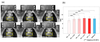 |
108 |
Knowledge Distillation Enables Efficient Neural Network for
Better Generalizability in MR Image Denoising and Super
Resolution
Qinyang Shou1,
Zechen Zhou2,
Kevin Blansit2,
Praveen Gulaka2,
Enhao Gong2,
Greg Zaharchuk2,
and Ajit Shankaranarayanan2
1University of Southern California, Los Angeles, CA, United States, 2Subtle Medical Inc., Menlo Park, CA, United States Keywords: Machine Learning/Artificial Intelligence, Machine Learning/Artificial Intelligence, Knowledge Distillation In this work, knowledge distillation (KD) is investigated to improve model generalizability for image enhancement tasks. KD can allow a 35× faster convolutional network to achieve similar performance as a Transformer based model in image denoising tasks. In addition, KD can enable a single image enhancement model for both denoising and super-resolution tasks that outperforms the conventional multi-task model trained with mixed data. KD potentially allows efficient image enhancement models to achieve better generalization performance for clinical translation. |
|
4195.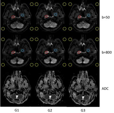 |
109 |
Clinical assessment of a deep learning-based denoising method
for DWI of Nasopharyngeal Cancer
Tiebao Meng1,
Haibin Liu1,
Haoqiang He1,
Jialu Zhang2,
and Long Qian2
1Department of Radiology, Sun Yat-Sen University Cancer Center, Guangzhou, China, 2MR Research, GE Healthcare, Beijing, China Keywords: Machine Learning/Artificial Intelligence, Diffusion/other diffusion imaging techniques Deep Learning reconstruction (DLR) has the potential to reduce MRI scan time while improving signal-to-noise ratio (SNR) and maintaining spatial resolution. This study evaluated results of DLR in 66 patients undergoing clinical DWI of nasopharyngeal cancer (NPC). To assess the image quality of the DWI with DLR, each patient underwent three different protocols: conventional DWI without DLR, fast DWI without DLR and fast DWI with DLR. The image quality was evaluated among these groups. Preliminary results suggested the feasibility of fast DWI with DLR in the diagnosis of NPC with reduced scan time. |
|
4196.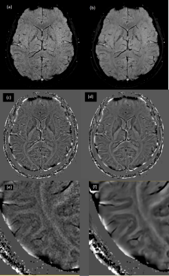 |
110 |
Improving SNR of high resolution multi-echo SWI using complex
domain DL based denoising
Florintina C1,
Sajith Rajamani1,
Preetham Shankpal1,
Suresh Joel1,
Sudhanya Chatterjee1,
Rohan Patil1,
Ramesh Venkatesan1,
Raja Sundaresan1,
and Harsh Agarwal1
1GE Healthcare, Bengaluru, India Keywords: Machine Learning/Artificial Intelligence, Machine Learning/Artificial Intelligence SWI is a high resolution MRI sequence, particularly sensitive to compounds which distort the local magnetic field making it useful in detecting blood products, calcium, etc. 3 and is used as part of brain MR imaging. The phase images are high pass filtered to remove the slow varying susceptibility changes and this is important to differentiate between para and diamagnetic substances. When this filtered phase image is used to accentuate the directly observed signal loss in the magnitude image, it is raised to a higher power and noise gets magnified as well, imparting undesirable effects in the SWI image. |
|
4197.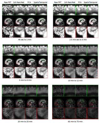 |
111 |
An MR-assisted Spatiotemporal Approach for 4D Dynamic Brain PET
Denoising
Hamed Yousefi1,
Chunwei Ying2,
Mahdjoub Hamdi2,
Richard Laforest2,
and Hongyu An2
1Imaging Science, Washington University in St.Louis, St Louis, MO, United States, 2Washington University in St.Louis, St Louis, MO, United States Keywords: Machine Learning/Artificial Intelligence, Brain In this study, dynamic brain PET denoising was done using registered MRI and reconstructed PET images reconstructed by the OSEM method, while maintaining TACs that were quite near to the original noisy data. The fundamental challenge to using the supervised learning approach is the lack of ground truth. The resulting spatiotemporal images improved the image quality according to various CNR, SNR, and CRC parameters while maintaining TACs that were similar to the original raw PET. This method only needs one trained network, one set of matrices for a statistical temporal PCA model, and 4D dynamic PET data as input. |
|
4198.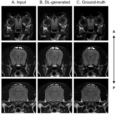 |
112 |
Improvement of canine brain FLAIR image using super-resolution
GAN
Yuseoung Son1,
Sumin Roh1,
Jae-Kyun Ryu1,
Won Beom Jung1,
Seok-Mn Lee2,
A-Rim Lee2,
Chuluunbaatar Otgonbaatar3,
Jaebin Lee4,
Ho-Jung Choi2,
Young-Won Lee2,
and Hackjoon Shim1,4
1Medical Imaging AI Research Center, Canon Medical Systems Korea, Seoul, Korea, Republic of, 2College of Veterinary Medicine, Chungnam National University, Daejeon, Korea, Republic of, 3College of Medicine, Seoul National University, Seoul, Korea, Republic of, 4Magnetic Resonance Business Unit, Canon Medical Systems Korea, Seoul, Korea, Republic of Keywords: Machine Learning/Artificial Intelligence, Machine Learning/Artificial Intelligence Most veterinary imaging has been achieved using human MRI scanners. Therefore, extensive averaging is required to obtain high-resolution images with high SNR for the animals, thereby leading to a long scan time. Veterinary MRI is typically performed under general anesthesia to minimize the level of stress and movement during image scanning. Therefore, long anesthetic conditions could affect animal normal physiology and be life-threatening, especially for patients in veterinary medical field. Here, we aimed to obtain higher image quality with short scanning time using super-resolution generative adversarial network (SRGAN) in the canine brain MRI. |
|
4199.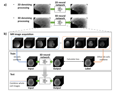 |
113 |
Coil2Coil 3D: Improved self-supervised MR image denoising using
phased-array coil images based on 3D structure information
Rokgi Hong1,
Juhyung Park1,
Hayeon Lee2,
Hongjun An1,
and Jongho Lee1
1Department of Electrical and Computer Engineering, Seoul National University, Seoul, Korea, Republic of, 2School of Electrical Engineering, Korea University, Seoul, Korea, Republic of Keywords: Machine Learning/Artificial Intelligence, Machine Learning/Artificial Intelligence
A deep-learning-based denoising method by extending
Coil2Coil method based on 3D network was proposed. This
method utilizes 3D information to conserve more tissue
structure details. When tested for the synthetic
noise-added images, the quantitative metrics were
improved than using 2D network (PSNR: 46.57 ± 2.50 for
3D vs. 45.96 ± 2.47 for 2D). For the real-world noise,
the proposed method also demonstrated qualitatively
improved denoising than 2D case by conserving the
structure details in MR images. Additionally, the
quantitative mapping experiment for R2* showed
the more accurate mapping results, suggesting
improvement of the quantification by 3D denoising.
|
|
4200.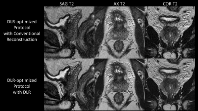 |
114 |
Increased Resolution and Shortened Scan Time of T2 weighted
Prostate MRI using Deep Learning Denoising Reconstruction at
1.5T
Hung Phi Do1,
Dawn Berkeley1,
Brian Tymkiw1,
Wissam AlGhuraibawi1,
and Mo Kadbi1
1Canon Medical Systems USA, Inc., Tustin, CA, United States Keywords: Prostate, Prostate In prostate MRI, high SNR images are desired for a better depiction of anatomical and pathological structures; however, it often requires a longer scan time, especially at 1.5T. Deep Learning Denoising Reconstruction (DLR) has been shown to effectively remove noise, allowing high SNR with higher resolution and shortened scan time simultaneously. This study demonstrates that DLR enables the acquisition of high SNR images with 29.48% scan time reduction and 31.51% increase in spatial resolution at 1.5T. |
|
4201.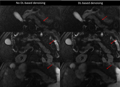 |
115 |
Multi-Shot M1-Nulled Pancreatic Diffusion Weighted Imaging with
Deep Learning-Based Denoising
Kang Wang1,
Matthew J. Middione1,
Andreas Markus Loening1,
Patricia Lan2,
Xinzeng Wang3,
Daniel B Ennis1,
and Ryan Lennex Brunsing1
1Radiology, Stanford University, Palo Alto, CA, United States, 2GE HealthCare, Menlo Park, CA, United States, 3GE HealthCare, Houston, TX, United States Keywords: Pancreas, Diffusion/other diffusion imaging techniques, Diffusion Weighted Imaging, Denoising, M1-nulled, ADC Multi-shot DWI (msDWI) may be improved by M1 motion-compensation, but at a penalty of longer TE and lower SNR. DL-based denoising has recently emerged as an option for DWI, which could help offset this TE penalty. Here we assess the impact of a commercially available DL-based denoising tool in the setting of motion-compensated msDWI. |
|
4202.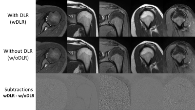 |
116 |
Deep Learning Denoising Reconstruction Enables up to 2X Scan
Time Reduction while Maintaining Image Quality for
Musculoskeletal Imaging
Hung Phi Do1,
Dawn Berkeley1,
Brian Tymkiw1,
Wissam AlGhuraibawi1,
and Mo Kadbi1
1Canon Medical Systems USA, Inc., Tustin, CA, United States Keywords: MSK, MSK Two-average (2NAQ) scans are often required to acquire high resolution images with adequate Signal-to-Noise Ratio (SNR). Deep Learning Denoising Reconstruction (DLR) effectively removes noise hence improving the SNR of reconstructed images. The SNR gain could be used to increase resolution and/or reduce scan time. This study demonstrates that the DLR-reconstructed one-average (1NAQ) images have similar image quality to those acquired with 2NAQ and reconstructed with conventional reconstruction. As a result, approximately 2X scan time reduction is achieved with DLR. |
|
4203.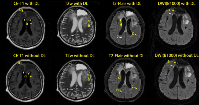 |
117 |
Assessment of Multi-modal MR Imaging for Glioma Based on a Deep
Learning Reconstruction Approach with the Denoising Method
Jun Sun1,
Yiding Guo1,
Zhizheng Zhuo1,
Siyao Xu1,
Min Guo1,
Li Chai1,
Junjie Li1,
Liying Qu1,
Minghao Wu1,
Juan Wei2,
Mingna Li3,
Tong Li3,
Jinyuan Weng4,
Xiaodong Gong5,
Yunyun Duan1,
Dabiao Zhou1,
and Yaou Liu1
1Capital Medical Universtiy, Beijing Tiantan Hospital, Beijing, China, 2MR Research, GE Healthcare, Beijing, China, 3BioMind Inc., Beijing, China, 4Department of Medical Imaging Product, Neusoft, Group Ltd., Shenyang, China, 5Department of Medical Imaging Product, Neusoft, Group Ltd., Beijing, China Keywords: Tumors, Brain Deep learning reconstruction (DLR) approach with denoising can improve image quality of magnetic resonance (MR) images. However, its applications on multi-modal glioma imaging have not been assessed.Multi-modal images of 107 glioma patients were evaluated by signal-to-noise ratio (SNR), contrast-to-noise ratio (CNR), edge sharpness, visual assessment, diagnosis accuracy and efficiency. Contrasted with conventionally reconstructed images, the DLR images showed higher tumor/residual tumor SNR, higher tumor to white/gray matter CNR, better results of the visual assessment, and a trend of improved diagnosis efficiency and comparable accuracy. DLR can improve image quality of multi-modal glioma images which should benefit the glioma diagnosis. |
|
4204.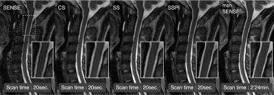 |
118 |
Fast MRI of the cervical spinal cord by combining the deep
learning-based denoising and Super-Resolution technique
Takuya Aoike1,
Noriyuki Fujima2,
Jihun Kwon3,
Masami Yoneyama3,
Satonori Tsuneta2,
Kinya Ishizaka1,
and Kohsuke Kudo4
1Department of Radiological Technology, Hokkaido University Hospital, Sapporo, Japan, 2Department of Diagnostic and Interventional Radiology, Hokkaido University Hospital, Sapporo, Japan, 3Philips Japan, Ltd., Tokyo, Japan, 4Department of Diagnostic Imaging, Hokkaido University Graduate School of Medicine, Sapporo, Japan; Global Center for Biomedical Science and Engineering, Faculty of Medicine, Hokkaido University, Sapporo, Japan Keywords: Spinal Cord, Spinal Cord SmartSpeed Precise Image (SSPI) is one of novel image reconstruction technique which combined the physics driven type deep learning-based denoising process and Super-Resolution techniques. We investigated the utility of SSPI-based single-shot turbo spin-echo sequence of the cervical spinal cord T2-weighted image with the short acquisition time. High quantitative signal-to-noise ratio and qualitative image sharpness were successfully demonstrated by using SSPI. This methodology can be useful for the better image quality and the short acquisition time in the assessment of patients with cervical spinal cord diseases. |
|
4205.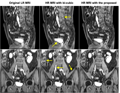 |
119 |
Patient-specific Self-supervised Resolution-enhancing Networks
for Synthesizing High-resolution Magnetic Resonance Images
Xiaofeng Yang1,
Sagar Mandava2,
Yang Lei1,
Huiqiao Xie1,
Tonghe Wang3,
Justin Roper1,
Tian Liu4,
and Hui Mao1
1Emory University, Atlanta, GA, United States, 2GE Healthcare, Atlanta, GA, United States, 3Memorial Sloan Kettering Cancer Center, New York, NY, United States, 4Icahn School of Medicine at Mount Sinai, New York, NY, United States Keywords: Quantitative Imaging, Data Processing This study aims to develop an efficient and clinically applicable method using patient-specific self-supervised resolution-enhancing network to synthesize the high-resolution information of MR images in the low-resolution direction to generate respective high-resolution MRI. |
|
 Back to Meeting Home
 Back to the Program-at-a-Glance
The International Society for Magnetic Resonance in Medicine is accredited by the Accreditation Council for Continuing Medical Education to provide continuing medical education for physicians.
 Back to Meeting Home
 Back to the Program-at-a-Glance
 View Presentation Video