Digital Poster
ML/AI for Acquisition & Reconstruction I
ISMRM & ISMRT Annual Meeting & Exhibition • 03-08 June 2023 • Toronto, ON, Canada

Digital Poster
ML/AI for Acquisition & Reconstruction I
| Computer # | |||
|---|---|---|---|
3701.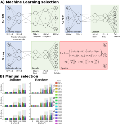 |
121 |
Comparison of data-driven and physics-informed learning
approaches for optimising multi-contrast MRI acquisition
protocols
Álvaro Planchuelo-Gómez1,2,
Maxime Descoteaux3,
Santiago Aja-Fernández2,
Jana Hutter4,
Derek K. Jones1,
and Chantal M.W. Tax1,5
1CUBRIC, Cardiff University, Cardiff, United Kingdom, 2Imaging Processing Laboratory, Universidad de Valladolid, Valladolid, Spain, 3SCIL, Université de Sherbrooke, Sherbrooke, QC, Canada, 4Centre for Medical Engineering, Centre for the Developing Brain, King's College London, London, United Kingdom, 5Image Sciences Institute, University Medical Centre Utrecht, Utrecht University, Utrecht, Netherlands Keywords: Machine Learning/Artificial Intelligence, Data Acquisition, Quantitative Imaging Multi-contrast MRI is used to assess the biological properties of tissues, but excessively long times are required to acquire high-quality datasets. To reduce acquisition time, physics-informed Machine Learning approaches were developed to select the optimal subset of measurements, decreasing the number of volumes by approximately 63%, and predict the MRI signal and quantitative maps. These selection methods were compared to a full data-driven and two manual strategies. Synthetic and real 5D-Diffusion-T1-T2* data from five healthy participants were used. Feature selection via a combination of Machine Learning and physics modelling provides accurate estimation of quantitative parameters and prediction of MRI signal. |
|
3702.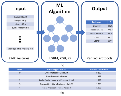 |
122 |
Predicting Abdominal MRI Protocols using Electronic Health
Records
Peyman Shokrollahi1,
Juan M. Zambrano Chaves1,
Avishkar Sharma1,
Jonathan P.H. Lam1,
Debashish Pal2,
Naeim Bahrami2,
Akshay S. Chaudhari1,
and Andreas M. Loening1
1Radiology, Stanford University, Stanford, CA, United States, 2GE Healthcare, Sunnyvale, CA, United States Keywords: Machine Learning/Artificial Intelligence, Modelling, MR Radiology Workflow Inaccurate selection of MRI protocols can impede diagnostics and therapeutic workflows, delay appropriate treatment, increase misdiagnosis likelihoods, and increase healthcare costs. Here we depict a machine-learning (ML) based system to accurately predict abdominal MR protocols, trained on the electronic medical records from 11,251 MR exam orders on 6,882 patients. The best model achieved a cumulative F1-score of 95.6% for top-three most-often-ordered protocols and a top-one F1 score of 78.5%. The proposed system can guide radiologists to appropriate protocol selections quickly, optimize workflows, and improve diagnostic accuracy, thereby by serving to support optimal patient outcomes. |
|
3703.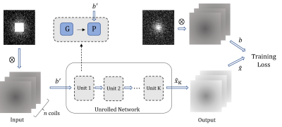 |
123 |
Theoretical guaranteed unfolding method for k-space
interpolation in a self-supervised manner
Chen Luo1,2,
Zhuoxu Cui2,
Huayu Wang1,2,
Qiyu Jin1,
Guoqing Chen1,
and Dong Liang2
1Inner Mongolia University, Hohhot, China, 2Shenzhen Institute of Advanced Technology,Chinese Academy of Sciences, Shenzhen, China Keywords: Machine Learning/Artificial Intelligence, Image Reconstruction Recently, iterative algorithm driven deep neural network unfolding methods have been successfully applied to MRI. However, the network replaces the original algorithm structure and mathematical properties such as interpretability and convergence of the original algorithm are not guaranteed. Fortunately, the k-space-filled Hankel low rank can naturally be associated with convolutional networks. Given this, we propose an unfolding method for k-space-filling, which guarantees convergence to the unique real MR image. Furthermore, we train this network in a self-supervised manner to cope with scenarios where fully sampled data are difficult to obtain. Finally, numerical experiments validate the effectiveness of the proposed method. |
|
3704.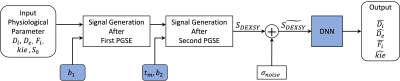 |
124 |
Deep learning-based acquisition protocol optimization and
parameter estimation for diffusion exchange spectroscopy
Zhaowei Cheng1,
Guangxu Han2,
Songtao Hu2,
Ke Fang1,
and Ruiliang Bai3
1College of Information Science and Electronic Engineering, Zhejiang University, Hangzhou, China, 2Key Laboratory of Biomedical Engineering of Ministry of Education, College of Biomedical Engineering and Instrument Science, Zhejiang University, Hangzhou, China, 3School of Medicine, Zhejiang University, Hangzhou, China Keywords: Machine Learning/Artificial Intelligence, Data Acquisition We propose a deep-learning-based method to optimize acquisition protocol and estimate parameter for diffusion exchange spectroscopy. A unified framework has been carefully designed to achieve both goals simultaneously. Using this framework, the acquisition protocol can be directly optimized with an objective function that minimizes the parameter estimation errors, regardless of its degree of freedom. The experimental results from Monte Carlo simulations show that the proposed method outperforms the existing methods in both accuracy and precision of parameters’ estimation. Besides, it speeds up the calculation by 480 times. |
|
3705.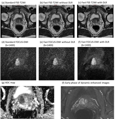 |
125 |
Feasibility of Deep Learning Reconstruction in Prostate
Multiparametric MRI: a Preliminary Prospective Study
Yichen Wang1,
Xinxin Zhang1,
Sicong Wang2,
Xinming Zhao1,
and Yan Chen1
1Radiology, National Cancer Center/National Clinical Research Center for Cancer/Cancer Hospital, Chinese Academy, Beijing, China, 2GE Healthcare, MR Research, Beijing, China Keywords: Prostate, Machine Learning/Artificial Intelligence, Deep learning reconstruction In this prospective study, feasibility of deep learning reconstruction (DLR) in axial FSE-T2WI and axial reduced-FOV DWI (FOCUS DWI) were evaluated compared with standard protocols. Fast protocol with DLR substantially reduced scanning time (axial FSE-T2WI: -32.1%; FOCUS-DWI: -36.8%). Fast FOCUS DWI with DLR showed the highest SNR and CNR for prostate PZ, TZ and lesion. Fast FSE-T2WI with DLR showed the highest SNR and CNR for prostate PZ and TZ. Moreover, fast FOCUS-DWI and FSE-T2WI with DLR demonstrated equivalent or better image quality than standard images. DLR may be useful in prostate multiparametric MRI protocol optimization and high-quality image acquisition. |
|
3706.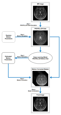 |
126 |
Deep Learning Augmented PROPELLER Reconstruction for Improved
MRI Motion Correction
Sixing Liu1,
Lifeng Mei1,
Shoujin Huang1,
and Mengye Lyu1
1College of Health Science and Environmental Engineering, Shenzhen Technology University, Shenzhen, China Keywords: PET/MR, Machine Learning/Artificial Intelligence, deep learning Applying the Periodically Rotated Overlapping Parallel Lines with Enhanced Reconstruction (PROPELLER) technique is one of the strategies to mitigate motion artifacts in MR images. However, due to technical limitations, existing method estimates motion parameters with unsatisfactory results, motion artifacts will still be presented in the final image. Deep learning algorithms are expected to optimize the motion parameter estimation part of PROPELLER technique. We develop a PROPELLER imaging technique incorporating a deep learning model that can provide accurate results and greatly shorten the elapsed time. |
|
3707. |
127 |
Deep learning reconstruction algorithm for 100-second rapid
Ischemic stroke imaging
Xin Fang1,
Ailian Liu1,
Qingwei Song1,
Guobin Li2,
and Shuheng Zhang2
1the First Affiliated Hospital of Dalian Medical University, Dalian, China, 2Shanghai United Imaging Healthcare Co., Ltd, Shanghai, China Keywords: Brain Connectivity, Brain Head Computed Tomography (HCT) is a common imaging method for the diagnosis of emergency cerebral infarction. However, it is easy to miss the diagnosis due to the poor performance of the ultra-early or early cerebral infarction lesions. Compared with CT, head Magnetic Resonance Imaging (MRI) has the characteristics of high tissue contrast, no ionizing radiation damage and can make functional imaging sequences, so it has significant advantages in the diagnosis of posterior fossa lesions and ultra-early cerebral infarction |
|
3708.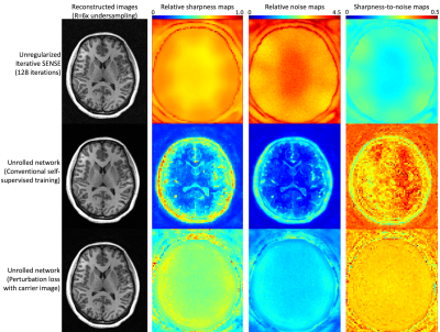 |
128 |
Perturbation loss with carrier image reconstruction: A loss
function for optimized point spread functions
R. Marc Lebel1,2
1GE Healthcare, Calgary, AB, Canada, 2Radiology, University of Calgary, Calgary, AB, Canada Keywords: Machine Learning/Artificial Intelligence, Image Reconstruction Deep learning (DL) assisted image reconstructions are becoming state-of-the art, producing better image quality and/or enabling higher acceleration rates than achievable with conventional methods. DL networks are used to mitigate noise amplification while retaining important signal characteristics. However, typical loss functions produce object-dependent noise alterations and non-uniform point-spread functions. Here we present a method for training networks that prioritizes maximizing the point spread function to ensure maximal detail retention. |
|
3709.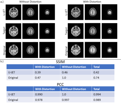 |
129 |
U-JET: Preliminary results of a convolutional neural network
approach for distortion-free image reconstruction of PROPELLER
data
Jörn Huber1,
Klaus Eickel1,2,
and Matthias Günther1,2,3
1Fraunhofer Institute for Digital Medicine MEVIS, Bremen, Germany, 2mediri GmbH, Heidelberg, Germany, 3Faculty 1 (Physics/Electrical Engineering), University of Bremen, Bremen, Germany Keywords: Machine Learning/Artificial Intelligence, Artifacts Arterial Spin Labeling has great potential in clinics as a non-invasive alternative to Dynamic Contrast Enhanced imaging. However, motion sensitivity needs to be tackled by readout techniques such as 3D GRASE PROPELLER, which unfortunately shows a high sensitivity to geometric distortion. Analytical separation of motion and distortion effects is computationally demanding and might fail in some situations. To this aim, a U-NET based convolutional neural network approach is demonstrated which might overcome this limitation. |
|
3710.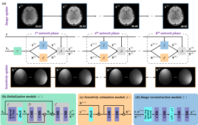 |
130 |
A Faithful Deep Sensitivity Estimation Makes High-quality MRI
Reconstruction
Zi Wang1,
Haoming Fang1,
Chen Qian1,
Boxuan Shi1,
Lijun Bao1,
Liuhong Zhu2,
Jianjun Zhou2,
Wenping Wei3,
Jianzhong Lin4,
Di Guo5,
and Xiaobo Qu1
1Department of Electronic Science, Biomedical Intelligent Cloud R&D Center, Fujian Provincial Key Laboratory of Plasma and Magnetic Resonance, National Institute for Data Science in Health and Medicine, Xiamen University, Xiamen, China, 2Department of Radiology, Zhongshan Hospital (Xiamen), Fudan University, Xiamen, China, 3Department of Radiology, Xiamen University, Xiamen, China, 4Department of Radiology, Zhongshan Hospital affiliated to Xiamen University, Xiamen, China, 5School of Computer and Information Engineering, Xiamen University of Technology, Xiamen, China Keywords: Machine Learning/Artificial Intelligence, Machine Learning/Artificial Intelligence Recent deep learning is superior in providing high-quality images and ultra-fast reconstructions in accelerated magnetic resonance imaging (MRI). Faithful coil sensitivity estimation is vital for MRI reconstruction. In this work, we propose a Joint Deep Sensitivity estimation and Image reconstruction network (JDSI). During the image artifacts removal, it gradually provides more faithful sensitivity maps, leading to greatly improved image reconstructions. Results on in vivo datasets and radiologist reader study demonstrate that, the proposed JDSI achieves the state-of-the-art performance visually and quantitatively, especially when the accelerated factor is high. Besides, JDSI also owns nice robustness to abnormal subjects. |
|
3711.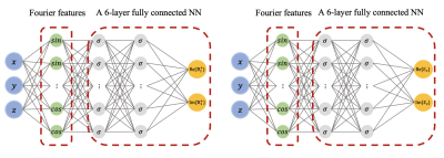 |
131 |
PIFN EPT: MR-Based Electrical Property Tomography Using
Physics-Informed Fourier Networks
Xinling Yu1,
Jose Serralles2,
Ilias Giannakopoulos3,4,
Ziyue Liu5,
Luca Daniel2,
Riccardo Lattanzi3,4,
and Zheng Zhang1
1Department of Electrical and Computer Engineering, University of California, Santa Barbara, Santa Barbara, CA, United States, 2Department of Electrical Engineering and Computer Science, Massachusetts Institute of Technology, Cambridge, MA, United States, 3Center for Advanced Imaging Innovation and Research (CAI2R), Department of Radiology, New York University Grossman School of Medicine, New York, NY, United States, 4Bernard and Irene Schwartz Center for Biomedical Imaging (CBI), Department of Radiology, New York University Grossman School of Medicine, New York, NY, United States, 5Department of Statistics and Applied Probability, University of California, Santa Barbara, Santa Barbara, CA, United States Keywords: Machine Learning/Artificial Intelligence, Electromagnetic Tissue Properties We introduce physics-informed Fourier networks (PIFNs) for Electrical Properties (EP) Tomography (EPT). Our novel deep learning-based method is capable of learning EPs globally from noisy magnetic resonance (MR) measurements, i.e, the magnitude of the magnetic transmit field and the transceive phase. Our proposed method also provides noise-free transmit field reconstructions. Two separate Fourier neural networks are used to efficiently estimate the transmit field and EPs at any location. We show that PIFN EPT accurately infers the EPs distribution of an inhomogeneous phantom from noisy simulated measurements. |
|
3712.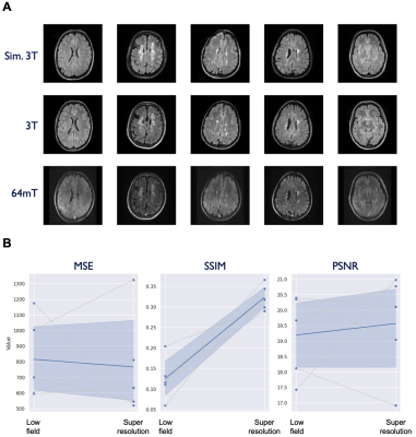 |
132 |
Super resolution imaging from low-field strength scanners using
generative adversarial networks
Thomas Campbell Arnold1,
Serhat V Okar2,
Danni Tu3,
Govind Nair2,
John T. Pitts4,
Megan E. Poorman4,
Karan D. Kawatra2,
Lisa M. Desiderio5,
Matthew K. Schindler6,
Brian Litt6,
Russell T. Shinohara3,
Daniel S. Reich2,
and Joel M. Stein5
1Bioengineering, University of Pennsylvania, Philadelphia, PA, United States, 2National Institute of Neurological Disorders and Stroke, Bethesda, MD, United States, 3Biostatistics, University of Pennsylvania, Philadelphia, PA, United States, 4Hyperfine, Guilford, CT, United States, 5Radiology, University of Pennsylvania, Philadelphia, PA, United States, 6Neurology, University of Pennsylvania, Philadelphia, PA, United States Keywords: Machine Learning/Artificial Intelligence, Low-Field MRI, super resolution High-field MRI provides superior imaging for diverse clinical applications, but cost and other factors limit availability in various healthcare and lower resource settings. Lower-field strength units promise to expand access but involve tradeoffs including reduced signal, longer scan times, and lower resolution. Here we develop super-resolution methods that can generate high-field quality images from low-field scanner inputs, thus increasing signal and resolution. We use generative adversarial networks to demonstrate image enhancement in T1, T2 and FLAIR sequences. |
|
3713.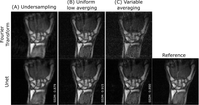 |
133 |
Impact of sampling strategies and residual U-net reconstruction
on preserving high spatial frequencies in accelerated low-field
MRI
Reina Ayde1,2,
Tobias Senft1,
Marco Fiorito1,
Mauro Spreiter1,
Najat Salameh1,2,
and Mathieu Sarracanie1,2
1Center for Adaptable MRI Technology (AMT center), Department of Biomedical Engineering, University of Basel, Allschwil, Switzerland, 2AMT center, Institute of Medical Sciences, School of Medicine, Medical Sciences & Nutrition, University of Aberdeen, Aberdeen, United Kingdom Keywords: Machine Learning/Artificial Intelligence, Image Reconstruction, undersampling, averaging, data sampling Low signal-to-noise (SNR) ratios inherent to low-field (LF) MRI challenge its relevance in clinical applications. Accelerating the acquisition by undersampling k-space followed by reconstruction techniques has already shown promising results. Yet, undersampling is usually done by skipping high-frequency information which can lead to misdiagnosis as small lesions can be missed. In this study, we exploited a specificity of low-SNR regimes, that is signal averaging, to explore different acceleration strategies without skipping crucial information in k-space. The DL-reconstructed images arising from those sampling schemes have been evaluated on acquired in-vivo and ex-vivo LF-MRI datasets, showcasing high-frequency preservation and potential for generalization. |
|
3714.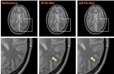 |
134 |
A deep learning approach for compressed sensing reconstruction
using adaptive shrinkage threshold
Yuan Lian1 and
Hua Guo1
1Center for Biomedical Imaging Research, Department of Biomedical Engineering, School of Medicine, Tsinghua University, Beijing, China Keywords: Machine Learning/Artificial Intelligence, Image Reconstruction Compressed Sensing is widely used for accelerating acquisitions of MR images. Deep learning methods have been introduced to CS-MRI reconstruction to improve image quality and computation speed. Here we introduce a new shrinkage function with adaptive threshold selection for Model-driven deep learning networks to suppress aliasing artifacts by utilizing the information on each feature map. We combine our adaptive threshold selection module with ISTA-Net, and demonstrate that the method can reduce reconstruction errors while preserving structural details effectively. |
|
3715.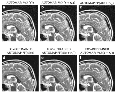 |
135 |
The role of training on the robustness of domain-transform
manifold learning
Danyal Bhutto1,2,
Bo Zhu2,
Jeremiah Zhe Liu3,4,
Stephen Cauley2,5,
Neha Koonjoo2,5,
Bruce R Rosen2,5,
and Matthew S Rosen2,5,6
1Biomedical Engineering, Boston University, Boston, MA, United States, 2Athinoula A. Martinos Center for Biomedical Imaging, Charlestown, MA, United States, 3Google, Mountain View, CA, United States, 4Biostatistics, Harvard University, Cambridge, MA, United States, 5Harvard Medical School, Boston, MA, United States, 6Physics, Harvard University, Boston, MA, United States Keywords: Machine Learning/Artificial Intelligence, Image Reconstruction Domain-transform manifold learning is a trained reconstruction approach where care needs to be taken to appropriately represent the forward encoding model during training, including for example the numerical properties of the source sensor data, phase relationship of complex sensor data, and field-of-view to prevent artifacts arising in the reconstruction. Here, we study the role that the training corpus and the numeral properties of the training have on the performance of the reconstruction of MRI data and demonstrate reconstruction artifacts that result from inference on out-of-training-distribution data if the training data is not augmented sufficiently. |
|
3716.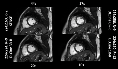 |
136 |
Relative noise variation with Unrolled Neural Networks for
Accelerated Cardiac Cine Reconstruction
Suryanarayanan Sivaram Kaushik1,
Xucheng Zhu2,
Robert Marc Lebel3,
Kavitha Manickam1,
Ke Li1,
Kailash Saravanan1,
Florian Wiesinger4,
and Martin Janich4
1GE Healthcare, Waukesha, WI, United States, 2GE Healthcare, Menlo Park, CA, United States, 3GE Healthcare, Calgary, AB, Canada, 4GE Healthcare, Munich, Germany Keywords: Machine Learning/Artificial Intelligence, Cardiovascular Deep Learning (DL) based reconstructions help alleviate the longer scan times seen in bSSFP Cine acquisitions by offering higher acceleration factors. This work analyzes the spatial and temporal variations in the noise as a function of acceleration factors in DL reconstructions of highly accelerated bSSFP Cine data. |
|
3717.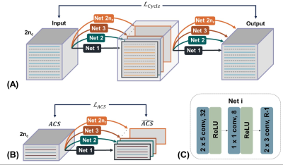 |
137 |
A noise robust image reconstruction deep neural network with
cycle interpolation
Jeewon Kim1,
Wonil Lee1,
Beomgu Kang1,
Seohee So2,
and HyunWook Park1
1Korea Advanced Institute of Science and Technology (KAIST), Daejeon, Korea, Republic of, 2Korea Institute of Science and Technology, Seoul, Korea, Republic of Keywords: Machine Learning/Artificial Intelligence, Parallel Imaging We propose a new Parallel Imaging scheme using a deep neural network which performs well with fewer ACS line in noisy environments. The proposed scheme includes ACS loss which is used in RAKI and cycle interpolation loss that we newly propose in our work. RAKI generalized GRAPPA in noisy environments by applying non-linear k-space interpolation with a deep neural network. However, it requires additional ACS lines to output satisfactory performance. Here, we suggest a new scheme to overcome the reconstruction performance in a noisy environment with fewer ACS lines. |
|
3718.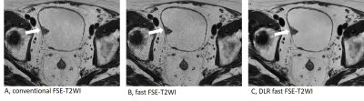 |
138 |
Feasibility of Deep Learning Reconstruction in the Clinical
Application of MRI for patients with Bladder Cancer: a
preliminary prospective study
Xinxin Zhang1,
Yichen Wang1,
Sicong Wang2,
Min Li2,
Yan Chen1,
and Xinming Zhao1
1National Cancer Center/National Clinical Research Center for Cancer/Cancer Hospital, Chinese Academy of Medical Sciences and Peking Union Medical College, Beijing, 100021, China, Beijing, China, 2GE Healthcare, MR Research China, Beijing, Beijing, China Keywords: Urogenital, Bladder, Deep Learning Reconstruction The application of DLR significantly shortened scan times and improved the overall image quality score and image artifacts score and SNR and CNR of FSE-T2WI. DLR fast FSE-T2WI demonstrated significantly higher SNR (256.7±102.9 VS 94.7±40.8, p < 0.05) and CNR (168.0±77.3 VS 59.6±29.8, p < 0.05) and overall image quality scores (median, 4.0 vs. 3.0 for reader1 and 4.0 vs. 3.5 for reader2) than those of conventional FSE-T2WI. DLR may be useful in reducing the acquisition time of bladder MRI without compromising image quality. |
|
3719.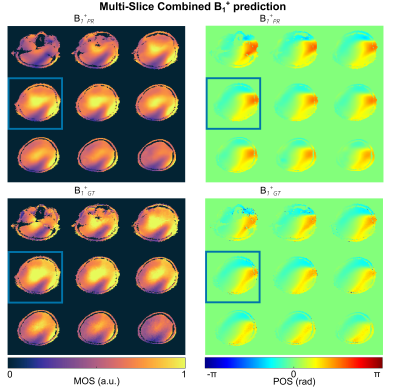 |
139 |
Rapid multi-slice whole-brain B1+-mapping at 7T using deep
learning
Felix Krueger1,
Christoph Stefan Aigner1,
Max Lutz1,
Layla Tabea Riemann1,
Katja Degenhardt1,
Bernd Ittermann1,
Tobias Schaeffter1,2,
Kerstin Hammernik3,4,
and Sebasian Schmitter1,5,6
1Physikalisch-Technische Bundesanstalt, Berlin and Braunschweig, Germany, 2Division of Imaging Sciences and Biomedical Engineering, King's College London, London, United Kingdom, 3Technical University of Munich, Munich, Germany, 4Imperial College London, London, United Kingdom, 5Medical Physics in Radiology, German Cancer Research Center (DKFZ), Heidelberg, Germany, 6Center for Magnetic Resonance Research, University of Minnesota, Minneapolis, MN, United States Keywords: RF Pulse Design & Fields, Parallel Transmit & Multiband In this work we utilize deep learning to estimate multi-slice whole-brain B1+-maps in sub-seconds from initial localizer scans at 7T. The investigated neural networks use the receive profiles of the individual coil elements of an 8Tx/8Rx transceiver head coil as input information. The networks are trained on seven volunteers and tested in 2 unseen subjects for transversal/coronal/sagittal slices by comparing the prediction with the acquired B1+-maps. Subsequently, the feasibility of using the DL-based B1+-maps in a subject-specific calibration pipeline is demonstrated. |
|
3720. |
140 |
Universal sequence-invariant deep learning image reconstruction
for cardiovascular MR Multitasking
Zheyuan Hu1,2,
Zihao Chen1,2,
Hsu-Lei Lee1,
Yibin Xie1,
Debiao Li1,2,
and Anthony Christodoulou1,2
1Biomedical Imaging Research Institute, Cedars-Sinai Medical Center, Los Angeles, CA, United States, 2Department of Bioengineering, University of California, Los Angeles, Los Angeles, CA, United States Keywords: Machine Learning/Artificial Intelligence, Quantitative Imaging Cardiovascular MR (CMR) Multitasking can quantify various parameter combinations without breath-holding or ECG monitoring. Clinically practical reconstruction time is viable using supervised deep subspace learning, but it depends on sequence-specific training. Here we explore whether universal, sequence-invariant CMR Multitasking deep learning reconstruction is practical by trading temporal awareness (breadth) for added depth in spatial domain. We evaluated the performance and generalizability of both strategies by training on T1 mapping data only and testing on two datasets: a) a matched-sequence T1 mapping data; and b) a novel-sequence T1-T2 mapping data. |
|
 Back to Meeting Home
 Back to the Program-at-a-Glance
The International Society for Magnetic Resonance in Medicine is accredited by the Accreditation Council for Continuing Medical Education to provide continuing medical education for physicians.
 Back to Meeting Home
 Back to the Program-at-a-Glance
 View Presentation Video