Digital Poster
Relaxometry & Diffusion I
ISMRM & ISMRT Annual Meeting & Exhibition • 03-08 June 2023 • Toronto, ON, Canada

| Computer # | |||
|---|---|---|---|
1604.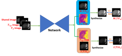 |
141 | Self-supervised Learning Based Liver Multi-parametric Mapping in a Single Breath-hold
Chaoxing Huang1,2, Yurui Qian1, Jian Hou1, Baiyan Jiang1,3, Queenie Chan4, Vincent Wai-Sun Wong5, Winnie Chiu-Wing Chu1,2, and Weitian Chen1,2
1Department of Imaging and Interventional Radiology, The Chinese University of Hong Kong, Shatin, Hong Kong, 2CUHK Lab of AI in Radiology (CLAIR), Shatin, Hong Kong, 3Illuminatio Medical Technology Limited, Hong Kong SAR, China, 4Philips Healthcare, Hong Kong SAR, China, 5Department of Medicine and and Therapeutics, The Chinese University of Hong Kong, Shatin, Hong Kong Keywords: Quantitative Imaging, Liver A self-supervised learning based multiparametric mapping method is proposed to map T1ρ and T2 simultaneously, by utilising the relaxation constraint in the learning process. The method was examined on a dataset of 52 patients with non-alcoholic fatter liver disease. Results showed that the proposed method can produce comparable parametric maps to the traditional fitting method, with reduced number of images, and reduced scan time. |
|
1605.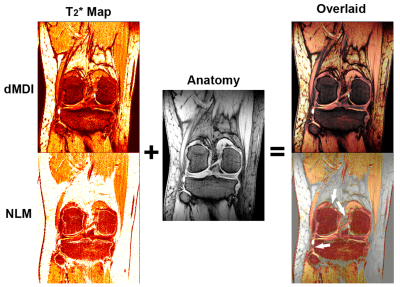 |
142 | Mask-free T2* and R2* mapping with dual multi-dimensional integration (dMDI)
Yongquan Ye1, Jian Xu1, Zhongqi Zhang1, Yan Zhang2, Qiang Zhao3, Jiajia Xu3, and Huishu Yuan3
1United Imaging, Houston, TX, United States, 2Beijing United Imaging Intelligent Imaging Technology Research Institute, Beijing, China, 3Radiology, Peking University Third Hospital, Beijing, China Keywords: Quantitative Imaging, Quantitative Imaging A dual MDI strategy was developed and demonstrated to achieve T2* and R2* mapping that are both highly delineated between noise backgrounds and normal tissues as well as spikes free, thus eliminating the need for routine noise masking or manual segmentation. |
|
1606.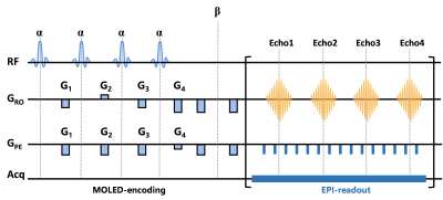 |
143 | Diagnostic value of ultrafast quantitative T2 imaging in the preoperative differentiation of meningioma WHO grades
Zongye Li1, Yijie Yang2, Yue Zhang1, Yanhong Lin2, Xiao Wang1, Yuchuan Zhuang3, Qinqin Yang2, Eryuan Gao1, Yanan Ren1, Yong Zhang1, Shuhui Cai2, Zhong Chen2, Congbo Cai2, Jingliang Cheng1, and Jianfeng Bao1
1Department of Magnetic Resonance Imaging, the First Affiliated Hospital of Zhengzhou University, Zhengzhou, China, 2Department of Electronic Science, Xiamen University, Xiamen, China, 3Department of Imaging Sciences, University of Rochester, Rochester, NY, United States Keywords: Quantitative Imaging, Neurography We assess the ability of a novel single-shot quantitative T2 magnetic resonance imaging (MRI) sequence and apparent diffusion coefficient (ADC) maps in predicting the WHO grade in meningiomas. We used histogram analysis of T2 maps and ADC maps to compare different grades of meningioma. ADC P10 proved to be the best predictor among all histogram parameters of ADC. While histogram analysis of T2 maps did not receive satisfactory results, the combination of T2 kurtosis and ADC P10 proved to be the best predictor. It is worthy of consideration to try another method of ROI placement and increase the sample size. |
|
1607.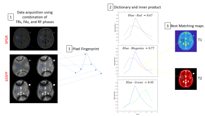 |
144 | Insensitive to Off-Resonance 3D T1/T2 Mapping SPGR/bSSFP using Dictionary-Matching: Application to the Prostate at 3T
Ronal Coronado1,2, Carlos Castillo-Passi3,4, Cecilia Besa2,5, and Pablo Irarrazaval2,6
1Biomedical Imaging Center-Universidad Catolica de Chile, Santiago, Chile, 2Millenium Institute for Intelligent Healthcare Engineering, Santiago, Chile, 3Institute for Biological and Medical Engineering, Pontificia Universidad Católica de Chile, Santiago, Chile, Santiago, Chile, 4King's College London, London, United Kingdom, 5Departamento de Radiologia-Universidad Católica de Chile, Santiago, Chile, 6Department of Electrical Engineering, Pontificia Universidad Católica de Chile, Santiago, Chile Keywords: Quantitative Imaging, Prostate Balanced Steady State Free Precession (bSSFP) has been successfully used to estimate T2 maps (DESPOT2), but bSSFP is prone to off-resonance artifacts. These artifacts affect the reconstruction of the T2 maps. We propose a dictionary-matching reconstruction using the signal model of bSSFP and spoiled gradient echo (SPGR). We tested this approach with simulation, 3D phantom and 3D in-vivo prostate acquisitions. In all of them, the off-resonance banding artifacts are greatly reduced, which is reflected in reduced NRMSE values. This method allows using a faster sequence with high SNR to quantify complicated body regions like the prostate. |
|
1608.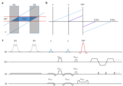 |
145 | T2 mapping with ZOOM-OLED on the kidney
Jian Wu1, Taishan Kang2, Simin Li1, Weikun Chen1, Zhigang Wu3, Congbo Cai1, and Shuhui Cai1
1Department of Electronic Science, Xiamen University, Xiamen, China, 2Department of Radiology, Zhongshan Hospital of Xiamen University, School of Medicine, Xiamen University, Xiamen, China, 3Clinical & Technical Solutions, Philips Healthcare, Shenzhen, China Keywords: Quantitative Imaging, Kidney The overlapping-echo detachment (OLED) imaging can capture a T2 map within about 150 ms. However, when the region of interest (ROI) is smaller than the object, it is inefficient to acquire the whole object because many unnecessary phase-encoding steps should be acquired. The zonal oblique multislice (ZOOM) is a method to reduce the field of view (FOV) by using two pulses to excite two slabs that have a specific angle between them. This study combined ZOOM with OLED to capture the T2 map with reduced FOV. The ZOOM-OLED can reduce geometric distortion and improve image resolution. |
|
1609.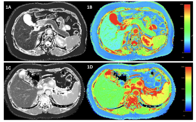 |
146 | T1 mapping of the pancreas in patients with pancreatic cancer—does pancreatic extracellular volume fraction increase the diagnostic value?
Hai-Yan Chen1, Kai Li1, Yi-Shi Wang2, and Lei Shi1
1The Cancer Hospital of the University of Chinese Academy of Sciences (Zhejiang Cancer Hospital), Institute of Basic Medicine and Cancer (IBMC), Chinese Academy of Sciences, Hangzhou, China, 2Philips Healthcare, Beijing, China Keywords: Quantitative Imaging, Quantitative Imaging There was no related study focused on pancreatic cancer, and we have no idea whether T1 mapping and ECV fraction could be used in such patients. Our study was to evaluate whether the T1 relaxation time of the pancreas can detect parenchymal changes in pancreatic cancer (PC) patients and whether extracellular volume (ECV) fraction improves the diagnostic value. We found that native T1 relaxation time of the pancreas in patients with PCs was significantly higher than patients with Non-PCs and volunteers, respectively. The pancreatic ECV was failed to show a significant difference among three groups. |
|
1610.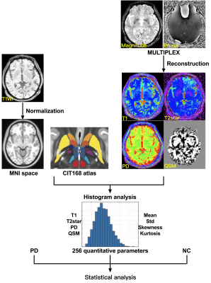 |
147 | Clinical application of MULTIPLEX as a rapid multi-parametric brain imaging method in early Parkinson’s disease
Tong Fu1, Rongrong Pan2, Youyong Tian2, Qing Gao2, Xinying Wu 1, Lindong Liu1, Hai Lin3, and Yongming Dai3
1Department of Radiology, Nanjing first hospital,Nanjing Medical University, Nanjing, China, 2Department of Neurology, Nanjing first hospital,Nanjing Medical University, Nanjing, China, 3Central Research Institute, United Imaging Healthcare, Shanghai, China Keywords: Quantitative Imaging, Multi-Contrast Quantitative MRI can measure a variety of physiological tissue parameters, such as longitudinal T1 value, transverse T2 value and proton density, iron content and fat content. We tried to apply a multi-parametric MR imaging technique of MULTIPLEX that provides the maps of T1, T2*, proton density and quantitative susceptibility mapping into the diagnosis of early Parkinson’s disease (PD). We found that MULTIPLEX could provide comprehensive and quantitative assessment of Parkinson’s disease-related subcortical nucleus and dopaminergic midbrain regions captured in multiple MR imaging parameters, which might assist in the diagnosis and better understanding of early Parkinson’s disease under tight clinical time-constraints. |
|
1611. |
148 | Establishment of SPGR-based MOLLI T1 mapping for the calculation of extracellular volume fraction (ECV) in Gd-EOB-DTPA-enhanced MRI
Ryotaro Jingu1, Keisuke Sato2, Atsushi Nozaki3, Tetsuya Wakayama3, Ryuji Nakamuta1, and Kengo Yoshimitsu2
1Radiology center, Fukuoka University Hospital, Fukuoka city, Japan, 2Department of Radiology, Faculty of Medicine, Fukuoka University, Fukuoka city, Japan, 3GE Healthcare, Hino city, Japan Keywords: Quantitative Imaging, Quantitative Imaging, T1 mapping In phantom study, SPGR-MOLLI acquisition scheme was optimized for abdominal T1 mapping in EOB-MRI, which was applied to clinical patients to measure ECV of the spleen and paraspinal muscle obtained 4 min and 25 min after the contrast administration. ECV obtained from contrast-enhanced CT within 3 months from EOB-MRI was used as reference standard. ECVs calculated from EOB-MRI showed significant agreements with those obtained from CT with ICCs of 0.9 and 0.74 for 4 min and 25 min, respectively (p<0.0001), which suggested that our T1 mapping protocol is appropriate and applicable to ECV measurement in EOB-MRI similarly with contrast-enhanced CT. |
|
1612. |
149 | Quantitative MR evaluation of pancreas parenchyma in patients with IPMN: association with tumor progression
Kiyoka Maeba1, Akihiko Kanki1, Yoshihiko Fukukura1, Hidemitsu Sotozono1, Akira Yamamoto1, and Tsutomu Tamada1
1Kawasaki Medical School Hospital, Kurashiki, Japan Keywords: Quantitative Imaging, Pancreas This study focused on the feasibility of quantitative MR evaluation of the pancreas parenchyma in patients with IPMN as a non-invasive tool for predicting the risk of malignancy. Our study showed that increased signal intensity on T2WI and longer T1 relaxation time were associated with a higher risk of malignant IPMN. These results suggested that quantitative MR evaluation of the underlying pancreas parenchyma in patients with IPMN can be a surrogate marker for predicting malignant IPMN. |
|
1613.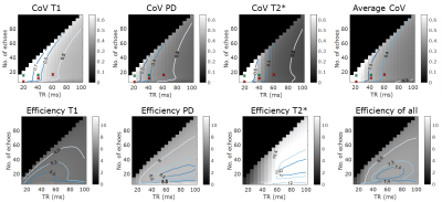 |
150 | Simulating the efficiency of Variable Flip Angle (VFA) multi-parametric mapping of T1, PD, and T2* at 7T suggests longer TRs may be optimal
Jyoti Mangal1,2, Ayse Sila Dokumaci1,2, David Leitao1,2, Raphael Tomi-Tricot1,2,3, Pip Bridgen1,4, Joseph V Hajnal2,5, Claudia Prieto1, Shaihan Malik1,2, and David W Carmichael1,2
1Biomedical Engineering Department, School of Biomedical Engineering and Imaging Sciences, King's College London, London, United Kingdom, 2London Collaborative Ultra high field System (LoCUS), London, United Kingdom, 3MR Research Collaborations, Siemens Healthcare Limited, Frimley, United Kingdom, 4London Collaborative Ultra high field System (LoCUS), Lodnon, United Kingdom, 5Centre for the Developing Brain, School of Biomedical Engineering and Imaging Sciences, King's College London, London, United Kingdom Keywords: Quantitative Imaging, Neuro, MPM, sequence optimization Quantitative MRI at 7T may have important clinical applications owing to the potential for enhanced resolution and uniform tissue characterisation. Current protocols, often for pragmatic reasons, use short TRs(~20ms) and a handful of echo times but this might not be optimal for T2* estimation. We simulated the optimal TR and echo number for dual flip angle mapping of T1, PD, and T2* for realistic 7T tissue and sequence parameters. The optimal efficiency was found to be at 65ms with 20 echoes. The simulations suggested that longer TRs of 40-60ms would be optimal. Preliminary in-vivo data only partially supported these findings |
|
1614.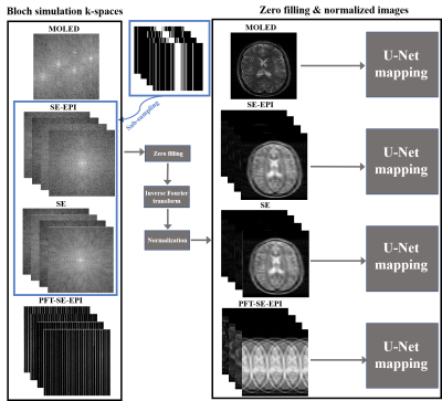 |
151 | Compressed sensing vs multiple overlapping echo detachment in acceleration efficiency of DL-based quantitative parametric mapping
Qingdang Qin1, Jiechao Wang1, Zhigang Wu2, Shuhui Cai1, and Congbo Cai1
1Department of Electronic Science, Xiamen University, Xiamen, China, 2MSC Clinical & Technical Solutions, Philips Healthcare, ShenZhen, China Keywords: Quantitative Imaging, Quantitative Imaging Compressed sensing (CS) relies on random under-sampling measurements and has been the primary technique to accelerate quantitative parametric mapping (QPM). The multiple overlapping-echo detachment (MOLED) technique proposed recently is a novel technique to realize quantitative parametric mapping in a single shot within around 100 milliseconds. However, the two techniques still lack a systematic comparison of acceleration performance in QPM. For the first time, we analyze and compare the performance of these two techniques under the same conditions via numerical experiments. The results show that MOLED outerperforms one-dimensional under-sampling CS acceleration technique in T2 quantification. |
|
1615.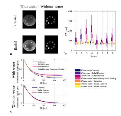 |
152 | Over-estimation of T2 with radial sampling: Identification, understanding and strategies to mitigate the bias.
Nadège Corbin1,2, Aurélien J. Trotier1, Sylvain Miraux1, and Emeline J. Ribot1
1Centre de Résonance Magnétique et Systèmes Biologiques UMR5536 CNRS/University of Bordeaux, Bordeaux, France, 2Wellcome Centre for Human Neuroimagin, UCL Queen Square, Institute of Neurology, University College of London, London, United Kingdom Keywords: Quantitative Imaging, Relaxometry, radial sampling Quantitative MRI protocols can benefit from radial trajectory sampling to reduce the acquisition time. However we identified a bias in T2 relaxometry studies conducted with radial imaging. After in-depth analysis aiming at further our understanding of the underlying mechanisms, we propose some guidelines to mitigate this bias. The error depends on the range of T2 present in the object of interest and its geometry, the number of spokes and the trajectory of the sequence. Parallel imaging and compressed sensing help reducing the bias. |
|
1616.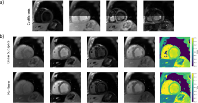 |
153 | Myocardial T1 Mapping Using Single-Shot Inversion-Recovery Radial FLASH: Comparison of Subspace and Nonlinear Model-Based Reconstruction
Martin Schilling1, Moritz Blumenthal2, Xiaoqing Wang3,4, and Martin Uecker2
1University Medical Center Göttingen, Göttingen, Germany, 2Institute for Biomedical Imaging, TU Graz, Graz, Austria, 3Athinoula A. Martinos Center for Biomedical Imaging, Massachusetts General Hospital, Charlestown, MA, United States, 4Department of Radiology, Harvard Medical School, Boston, MA, United States Keywords: Quantitative Imaging, Myocardium Linear subspace-based models are a powerful method for reconstruction in quantitative magnetic resonance imaging (qMRI). Compared to nonlinear model-based reconstructions, they are computationally more efficient and avoid partial volume effects. Both methods have not yet been compared with respect to myocardial T1 mapping using single-shot inversion-recovery radial FLASH. This work quantitatively compares the two methods for different regularization parameters. Reconstruction quality and T1 maps of both methods are comparable. |
|
1617.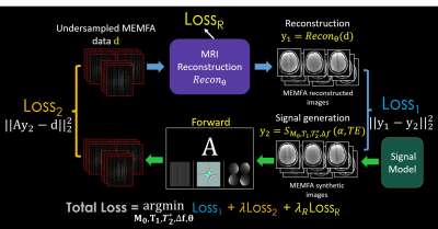 |
154 | Joint MR T1 and T2* Parameter Mapping with Scan Specific Unsupervised Networks
Amir Heydari1, Tae Hyung Kim2,3,4, Abbas Ahmadi1, and Berkin Bilgic3,4
1Industrial Engineering and Management Systems, Amirkabir University of Technology, Tehran, Iran (Islamic Republic of), 2Department of Computer Engineering, Hongik University, Seoul, Korea, Republic of, 3Athinoula A. Martinos Center for Biomedical Imaging, Massachusetts General Hospital, Charlestown, MA, United States, 4Radiology, Harvard Medical School, Boston, MA, United States Keywords: Quantitative Imaging, Quantitative Imaging, Image Reconstruction We introduce a new method for joint T1 and T2* MR parameter mapping in MAPLE framework which takes advantage of multi-echo multi-flip angle (MEMFA) gradient echo data. It combines joint reconstruction of MEMFA data with a joint relaxation signal model to improve parameter estimation. The proposed method estimates T1 and T2* parameters jointly with enough flexibility to incorporate different standard and state-of-the-art reconstruction methods, including zero-shot self-supervised learning (ZS-SSL) reconstruction. It improves the reconstruction performance of the methods with learnable parameters by offering a physics-based additional regularization into their optimization process and exploiting spatio-temporal correlations. |
|
1618.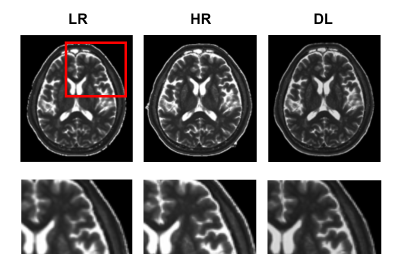 |
155 | Feasibility of Using Deep Learning to Reduce Bias in Quantitative Values: A Study Based on fast Multidynamic Mutiecho Imaging
Yawen Liu1, Pengling Ren2, Hongxia Yin3, Yi Zhu4, Rong Wei5, Linkun Cai1, Haijun Niu1, and Zhenchang Wang1,2
1School of Biological Science and Medical Engineering, Beihang University, Beijing, China, 2Department of Radiology, Beijing Friendship Hospital, Capital Medical University, Beijing, China, 3Department of Medical Engineering, Beijing Friendship Hospital, Capital Medical University, Beijing, China, 4Philips Healthcare, Beijing, China, 5Peking university Academy for Advanced Interdisciplinary Studies, Beijing, China Keywords: Quantitative Imaging, Machine Learning/Artificial Intelligence Rapid quantitative magnetic resonance imaging (qMRI) is the trend of MR development and has essential diagnostic value. However, reducing acquisition time will come at the expense of image quality and will also affect the accuracy of quantitative values. Here we propose a method for reconstructing fast low-resolution qMRI images using deep learning, aiming to improve image quality while reducing bias in quantitative values. The research results show that after deep learning, the image quality is comparable to that of conventional high-resolution scanning images, and quantitative values are also more stable. |
|
1619.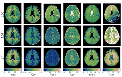 |
156 | B0 Dependence of Magnetization Transfer and Spin Relaxation Times
Sebastian Flassbeck1, Andrew Mao1, and Jakob Assländer1
1Dept. of Radiology, NYU Langone, New York, NY, United States Keywords: Quantitative Imaging, CEST & MT, MR Fingerprinting, qMT, Low field, B0-field dependance We demonstrate an approach to quantify unconstrained MT parameter mapping and utilized this approach to study the magnetic field dependence of the relaxation and MT parameters. The largest field dependence was observed in the longitudinal relaxation rate of the semi-solid spin pool. |
|
1620.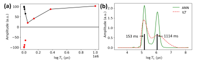 |
157 | T1 Spectrum Analysis with Reduced Number of Datapoints Using Neural Networks
Tristhal Parasram1 and Dan Xiao1
1University of Windsor, Windsor, ON, Canada Keywords: Quantitative Imaging, Relaxometry Quantitative analysis of T1 spectra could reveal microscopic properties and have been used to study biological tissues such as the heart, brain and related disease. It is challenging to determine the relaxation times from magnetic resonance signals particularly with multicomponent continuous spectra as it is an inherently ill-posed exponential analysis problem even with a large number of input data points. Artificial neural networks have been trained to determine T1 spectra with 8 to 4 logarithmically spaced data points. Improved performance over a larger range of T1 values and faster processing time compared to traditional methods have been achieved. |
|
1621.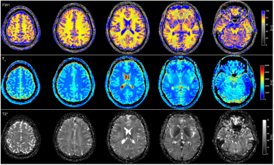 |
158 | Multiparametric quantitative MRI mapping using ultrahigh-field strength
Zaheer Abbas1, Markus Zimmermann1, Dominik Ridder1, Ana-Maria Oros-Peusquens1, and N. Jon Shah1,2,3,4
1Institute of Neuroscience and Medicine 4, Jülich, Germany, 2Institute of Neuroscience and Medicine 11, INM-11, JARA, Forschungszentrum Jülich, Jülich, Germany, 3JARA - BRAIN - Translational Medicine, Aachen, Germany, 4Department of Neurology, RWTH Aachen University, Aachen, Aachen, Germany Keywords: Quantitative Imaging, Brain Measurement of quantitative, tissue-specific MR properties such as water content or relaxation times using quantitative-MRI at clinical field strength (1.5T and 3T) is a well-explored topic. Established methods for water content mapping are based on the variable flip-angle (VFA) approach. However, transition to ultrahigh field strength remains challenging due to significantly increased RF field inhomogeneity. Here, we demonstrate a novel VFA method to acquire quantitative water content and relaxation times at 7T with full brain coverage and 1x1x1.5 mm resolution within 7 min. Accuracy and precision of the parametric maps is demonstrated by comparison to already reported results. |
|
1622.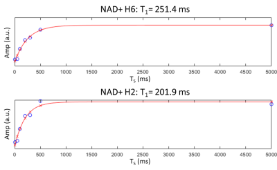 |
159 | Measurement of NAD+ T1 relaxation time at 7T in the human brain using saturation recovery downfield MRS
Neil Wilson1, Sophia Swago2, Mark Elliott1, Ravi Prakash Reddy Nanga1, Ravinder Reddy1, and Walter Witschey1
1Radiology, University of Pennsylvania, Philadelphia, PA, United States, 2Bioengineering, University of Pennsylvania, Philadelphia, PA, United States Keywords: Quantitative Imaging, Spectroscopy Here we present a method based on spectrally selective saturation recovery 1H spectroscopy to measure the apparent T1 values of NAD+ in the downfield at 7T for the first time. With spectrally selective saturation and excitation, NAD+ exhibits enhanced T1 recovery do to cross relaxation with water, and we report values in three subjects between 200 and 300 ms. Measuring apparent T1 is crucial as a correction factor for quantitative spectroscopy of NAD+. |
|
1623.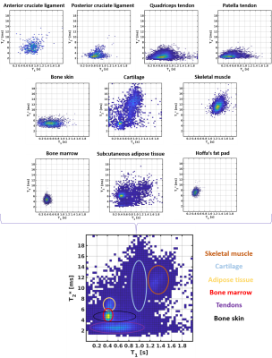 |
160 | In vivo T1 and T2* relaxation times of fast relaxing tissues of the healthy knee
Maik Rothe1, Selina Riedel1, Andreas Deistung1, Brill Richard1, Walter Alexander Wohlgemuth1, and Alexander Gussew1
1University Clinic and Outpatient Clinic for Radiology, University Hospital Halle (Saale), Halle (Saale), Germany Keywords: Quantitative Imaging, Relaxometry In this study, we used a method for fast in vivo T1 and T2* mapping of fast relaxing tissues of the knee using a high resolution 3D spoiled gradient ultrashort echo time research application sequence with spiral read out to quantitatively describe different tissues. We manually segmented 10 different tissues in five healthy young volunteers and found good agreement with literature values for fast relaxing tissues. The bivariate histograms showed tissue specific clusters, which can be used for tissue differentiation. Additional information, such as phase images, could further improve bivariate histogram-based tissue differentiation and should be explored in future studies. |
|
 Back to Meeting Home
 Back to the Program-at-a-Glance
The International Society for Magnetic Resonance in Medicine is accredited by the Accreditation Council for Continuing Medical Education to provide continuing medical education for physicians.
 Back to Meeting Home
 Back to the Program-at-a-Glance
 View Presentation Video