Digital Poster
Phantoms & Repeatability I
ISMRM & ISMRT Annual Meeting & Exhibition • 03-08 June 2023 • Toronto, ON, Canada

| Computer # | |||
|---|---|---|---|
5083.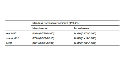 |
141 |
Improved diagnosis performance of CAD using single-bolus
quantitative stress perfusion CMR imaging: a pilot study of
reproducibility
Zhaoxia Yang1,
Chunlin Xiang1,
Weiyin Vivian Liu2,
Haonan Wang3,
Lu Huang1,
and Liming Xia1
1Tongji Hospital,Huazhong University of Science and Technology, wuhan, China, 2MR Research, GE Healthcare, Beijing, China, 3MR Research, General Electric Healthcare, Chicago, IL, United States Keywords: Quantitative Imaging, Ischemia, stress cardiac magnetic resonance imaging Automated CMR perfusion maps enable quantification of MBF for detection of myocardial ischaemia rapidly within a clinical workflow. Stress perfusion CMR derived quantitative parameters had good to excellent reproducibility, as well as quantitative MPR was moderately correlated with semi-quantitative MPR. Quantitative myocardial perfusion CMR provides objective indices (MBF and MPR), which could better identify disease extent and detect coronary microvascular disease than visual interpretation. |
|
5084.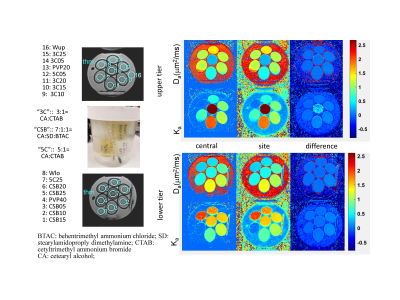 |
142 |
Reproducibility of diffusion kurtosis parameters for a
quantitative phantom in a multi-system study
Dariya Malyarenko1,
Scott D Swanson1,
Ramesh Paudyal2,
Amaresh Konar2,
Eddy Solomon3,
Eric E Sigmund4,
Stephen E Russek5,
Sungheon Gene Kim3,
Amita Shukla-Dave2,6,
and Thomas L Chenevert1
1Radiology, University of Michigan Health System, Ann Arbor, MI, United States, 2Medical Physics, Memorial Sloan Kettering Cancer Center, New York, NY, United States, 3Radiology, MRI Research Institute, Weill Cornell Medical College, New York, NY, United States, 4Radiology, Center for Biomedical Imaging, NYU Langone Health, New York, NY, United States, 5National Institute of Standards and Technology, Boulder, CO, United States, 6Radiology, Memorial Sloan Kettering Cancer Center, New York, NY, United States Keywords: Quantitative Imaging, Diffusion/other diffusion imaging techniques, diffusion kurtosis, multi-system reproducibility, quantitative phantom This work studied reproducibility of apparent diffusion (Da) and kurtosis (Ka) coefficients for three families of lamellar-vesicle materials in a quantitative phantom scanned at ambient temperature in a multi-site setting. Inter-system reproducibility of Ka was improved by maximum b-value fit-constraints for phantom materials with higher solid concentration, while temperature calibration improved Da parameter consistency predominantly for materials with low solid concentration and containing longer-chain alcohols. After correction for temperature-dependence, these materials exhibited apparent diffusion reproducibility comparable to that of standard Gaussian diffusion controls. |
|
5085.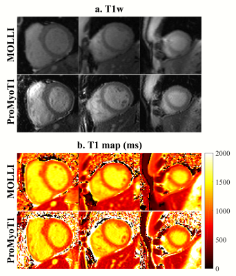 |
143 |
Repeatability of ProMyoT1: an open-source inversion recovery
myocardial T1 mapping sequence for fast prototyping
Andreia S Gaspar1,
Nuno A Silva2,
António M Ferreira3,4,
and Rita G Nunes1
1Institute for Systems and Robotics - Lisboa and Department of Bioengineering, Instituto Superior Técnico – Universidade de Lisboa, Lisbon, Portugal, 2Hospital da Luz Learning Health, Luz Saúde, Lisbon, Portugal, 3Serviço de Cardiologia, Hospital de Santa Cruz, Centro Hospitalar Lisboa Ocidental, Lisbon, Portugal, 4Unidade de Imagiologia Cardíaca Avançada, Hospital da Luz, Lisbon, Portugal Keywords: Quantitative Imaging, Heart, T1 mapping The open-source method Prototype of Myocardial T1 mapping (ProMyoT1) created with Pulseq was tested in vivo in healthy subjects to evaluate its repeatability and compare its performance with that of the clinical MOLLI method. ProMyoT1 provided myocardium T1 values similar to those obtained with MOLLI. The precision was better for MOLLI, but the repeatability measures (Coefficient of Variation and Repeatability Coefficient) were similar in both methods. The developed ProMyoT1 method was shown to be reliable and should improve the accessibility to T1 mapping, while also enabling fast sequence prototyping so that further improvements can easily be tested. |
|
5086.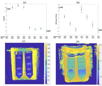 |
144 |
Hydrogel-based quantitative multi-parametric MRI phantom with
controlled T2, diffusion and kurtosis
Scott D. Swanson1,
Ted Lynch2,
Shigeto Ono2,
and Dariya I. Malyarenko1
1Department of Radiology, University of Michigan, Ann Arbor, MI, United States, 2CIRS/Mirion Inc., Norfolk, VA, United States Keywords: Quantitative Imaging, Phantoms, Diffusion, Kurtosis, Transverse Relaxation A set of materials with tunable T2, diffusion, and kurtosis were assembled to create quantitative biomimetic MRI phantoms. T2 is controlled with variable agarose concentration, mono-exponential diffusion by polyvinylpyrrolidone, and kurtosis by addition of lamellar vesicles. The phantoms are mechanically stabilized by polyacrylamide gels to allow biomimetic morphologies. These nanostructured systems provide an ideal platform for moldable multiparametric MRI phantoms that are useful for pulse sequence design and protocol standardization for multi-site multi-vendor imaging trials, as well as for refinement of emerging AI analysis methods. |
|
5087.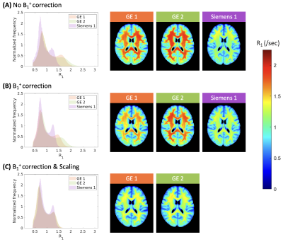 |
145 |
Inter-site variability observed in R1 maps of the brain
generated from two-point inversion-recovery MRI
Stella Heo1,
Christopher Rowley2,
Christine Tardif2,
and Nicholas Bock1
1Department of Psychology, Neuroscience & Behaviour, McMaster University, Hamilton, ON, Canada, 2McConnell Brain Imaging Centre, Montreal Neurological Institute and Hospital, McGill University, Montreal, QC, Canada Keywords: Quantitative Imaging, Challenges, Reproducibility The longitudinal relaxation rate (R1) is deemed to be a suitable quantitative imaging metric for multi-site investigations as it describes a quantitative property of the tissue that can be measured, reproduced, and compared across sites when differences in hardware and acquisition settings are accounted for. Here, we present inter- and intra-vendor variability observed in whole-brain R1 maps generated from two-point inversion-recovery MRI data after accounting for variations in pulse sequences and B1+ field maps. We posit that this inter-site variability may be due to differential intensity scaling applied at acquisition and suggest a potential correction method using site-specific scaling factors. |
|
5088.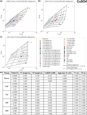 |
146 |
Modeling and selection of T1 and T2 tissue mimics for 0.0065T,
0.064T, 0.55T MRI using agarose with manganese, gadolinium,
copper, or nickel
Kalina V Jordanova1,
Ye Tian2,
Sheng Shen3,
Michele N Martin1,
Megan E Poorman4,
Rui Pedro Teixeira4,
Krishna S Nayak2,
Matthew S Rosen3,5,
and Kathryn E Keenan1
1NIST: National Institute of Standards and Technology, Boulder, CO, United States, 2Ming Hsieh Department of Electrical and Computer Engineering, University of Southern California, Los Angeles, CA, United States, 3Athinoula A. Martinos Center for Biomedical Imaging, Massachusetts General Hospital and Harvard Medical School, Boston, MA, United States, 4Hyperfine, Inc., Guilford, CT, United States, 5Department of Physics, Harvard University, Cambridge, MA, United States Keywords: Quantitative Imaging, Relaxometry There is re-emerging interest in MRI fields ≤0.55T as well as quantitative MRI (qMRI) methods. Physiologically relevant reference objects are needed to adapt qMRI techniques to lower fields.
We investigate materials as tissue mimics for brain imaging at 0.0065T, 0.064T, 0.55T, for white matter, gray matter, fat, cerebrospinal fluid, and blood. We create samples composed of agarose and paramagnetic salts and measure relaxation across field strengths. Samples suitable to mimic each tissue are presented for each field strength. This work will facilitate qMRI development for fields ≤0.55T by providing accessible mimic compositions to the community. |
|
5089.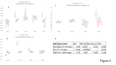 |
147 |
Repeatability, Distortion and SNR comparison of
diffusion-weighted imaging techniques on an MR-Linac
Prashant Prabhakaran Nair1,
Robin J.M. Navest2,
Rosie Goodburn1,
Bastien Lecoeur1,
Uwe Oelfke1,
and Andreas Wetscherek1
1Radiotherapy and Imaging, The Institute of Cancer Research, London, United Kingdom, 2Netherlands Cancer Institute, Amsterdam, Netherlands Keywords: Quantitative Imaging, Precision & Accuracy, MR-Linac DW-acquisitions with high ADC accuracy, precision and low geometric distortion are necessary for using ADC maps for treatment planning and response assessment in MR-guided radiotherapy on MR-Linacs. We measured SNR, ADC bias, repeatability coefficient and marker distances on the NIST phantom for DW-EPI, DW-TSE and SPLICE protocols. Two volunteers were scanned in the head and neck region. The investigated TSE-based protocols showed ADC bias and DW-EPI demonstrated better repeatability. Spectral fat-suppression techniques led to higher SNR than using STIR. SPLICE DWI could be an alternative to diffusion EPI with reduced distortion and comparable SNR, if low-high profile order is used. |
|
5090.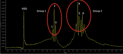 |
148 |
Establishing absolute temperature scale and diffusion metrics in
PVP phantoms for cross-center standardization
Neville D Gai1,
Ruifeng Dong1,
Jeffrey Hopkins2,
and Carlo Pierpaoli1
1Laboratory of Quantitative Imaging, National Institute of Biomedical Imaging and Bioengineering, Bethesda, MD, United States, 2MR Division, GE Healthcare, Waukesha, WI, United States Keywords: Quantitative Imaging, Diffusion Tensor Imaging, PVP phantom, spectroscopy, temperature mapping Clinical acceptance of quantitative diffusion MRI faces difficulties due to lack of standardization. Quality control across platforms and centers is vital for multicenter efforts. Polyvinylpyrrolidone(PVP) phantoms provide excellent properties suitable for cross-center validation. However, diffusion metrics vary with temperature. Measuring phantom temperature along with diffusion metrics is necessary. Spectroscopic temperature mapping of the PVP phantom and ADC values were investigated on two different vendor platforms. It is shown that the water peak moves relative to easily identifiable stable peaks in PVP allowing intrinsic temperature measurement. Excellent agreement for temperature and ADC mapping across both platforms should make cross-site standardization possible. |
|
5091.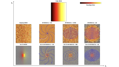 |
149 |
Increasing the repeatability of DESS 3D brain T2 mapping with
optimized k-space sampling order
Emile Clements Kadalie1,
Aurélien J Trotier1,
Nadège Corbin1,
Sylvain Miraux1,
and Emeline J Ribot1
1Centre de Résonance Magnétique des Systèmes Biologiques, UMR5536, CNRS / Université de Bordeaux, Bordeaux, France Keywords: Quantitative Imaging, Brain, T2 mapping While recent works have determined that the DESS sequence has the ability to produce 3D T2 mapping of the brain, it remains sensitive to B0-related variation due to respiration. Multiple encoding strategies, using cartesian-spiral trajectories with different lengths combined with a variable density Poisson undersampling mask and Compressed-Sensing reconstruction, were consequently employed and compared in order to suppress respiration artifacts as well as to establish accurate and repeatable 3D T2 maps of the brain at 3T. |
|
5092.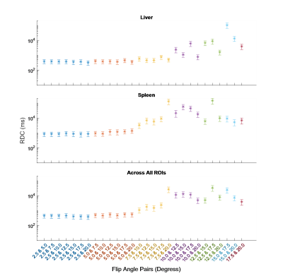 |
150 |
Reproducibility of Whole-Body Variable Flip Angle T1 Mapping
Using Only Two Flip Angles
Alistair Lamb1,
David Atkinson2,
Shonit Punwani2,
Hui Zhang3,
and Anna Barnes4
1Department of Medical Physics & Biomedical Engineering, University College London, London, United Kingdom, 2Centre for Medical Imaging, University College London, London, United Kingdom, 3Centre for Medical Image Computing, University College London, London, United Kingdom, 4King's Technology Evaluation Centre, King's College London, London, United Kingdom Keywords: Quantitative Imaging, Whole Body, T1-mapping We investigate the feasibility of whole-body (WB) variable flip angle (VFA) T1 mapping using linear least squares fitting with only two flip angles (FAs) in order to obtain WB T1 maps within a clinically viable timeframe. This could enable its use as an imaging biomarker in metastatic cancer. We assessed the agreement across eight subjects in a variety of abdominal tissues between T1 estimates fitted using eight FAs and just 2 FAs. We found that VFA T1 mapping can be achieved by acquiring only two FAs with minimal loss to precision, providing the lower FA is between 2.5° and 7.5°. |
|
5093.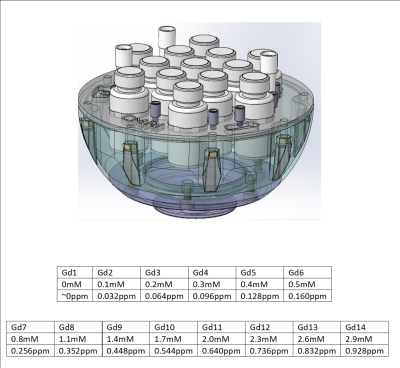 |
151 |
Long-term QSM reproducibility phantom from a commercially
available design
Eric Y. Pierre1,
David N. Vaughan1,2,
Warda T. Syeda3,
Bahman Tahayori1,
Heath R. Pardoe1,
David F. Abbott1,
and Graeme D. Jackson1
1Imaging and Epilepsy, The Florey Institute of Neuroscience and Mental Health, Melbourne, Australia, 2Department of Neurology, Austin Health, Melbourne, Australia, 3Department of Psychiatry, The University of Melbourne, Melbourne, Australia Keywords: Quantitative Imaging, Quantitative Susceptibility mapping, Reproducible Research We propose the customization of a commercially-available phantom design to be used for QSM reproducibility research with long-term stability, allowing reproduction of the phantom itself commercially. The proposed phantom is based on master-dilution of Gadolinium in pure water vials, offering long-term stability, with theoretical susceptibility range chosen to match normal tissue and common pathologies in the brain. We evaluate the suitability of the phantom for QSM reproducible research for different sequences, orientation within the scanner and regularization algorithms. |
|
5094.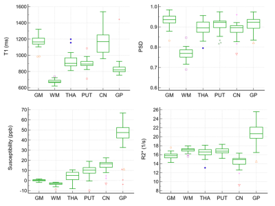 |
152 |
Test-Retest Repeatability of Smart BrainQuant (STAGE) of the
healthy brain at 1.5T
Yuting Ling1,
Jianjun Zhang2,
Jiangwei Xiang2,
Yu Wang1,
Zhenzhuang Miao1,
Xingxing Zhang1,
Xiaoyun Liang1,
and Feng Huang1
1Neusoft Medical Systems Co., Ltd., Shanghai, China, 2Department of Radiology, Affiliated Zhejiang Hospital, Zhejiang University School of Medicine, Hangzhou, China Keywords: Stroke, Screening Smart BrainQuant, adapted from strategically acquired gradient echo (STAGE) method, is capable to provide 10 multi-contrast and multi-quantitative images with high image quality in 4:21 minutes at 1.5T. A preclinical validation work was introduced in this study with the aim to assess the test-retest repeatability of T1, proton spin density (PSD), susceptibility, and R2* by recruiting 18 healthy volunteers. Coefficient of variation of T1, PSD, and R2* ranged from 3.3% to 4.1%. Bland-Altman demonstrated that the line of equality is in the 95% confidence interval (CI) of the mean difference for the four maps. |
|
5095.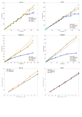 |
153 |
Multicenter and multi-vendor reproducibility of T1, T2 and ADC
phantom data on 1.5T and 3T MRI scanners
Anna Caroli1,
Siria Pasini1,
Tau Vandelboe2,
Anish Raj3,4,
Leyre Garcia-Ruiz5,
Anika Strittmatter3,4,
Rebeca Echeverria-Chasco5,
Giulia Villa1,
Paolo Brambilla6,
Esben Søvsø Szocska Hansen2,
Steffen Ringgaard2,
Frank G Zoellner3,4,
Maria Fernandez-Seara5,
Susan Francis7,
and Christoffer Laustsen2
1Bioengineering Department, Istituto di Ricerche Farmacologiche Mario Negri IRCCS, Ranica (BG), Italy, 2The MR Research Centre, Aarhus University, Aarhus, Denmark, 3Computer Assisted Clinical Medicine, Medical Faculty Mannheim, Heidelberg University, Mannheim, Germany, 4Mannheim Institute for Intelligent Systems in Medicine, Medical Faculty Mannheim, Heidelberg University, Mannheim, Germany, 5Clínica Universidad de Navarra, Pamplona, Spain, 6Unit of Radiology, ASST Papa Giovanni XXIII, Bergamo, Italy, 7University of Nottingham, Nottingham, United Kingdom Keywords: Quantitative Imaging, System Imperfections: Measurement & Correction This multicentre study aimed at assessing multi-vendor accuracy and reproducibility of typical MR biomarkers on 3T and 1.5T scanners from 5 clinical centers. MRI acquisitions were performed using NIST protocols, on Essential System and Diffusion NIST phantoms. T1 was measured by 3-parameter fitting, while T2 and ADC by mono-exponential fitting, using PhantomViewer software. When compared with reference values, non-negligible discrepancies were found across vendors, especially for T1 and T2 measurements. Reproducibility was vendor-dependent, and higher on 3T than on 1.5T scanners. ADC and T1 showed highest reproducibility. Corrections are likely needed to account for possible temperature and scanner differences. |
|
5096.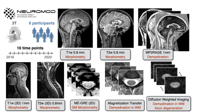 |
154 |
Longitudinal stability of brain and spinal cord structural
quantitative MRI measures
Mathieu Boudreau1,
Agah Karakuzu1,
Arnaud Boré2,3,
Kiril Zelenkovski4,
Basile Pinsard2,3,
Eva Alonso-Ortiz1,
Julie Boyle2,3,
Pierre Bellec2,3,5,
and Julien Cohen-Adad1,3,6,7
1NeuroPoly, Polytechnique Montreal, Montreal, QC, Canada, 2Centre de Recherche de l’Institut Universitaire de Gériatrie de Montréal (CRIUGM), Montreal, QC, Canada, 3Unité de Neuroimagerie Fonctionnelle (UNF), Centre de Recherche de l’Institut Universitaire de Gériatrie de Montréal (CRIUGM), Montreal, QC, Canada, 4Faculty of Computer Science and Engineering (FINKI), Skopje, Macedonia, 5Psychology Department, Université de Montréal, Montreal, QC, Canada, 6Mila - Quebec AI Institute, Montreal, QC, Canada, 7Centre de recherche du CHU Sainte-Justine, Université de Montréal, Montreal, QC, Canada Keywords: Quantitative Imaging, Quantitative Imaging The stability of quantitative MRI measures of microstructure in the brain and spinal cord was quantified longitudinally over three years. Six healthy subjects were scanned approximately four times per year with an structural quantitative imaging protocol (T1w, T2w, T2*w, DWI, MP2RAGE, MTsat, and B1). The intra-subject COV indicated good stability of all quantitative metrics measured in the brain (< 2.3% in WM, < 3.1% in GM). The spinal cord resulted in slightly higher COVs (3.9% - 9.5%). This work is part of a larger project, the Courtois project on neural modelling (CNeuroMod). |
|
5097.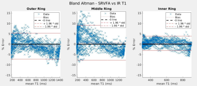 |
155 |
Validation of the Single Reference Variable Flip Angle T1
mapping method in an innovative phantom
Michael Malmberg1,
Henrik L Odéen2,
and Dennis L Parker2
1Biomedical Engineering, University of Utah, Salt Lake City, UT, United States, 2Radiology and Imaging Sciences, University of Utah, Salt Lake City, UT, United States Keywords: Quantitative Imaging, Quantitative Imaging, T1 mapping An innovative cylindrical phantom is used to mimic a dynamic acquisition with temperature-induced changes in T1. Single reference (SR) variable flip angle (VFA) T1 maps are acquired at several rotational positions of this phantom, and are compared to corresponding T1 values acquired with the dual-angle variable flip angle and inversion recovery T1 mapping methods. The SR-VFA method of T1 mapping is shown to produce T1 accuracy within 4-10% of both inversion recovery and dual-angle VFA T1 measurements. |
|
5098.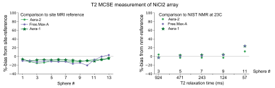 |
156 |
T1 and T2 measurement variability across commercial and
prototype 0.55T systems
Kathryn E Keenan1,
Bilal Tasdelen2,
Adrienne E Campbell-Washburn3,
Harish Sharma4,
Ahsan Javed3,
Rajiv Ramasawmy3,
Michele N Martin1,
Nicole Seiberlich4,
and Krishna Nayak2
1NIST, Boulder, CO, United States, 2Ming Hsieh Department of Electrical and Computer Engineering, University of Southern California, Los Angeles, CA, United States, 3Cardiovascular Branch, Division of Intramural Research, National Heart, Lung, and Blood Institute, National Institutes of Health, Bethesda, MD, United States, 4Department of Radiology, University of Michigan, Ann Arbor, MI, United States Keywords: Quantitative Imaging, Relaxometry We compare T1 and T2 measurements between the Free.Max and ramped-down Aera using the ISMRM/NIST system phantom at multiple sites. All 0.55T systems had comparable, to each other, measurements of T1 and T2 on both the NiCl2 and MnCl2 arrays. The T1 measurement of the NiCl2 array by VFA-GRE with B1+correction was overestimated compared to the site-reference by 1% to 92%. The rest of the measurements – MnCl2 T1, NiCl2 T2, and MnCl2 T2 – were within +/-20% of the site-reference and NMR-reference values. As adoption of 0.55T platforms increases, it is important to characterize and compare systems across sites. |
|
5099.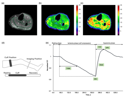 |
157 |
Blood Oxygen Level-Dependent Imaging in Peripheral Artery
Disease: Feasibility and Reproducibility of perfusion
measurements
Xiaoxi Yu1,
Zhaoxi Liu2,
Jianxun Qu3,
Fengdan Wang2,
Zhichao Lai1,
Xiaoyuan Fan2,
Luming Ye4,
Jiang Shao1,
Yan Zhang2,
Bao Liu1,
Zhengyu Jin2,
and Feng Feng2
1Vascular Surgery, Peking Union Medical College Hospital, Beijing, China, 2Radiology, Peking Union Medical College Hospital, Beijing, China, 3MR Collaboration, Siemens Healthineers Ltd., Beijing, China, 4Advanced Therapies Collaboration, Siemens Healthineers Ltd., Beijing, China Keywords: Quantitative Imaging, Perfusion This study used blood oxygen level-dependent (BOLD) imaging to assess the hemodynamics of the lower limbs among elder controls and patients with peripheral artery disease (PAD). The BOLD-derived perfusion parameters, such as time to peak and gradient during reactive hyperemia, showed good reproducibility and significantly differed between patients and controls. The results indicated decreased vasodilatation ability and perfusion levels in patients with PAD. Therefore, BOLD imaging can be a feasible perfusion assessment method for long-term noninvasive PAD monitoring. |
|
5100.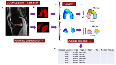 |
158 |
7T cross-vendor repeatability study of cartilage T2 values using
DOSMA on qDESS images
Jessica Lauren Asay1,
Krithika Balaji2,
Anthony A Gatti1,
Arjun D Desai1,
Michael Mendoza2,
Zimu Huo2,
Akshay S Chaudhari1,3,
Feliks Kogan1,
Peter J Lally4,5,
Neal K Bangerter2,
and Garry E Gold1
1Radiology, Stanford University, Stanford, CA, United States, 2Bioengineering, Imperial College London, London, United Kingdom, 3Biomedical Data Science, Stanford University, Stanford, CA, United States, 4Brain Sciences, Imperial College London, London, United Kingdom, 5UK Dementia Research Institute Centre for Care Research and Technology, London, United Kingdom Keywords: Quantitative Imaging, Data Processing, Repeatability, cartilage, knee Cartilage T2 relaxation times (T2), used to detect early knee osteoarthritis, lack standardization in acquiring and processing data, making comparisons between studies difficult. Standardizing image post-processing could possibly control for biases. Here, we assess qDESS cartilage T2 repeatability across two different sites and 7T scanner vendors with identical automatic segmentation and T2 mapping software. Within-site repeatability was good (ICC≥ 0.75) for most cartilage regions, while cross-vendor repeatability was good for the tibial and femoral posterior cartilage. This preliminary study shows standardizing acquisition and post-processing can lead to repeatable T2 values across different vendors. |
|
5101.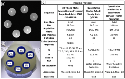 |
159 |
Phantom T1rho and T2 relaxation times demonstrate good
repeatability across sequences and scanner position
Yael Vainberg1,
Valentina Mazzoli1,
and Feliks Kogan1
1Stanford University, Stanford, CA, United States Keywords: Quantitative Imaging, Cartilage, Repeatability T2 and T1rho repeatability is essential for evaluating the small changes that occur during early osteoarthritis disease progression. In this study, we acquired phantom data on T2 and T1rho relaxation times across 6 time points as well as 3 distinct locations along the scanner for T2/T1rho 3D MAPSS and 3D qDESS with varying resolution for T2 mapping. We observed repeatability of the 4 quantitative MRI sequences that were well within the QIBA benchmark for phantom scans of 4-5%. Further, our data suggests that phantom positioning within the scanner bore did not affect repeatability or quantitative values. |
|
5102.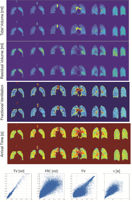 |
160 |
Repeatability of Hyperpolarized Xenon-129 4D Imaging
Hooman Hamedani1,
Stephen Kadlecek2,
Faraz Amzajerdian2,
Ryan Baron2,
Kai Ruppert2,
Mostafa Ismail2,
Luis Loza2,
Duncan Ian2,
and Rahim Rizi2
1Radiology, University of Pennsylvania, Philadelphia, PA, United States, 2University of Pennsylvania, Philadelphia, PA, United States Keywords: Quantitative Imaging, Lung, Hyperpolarized MRI In order to image function in the free breathing lung with high temporal and special resolution, we have developed a methodology that can be used in combination with a non-rigid image registration technique to generate comprehensive quantitative maps of lung ventilation and gas exchange. For these HP MRI-derived regional parameters to produce meaningful information that can be used to characterize lung disorders, a reproducibility study that systematically assesses the variability of these parameters is needed. Here, we assess the reproducibility of our previously reported dynamic hyperpolarized imaging measurements between two sessions separated by minutes. |
|
 Back to Meeting Home
 Back to the Program-at-a-Glance
The International Society for Magnetic Resonance in Medicine is accredited by the Accreditation Council for Continuing Medical Education to provide continuing medical education for physicians.
 Back to Meeting Home
 Back to the Program-at-a-Glance
 View Presentation Video