Digital Poster
Neurodegeneration IV
ISMRM & ISMRT Annual Meeting & Exhibition • 03-08 June 2023 • Toronto, ON, Canada

| Computer # | |||
|---|---|---|---|
2155.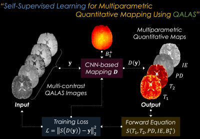 |
161 | SSL-QALAS: Self-Supervised Learning for Multiparametric Quantitative MRI Using QALAS
Yohan Jun1,2, Jaejin Cho1,2, Xiaoqing Wang1,2, Michael Gee2,3, P. Ellen Grant2,4, Berkin Bilgic1,2,5, and Borjan Gagoski2,4
1Athinoula A. Martinos Center for Biomedical Imaging, Charlestown, MA, United States, 2Department of Radiology, Harvard Medical School, Boston, MA, United States, 3Department of Radiology, Massachusetts General Hospital, Boston, MA, United States, 4Fetal-Neonatal Neuroimaging & Developmental Science Center, Boston Children’s Hospital, Boston, MA, United States, 5Harvard/MIT Health Sciences and Technology, Cambridge, MA, United States Keywords: Quantitative Imaging, Quantitative Imaging The 3D-quantification using an interleaved Look-Locker acquisition sequence with T2 preparation pulse (3D-QALAS) has been developed and used for acquiring high-resolution T1, T2, and PD maps from five measurements. The dictionary matching method can be used for generating quantitative maps from the acquired multi-contrast images; however, it requires an external dictionary, which needs to be pre-calculated and voxel-by-voxel fitting is computationally demanding. In this study, we propose to generate multiple quantitative maps including T1, T2, PD, and inversion efficiency (IE) maps using self-supervised learning from 3D-QALAS measurements (i.e., SSL-QALAS) for rapid, accurate, and dictionary-free multiparametric fitting. |
|
2156.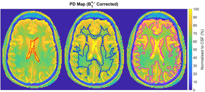 |
162 | Investigating the Accuracy of PD Measurements at 3T
Zach Lun Pang1, Maria Schmidt2, Evanthia Kousi2, and Julie Hughes3
1Medical Physics, St George’s University Hospitals NHS Foundation Trust, London, United Kingdom, 2Joint Department of Physics, The Institute of Cancer Research and The Royal Marsden NHS Foundation Trust, London, United Kingdom, 3The Royal Marsden NHS Foundation Trust, London, United Kingdom Keywords: Phantoms, Quantitative Imaging We investigated the accuracy of clinical proton density (PD) mapping employing the variable flip-angle (VFA) method at 3T, considering the effects of B1+ and B1- fields. Following B1+ correction, the PD measurements were within 5% of the expected value for flip-angle variations up to 18%. Considerable discrepancies were observed for T1<300ms and flip-angle variations >25%. Using image intensity filters, considerable PD variations were found at the edges of the test object. Clinically, the B1+ and B1- corrected PD values of the brain were similar to values reported in the literature, rendering the VFA method a viable tool for PD mapping. |
|
2157.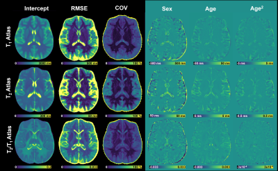 |
163 | Normative brain atlas of T2/T1 relaxometry ratio for detection of microstructural pathology
Veronica Ravano1,2,3, Samuele Caneschi1,2,3, Jan Krasensky4, Michaela Andelova5, Matej Kudrna4, Tomas Uher5, Gian Franco Piredda1,6,7, Jonathan Disselhorst1,2,3, Bénédicte Maréchal1,2,3, Jean-Philippe Thiran2,3, Jonas Richiardi2, Manuela Vaneckova4, Tom Hilbert1,2,3, and Tobias Kober1,2,3
1Advanced Clinical Imaging Technology, Siemens Healthineers International AG, Lausanne, Switzerland, 2Department of Radiology, Lausanne University Hospital and University of Lausanne, Lausanne, Switzerland, 3LTS5, Ecole Polytechnique Fédérale de Lausanne, Lausanne, Switzerland, 4Department of Radiology, First Faculty of Medicine, Charles University and General University Hospital, Prague, Czech Republic, 5Department of Neurology and Center of Clinical Neuroscience, First Faculty of Medicine, Charles University and General University, Prague, Czech Republic, 6Human Neuroscience Platform, Fondation Campus Biotech Geneva, Geneva, Switzerland, 7CIBM-AIT, Ecole Polytechnique Fédérale de Lausanne, Lausanne, Switzerland Keywords: Quantitative Imaging, Tissue Characterization, Neuroinflammation; Neurodegeneration Quantitative MRI allows the characterization and comparison of microstructural tissue properties and, by defining normative ranges for healthy tissue, detection of pathology. The simultaneous evaluation of multiple qMRI measures facilitates the understanding of what biological mechanisms underlie the observed deviations in relaxometry. Here, we introduce the ratio between quantitative T2 and T1 maps (T2/T1) as a microstructural property whose deviations from normative values could be predictive or characteristic of brain pathologies. A T2/T1 voxel-wise normative atlas was created and allowed characterization of tissue changes in two multiple sclerosis patients, providing complementary information to the existing T1 and T2 atlases. |
|
2158. |
164 | Free-water Investigation In Parkinsonian Disorders Using Diffusion-weighted Imaging
Apurva Shah1, Jitender Saini2, Madhura Ingalhalikar1, Shweta Prasasd3, and Pramod Pal3
1Symbiosis Centre for Medical Image Analysis, Symbiosis International (Deemed University), Pune, India, 2Department of Radiology, National Institutes of Mental Health and Neuroscience, Bengaluru, India, 3Department of Neurology, National Institutes of Mental Health and Neuroscience, Bengaluru, India Keywords: Neuroinflammation, Parkinson's Disease, Parkinsonism, Free water Parkinson's disease(PD), multiple system atrophy(MSA), and progressive supranuclear palsy(PSP) are neurodegenerative disorders with parkinsonism as a central feature. Although MSA and PSP are atypical parkinsonian disorders, they may be difficult to differentiate from PD particularly in the early stages. Multiple studies have demonstrated that patients with Parkinsonian disorders have higher freewater(FW) in the substantia-nigra-pars-compacta(SNc) and basal ganglia(BG), and FW was shown to reliably distinguish between these patients, making it a potential biomarker. Our investigation into the alterations of FW in PD and atypical parkinsonian disorders, we attempt to evaluate the alterations of freewater in the SNc and basal ganglia structures. |
|
2159.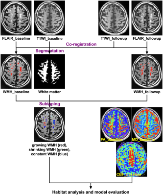 |
165 | Habitat analysis by multi-parametric MRI predicts progressive white matter hyperintensities in cerebral small vessel disease
Xu Han1, Xiyao Gu2, and Yan Zhou1
1Radiology, Renji Hospital, Shanghai Jiao Tong University School of Medicine, shanghai, China, 2Anesthesiology, Renji Hospital, Shanghai Jiao Tong University School of Medicine, shanghai, China Keywords: Vessels, Dementia White matter hyperintensities (WMH) are commonly seen in cerebral small vessel disease (CSVD) and are associated with an risk of cognitive impairment. However, the mechanism by which WMH develop is incompletely characterized. In this study, we conducted habitat analysis based on physiologic MRI parameters to investigate whether WMH habitat can predict progressive WMH in CSVD. We found that the physiologic MRI habitat with lower fractional anisotropy and cerebral blood flow, higher mean diffusivity, axial diffusivity and radial diffusivity was highly overlapping with the growing WMH in patients after one-year followup, and thus could predict progressive WMH in CSVD. |
|
2160.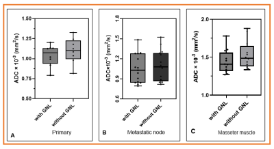 |
166 | Implementation of gradient nonlinearity correction for ADC measurements in head and neck cancer
Ramesh Paudyal1, Akash Deelip Shah2, Amaresha Shridhar Konar1, Victoria Yu1, Dariya I Malyarenko3, Nancy Lee4, Thomas L Chenevert3, and Amita Shukla-Dave1,2
1Medical Physics, Memorial Sloan Kettering Cancer Center, New York, NY, United States, 2Radiology, Memorial Sloan Kettering Cancer Center, New York, NY, United States, 3Radiology, University of Michigan, Ann Arbor, MI, United States, 4Radiation Oncology, Memorial Sloan Kettering Cancer Center, New York, NY, United States Keywords: Quantitative Imaging, Tumor The present study evaluated the correction efficacy for ADC values bias due to the gradient nonlinearity of the MRI system on primary tumors, neck nodal metastases, and masseter muscle in the head and neck region using a vendor-provided LOw VAriance (LOVA) ADC technique. The LOVA ADC technique was developed and implemented to compensate for gradient linearity errors that may allow consistent ADC values in large FOV independent of the offset from the scanner isocenter. Our results confirmed the potential of the LOVA technique to improve ADC accuracy independent of location and scanner gradient characteristics. |
|
2161.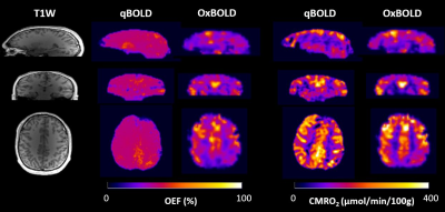 |
167 | Comparison of Constrained Quantitative BOLD with Dual-Gas Calibrated BOLD for 3D Mapping of Cerebral Oxygen Extraction Fraction
Hyunyeol Lee1,2, Jing Xu2, Maria A Fernandez-Seara2,3, and Felix W Wehrli2
1School of Electronics Engineering, Kyungpook National University, Daegu, Korea, Republic of, 2Department of Radiology, University of Pennsylvania, Philadelphia, PA, United States, 3Department of Radiology, Clinica Universidad de Navarra, Pamplona, Spain Keywords: Quantitative Imaging, Metabolism Recent advances of the cBOLD technique by means of dual-gas calibration have shown its ability in producing baseline OEF and CMRO2 in absolute physiologic units. A recently proposed constrained qBOLD method has shown its feasibility in proper separation of numerous confounders, yielding physiologically plausible values for both Yv and DBV across the entire brain. The purpose of this work was to compare the two oximetric techniques, dual-gas cBOLD versus constrained qBOLD, in terms of measured OEF and CMRO2 at baseline. Results suggest that the two methods yield statistically insignificant differences in OEF and CMRO2 quantifications for GM regions. |
|
2162.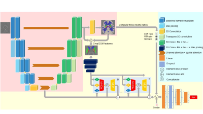 |
168 | A Brain Age Estimation Network based on QSM using the Segment Transformer
Yiqing Wang1, Yuting Shi1, and Hongjiang Wei1
1School of Biomedical Engineering, Shanghai Jiao Tong University, Shanghai, China Keywords: Quantitative Imaging, Quantitative Susceptibility mapping Quantitative Susceptibility Mapping (QSM) reflects various biological procedures, e.g., iron accumulation in deep gray matter, which is tightly associated with brain development and highly related to various neurodegenerative diseases. Nowadays, deep-learning-based methods have achieved great success in medical image segmentation due to their ability to extract multi-scale features. We create a novel network to segment key brain areas on QSM images to improve brain age prediction. Our network further integrates multi-level features by utilizing a variant of Vision Transformer, termed Segment Transformer. Results show that our method can improve brain age estimation compared to previous studies based on T1w MRI. |
|
2163.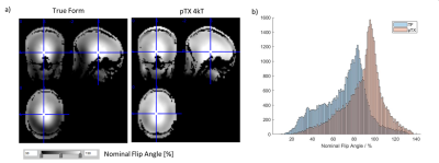 |
169 | Parallel transmit (pTx) kT-points pulses improve 500µm resolution quantitative multi-parameter mapping (MPM) at 7T
Kerrin J Pine1, Nicolas Groß-Weege1,2, Luke J Edwards1, Tobias Leutritz1, Patrick Freund1,3, and Nikolaus Weiskopf1,4
1Department of Neurophysics, Max Planck Institute for Human Cognitive and Brain Sciences, Leipzig, Germany, 2Siemens Healthcare GmbH, Erlangen, Germany, 3Spinal Cord Injury Center Balgrist, University Hospital Zurich, University of Zurich, Zurich, Switzerland, 4Felix Bloch Institute for Solid State Physics, Faculty of Physics and Earth Sciences, Leipzig University, Leipzig, Germany Keywords: Quantitative Imaging, Parallel Transmit & Multiband, Neuro Quantitative multi-parameter mapping (MPM) at 7T can provide detailed information about brain microstructure. However, it is limited by B1+ radio-frequency (RF) transmit field inhomogeneities leading to bias and dropouts. We implemented a kt-points RF pulse approach in 500µm resolution multi-echo spoiled GRE acquisitions and AFI B1+ mapping used for MPM. It was optimized for a homogeneous non-selective excitation with short RF pulse durations, SAR compliance and high usability. In a group of 8 volunteers, bias and shading artifacts were significantly reduced. The approach is integrated in the routine scanning workflow, improving whole brain ultra-high resolution MPM at 7T. |
|
2164.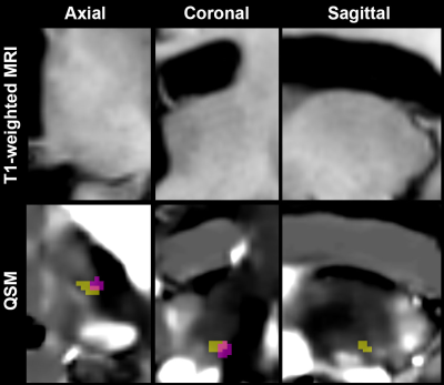 |
170 | Finding Precise, Direct, and Personalized Landmarks of Deep Brain Stimulus Tremor Targeting on Advanced MR imaging
Sohae Chung1,2, Ha Neul Song3, Yvonne W. Lui1,2, Brian H. Kopell3, and Ki Sueng Choi3
1Center for Advanced Imaging Innovation and Research (CAI2R), Department of Radiology, New York University Grossman School of Medicine, New York, NY, United States, 2Bernard and Irene Schwartz Center for Biomedical Imaging, Department of Radiology, New York University Grossman School of Medicine, New York, NY, United States, 3Nash Family Center for Advanced Circuit Therapeutics, Ichan School of Medicine at Mount Sinai, New York, NY, United States Keywords: Quantitative Imaging, Quantitative Susceptibility mapping, Tremor Deep brain stimulation (DBS) is an efficacious treatment for tremors. The clinical outcome is critically dependent on precisely personalized targeting, yet localization of DBS target remains challenging owing to difficulties in resolving individual thalamic nuclei or dentato-rubro-thalamic (DRT) pathways. Here we investigated retrospectively to identify neurosurgical landmarks of DBS targeting by examining the volume overlap and centroid distance between DBS contacts and DRT delineated from quantitative susceptibility imaging. Our findings show a significantly higher volume overlap of DBS contacts with DRT in the good-response tremor group, showing potential of our approach as a direct and personalized neurosurgical guidance tool. |
|
2165.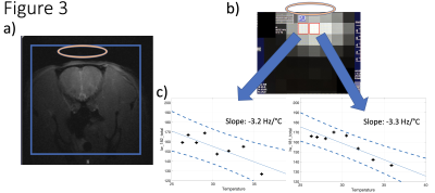 |
171 | Mapping of relative temperature changes through quadrupolar transverse H217O relaxivity in phantom and validated in rat at 7T and 16T UHF
Hannes M. Wiesner1, Tao Wang1, Xin Li1, Wei Zhu1, Kelsey Haney1, Xiao-Hong Zhu1, and Wei Chen1
1CMRR, Department of Radiology, University of Minnesota, Minneapolis, MN, United States Keywords: Quantitative Imaging, Thermometry In this study we evaluate the application of linewidth thermometry using the transverse relaxation of natural abundance 17O on leading human and preclinical high-field MRI platforms. It could be beneficial as a simple and fast alternative approach compared to the phase method to assess temperature. We validated it in post-mortem rodents after preparations on phantoms. Furthermore, due to the lower operating frequency some high-field challenges could be ameliorated with the right choice of volume to signal acquisition ratio. |
|
2166.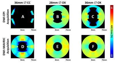 |
172 | Mapping the Accuracy of Diffusion Weighted Imaging Near Total Hip Implants
John Neri1, Matthew Koff1, Kevin Koch2, and Ek Tsoon Tan1
1Hospital for Special Surgery, New York, NY, United States, 2Medical College of Wisconsin, Milwaukee, WI, United States Keywords: Quantitative Imaging, Diffusion/other diffusion imaging techniques Multi-acquisition with variable resonance image combination (MAVRIC) with DWI can reduce artifacts associated with metal implants to characterize synovial reactions near total hip replacements. The purpose of this study was to evaluate the quantitative accuracy of DWI-MAVRIC near actual hip arthroplasty components and demonstrate the feasibility of spatially mapping ADC biases. DWI-EPI and DWI-MAVRIC images were acquired using a diffusion phantom in the presence of femoral heads of different material composition. It was found that cobalt-chromium femoral heads cause more ADC error than oxinium femoral heads. Spatial mapping of ADC accuracy near total hip implants was found to be feasible. |
|
2167.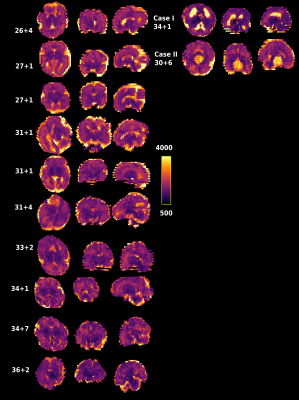 |
173 | Efficient fetal brain and placental T1 mapping at 0.55T
Jordina Aviles Verdera1,2, Raphael Tomi-Tricot1,2,3, Lisa Story1,4, Joseph V Hajnal 1,2, Mary A Rutherford1,2, Shaihan Malik1,2, and Jana Hutter1,2
1Center for the Developing Brain, School of Biomedical Engineering & Imaging Sciences, King's College London, London, United Kingdom, 2Biomedical Engineering Department, School of Biomedical Engineering & Imaging Sciences, King's College London, London, United Kingdom, 3MR Research Collaborations, Siemens Healthcare Limited, Frimley, United Kingdom, 4Women's Health, Guy's and St. Thomas' NHS Foundation Trust, London, United Kingdom Keywords: Quantitative Imaging, Low-Field MRI Fetal MRI provides crucial anatomical and functional information. At low field it could benefit from reduced distortion, increased bore size and increased B1 homogeneity. T1 contrast and mapping in the fetal brain provides complementary information for example for haemorrhages, infarcts and in placental disease. Here, a low-field (0.55T) fetal T1 inversion-recovery EPI-based sequence benefiting from the lower T1 and hence higher efficiency is demonstrated for placenta and brain in a cohort of pregnant participants including two clinical cases. Significant correlation with gestational age is demonstrated (p<0.05) and high quality maps allowing sub-regions analysis in the brain are shown. |
|
2168.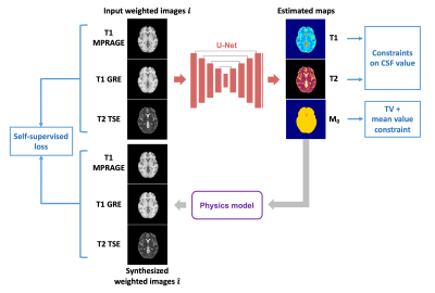 |
174 | Physics-guided self-supervised learning for retrospective T1 and T2 mapping from conventional weighted brain MRI
Shihan Qiu1,2, Anthony G. Christodoulou1,2, Pascal Sati1,3, Yibin Xie1, and Debiao Li1,2
1Biomedical Imaging Research Institute, Cedars-Sinai Medical Center, Los Angeles, CA, United States, 2Department of Bioengineering, UCLA, Los Angeles, CA, United States, 3Department of Neurology, Cedars-Sinai Medical Center, Los Angeles, CA, United States Keywords: Quantitative Imaging, Relaxometry Quantitative MRI directly measures tissue physical parameters, but has limited clinical adoption due to additional scan time and specialized sequence requirements. Supervised deep learning methods were developed to estimate relaxation maps from conventional weighted images. However, paired weighted images and quantitative maps required for training are hard to obtain. In this work, a physics-guided self-supervised learning approach was developed to estimate T1 and T2 maps from conventional weighted images. Using the Bloch equations to decode the estimated maps back to weighted images and enforcing similarity in the image space, the approach realized label-free training and provided maps comparable to references. |
|
2169.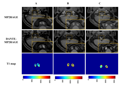 |
175 | Three-dimensional black-blood T1 mapping of arterial plaque and venous thrombosis: a concept-proof study
Yuhui Nie1, Zeping Liu1, Anyan Gu1, Liping Liao2, Qizeng Ruan2, Zehe Huang2, Yi Sun3, and Guoxi Xie1
1School of Biomedical Engineering, Guangzhou Medical University, Guangzhou, China, 2The First People’s Hospital of Qinzhou, Qinzhou, China, 3Siemens Shenzhen Magnetic Resonance Ltd, Shenzhen, China Keywords: Quantitative Imaging, Cardiovascular, T1 mapping Quantitative T1 mapping have showed the potential to characterize arterial plaque and venous thrombotic components. However, owing to the interference of blood signals, it is a challenge for T1 mapping technique to combine with black-blood imaging, which may otherwise be a confounder for plaque and thrombus component. In this study, we present a black-blood T1 mapping technique based on DANTE black-blood preparation and MP2RAGE sequence. Experiment results demonstrated that BB-MP2RAGE provided accurate measurement of T1 relaxation time with effective blood suppression. The technique has the potential to be a quantitative tool for thrombosis and plaque characterization. |
|
| 2170. | WITHDRAWN | ||
2171.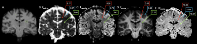 |
176 | Soma and neurite density imaging in multiple sclerosis reveals cortical cell body signal fraction loss associated with thalamic atrophy
Eva A. Krijnen1,2, Andrew W. Russo1, Elsa Salim Karam1, Hansol Lee3, Menno M. Schoonheim2, Susie Y. Huang3, and Eric C. Klawiter1
1Department of Neurology, Massachusetts General Hospital, Harvard Medical School, Boston, MA, United States, 2MS Center Amsterdam, Anatomy and Neurosciences, Amsterdam Neuroscience, Amsterdam UMC location VUmc, Amsterdam, Netherlands, 3Athinoula A. Martinos Center for Biomedical Imaging, Department of Radiology, Massachusetts General Hospital, Harvard Medical School, Charlestown, MA, United States Keywords: Multiple Sclerosis, Gray Matter, Diffusion/Other Diffusion Imaging Techniques Multiple sclerosis (MS) features complex microstructural changes in gray matter (GM) ranging from demyelinating lesions to neuronal loss. We hypothesize that GM microstructural metrics obtained from diffusion MRI can predict cortical and deep GM atrophy in MS. In this cross-sectional study of 41 people with MS, the soma and neurite density imaging (SANDI) compartment-based model was fitted to high-gradient diffusion MRI data acquired on the Connectome scanner. SANDI metrics were used to characterize MS-related microstructural pathology in cortical and deep GM. Cortical fsoma decreased with declining thalamic volume, suggesting a microstructural correlate of thalamic atrophy in MS. |
|
2172.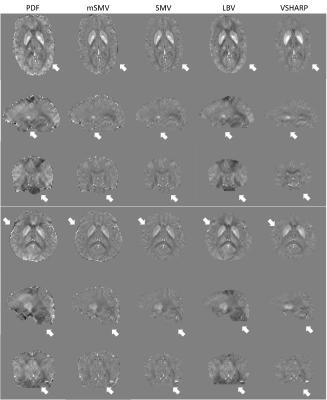 |
177 | Whole Brain Spherical Mean Value Filtering for Shadow Reduction in Quantitative Susceptibility Mapping
Alexandra Grace Roberts1,2, Pascal Spincemaille2, Thanh Nguyen2, and Yi Wang1,2
1Electrical Engineering, Cornell University, Ithaca, NY, United States, 2Radiology, Weill Cornell Medicine, New York, NY, United States Keywords: Quantitative Imaging, Artifacts Morphology Enabled Dipole Inversion (MEDI) is an iterative reconstruction algorithm for Quantitative Susceptibility Mapping (QSM) that is effective in suppressing streaking artifacts by exploiting the magnitude image as a morphological prior. However, contiguous areas of dipole incompatibility (such as noise) induce shadow artifacts whose spatial frequency components are not sufficiently regularized by the gradient-based regularization in MEDI. The harmonic quality allows the use of the maximum corollary of Green’s theorem to remove residual background field. This mSMV approach reduces shadows and preserves brain volume, comparing favorably to existing algorithms. |
|
2173. |
178 | Glymphatic system abnormality in systemic lupus erythematosus identified by diffusion tensor image analysis along the perivascular space
Jiaying Mo1, Kan Deng2, Xiangliang Tan1, and Yikai Xu1
1Department of Medical Imaging Center, Nanfang Hospital of Southern Medical University, Guangzhou, China, 2Philips Healthcare, Guangzhou, China Keywords: Head & Neck/ENT, Brain, Systemic lupus erythematosus DTI-ALPS is an non-invasive method to measure glymphatic clearance function in brain. Here, we aimed to utilize DTI-ALPS to evaluate the activity of glymphatic system and explore its relationship with grey matter volumes in patients with SLE. Our results demonstrated that the ALPS-index are lower in SLE patients than that in healthy control group. And there were significant correlations between lower ALPS-index and smaller gray matter volumes in some brain regions in SLE group. |
|
2174.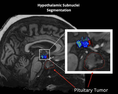 |
179 | Quantitative assessment of secondary white matter injury in hypothalamic subnuclei by pituitary adenomas utilizing 7-Tesla MRI
Mackenzie Langan1,2, Deborah Marshall3, Bradley Delman4, Raj Shrivastava5, and Priti Balchandani2,6,7
1Icahn School of Medicine at Mount Sinai, New York, NY, United States, 2Biomedical Engineering and Imaging Institute, Icahn School of Medicine at Mount Sinai, New York, NY, United States, 3Department of Radiation Oncology, Mount Sinai Hospital, New York, NY, United States, 4Diagnostic, Molecular and Interventional Radiology, Mount Sinai Hospital, New York, NY, United States, 5Department of Neurosurgery, Mount Sinai Hospital, New York, NY, United States, 6Diagnostic, Molecular and Interventional Radiology, Icahn School of Medicine at Mount Sinai, New York, NY, United States, 7Nash Family Department of Neuroscience, Icahn School of Medicine at Mount Sinai, New York, NY, United States Keywords: Tumors, Endocrine, Diffusion Tractograpy, hypothalamus Here we outline a preliminary analysis using a novel method that leverages UHF neuroimaging to measure detectable differences in volumetric, microstructural and tractographic measures in the hypothalamus in patients with pituitary tumors and matched controls. We uncovered significant detectable volumetric and differences within five hypothalamic subnuclei, and the whole left hypothalamus. We observed subtle differences in microstructural integrity, and although not statistically significant warrant further investigations in a larger cohort of pituitary adenoma patients. This may also facilitate exploration of microstructural integrity within other tumor types that may have effects on the hypothalamus. |
|
 Back to Meeting Home
 Back to the Program-at-a-Glance
The International Society for Magnetic Resonance in Medicine is accredited by the Accreditation Council for Continuing Medical Education to provide continuing medical education for physicians.
 Back to Meeting Home
 Back to the Program-at-a-Glance
 View Presentation Video