Digital Poster
New Quantitative Imaging Methods II
ISMRM & ISMRT Annual Meeting & Exhibition • 03-08 June 2023 • Toronto, ON, Canada

Digital Poster
New Quantitative Imaging Methods II
| Computer # | |||
|---|---|---|---|
1778.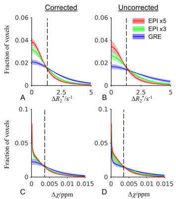 |
141 | Improved reliability of R2* and susceptibility quantification using 3D EPI acquisition
Yujia Huang1, Peter van Gelderen2, Lin Chen3,4, Jacco A. de Zwart2, Zeynep Demir2, Gael Saib5, Xu Li3,4, Jeff H. Duyn2, and Jiaen Liu1
1Advanced Imaging Research Center, UT Southwestern Medical Center, Dallas, TX, United States, 2Laboratory of Functional and Molecular Imaging , NINDS, NIH, Bethesda, MD, United States, 3F.M. Kirby Research Center for Functional Brain Imaging, Kennedy Krieger Institute, Baltimore, MD, United States, 4Department of Radiology and Radiological Sciences, Johns Hopkins University, Baltimore, MD, United States, 5NMR Research Center, NINDS, NIH, Bethesda, MD, United States Keywords: Quantitative Imaging, Quantitative Susceptibility mapping The quantification of decay rate R2* and susceptibility is useful in assessment of various neurological disorders manifesting iron accumulation and myelin loss. Conventional 3D GRE acquisition is sensitive to motion and B0 fluctuation due to its long scan time. In this study, we demonstrated improved reliability of multi-shot multi-echo 3D EPI when compared to GRE, showing reduced scan time and similar accuracy. |
|
1779.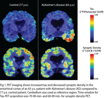 |
142 | Zooming In: MR and PET Synaptic Density Imaging Techniques in Neuropathology
Divya Ramakrishnan1, Takuya Toyonaga2, Behnaz Khazai3, Matthew Sala1, Jason Cai2, and Mariam Aboian1
1Department of Radiology, Yale School of Medicine, New Haven, CT, United States, 2Department of Radiology and Biomedical Imaging, Yale School of Medicine, New Haven, CT, United States, 3Department of Radiology, Norwalk Hospital, Norwalk, CT, United States Keywords: Quantitative Imaging, PET/MR, Synaptic Density, SV2A There are many different modalities to image synaptic density in the brain. The advent of a novel SV2A targeted PET tracer provides the ability to measure in-vivo synaptic density changes in neuropsychiatric disorders. In this education exhibit, we review the current MR and PET-based approaches to synaptic density imaging and present findings from recent animal and human studies of synaptic density changes in neurodegenerative disorders and epilepsy. Our aim is to provide clinicians with the most up-to-date knowledge about imaging techniques of synaptic density that can be integrated into clinical practice. |
|
1780.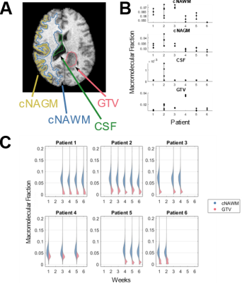 |
143 | Quantitative Magnetization Transfer Imaging in Glioblastoma Patients using Balanced Steady-state Free Precession on a 1.5 T MR-Linac
Brandon Thien Trong Tran1, Liam S. P. Lawrence1, Rachel W. Chan2, Chia-Lin Tseng3, Jay Detsky3, Hany Soliman3, Arjun Sahgal3, and Angus Z. Lau1,2
1Medical Biophysics, University of Toronto, Toronto, ON, Canada, 2Physical Sciences Platform, Sunnybrook Research Institute, Toronto, ON, Canada, 3Radiation Oncology, Sunnybrook Health Sciences Centre, Toronto, ON, Canada Keywords: Quantitative Imaging, Magnetization transfer Quantitative magnetization transfer (qMT) imaging could be used to assess tumor response on an MR-Linac. However, qMT protocols which use an off-resonance saturation pulse are slow and does not have the 3D coverage necessary to assess tumor response. We implemented a fast 3D balanced steady-state free precession qMT protocol on the MR-Linac. Six glioblastoma patients were scanned on the MR-Linac at weekly intervals during treatment. qMT parameter maps derived using non-linear least square fitting show similar contrast to other qMT protocols. Within-subject coefficient of variation was higher than values found in literature for off-resonance qMT protocols. |
|
1781.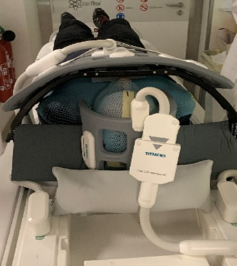 |
144 | An MRI protocol for RT planning: preliminary results for distortion, contrast and bulk density assignment
Laura Sayaque1, Benjamin Leporq1, Chloé Miran2, Olivier Hamelin3, Frank Pilleul1,3, Vincent Gregoire2, and Olivier Beuf1
1Univ Lyon, INSA‐Lyon, Université Claude Bernard Lyon 1, UJM-Saint Etienne, CNRS, Inserm, CREATIS UMR 5220, U1294, F‐69100, Lyon, France, 2CRLCC Léon Bérard – Département de Radiothérapie, Lyon, France, 3CRLCC Léon Bérard – Département de Radiologie, Lyon, France Keywords: Quantitative Imaging, Head & Neck/ENT, Multimodal The excellent contrast between soft tissues in MRI is of great interest for head and neck radiotherapy planning. This work presents an in vivo study aiming to evaluate a multimodal acquisition protocol for tumors and organs at risks delineation and synthetic CT reconstruction based on a density assignment method. Our results showed better contrast between the tumor and healthy tissues with injected T1 MRI compared to injected CT (44,7 +/- 30,5 vs 29,9 +/- 27,2) and acceptable geometric distortions (less than 2mm). Fat content mean absolute error between CT and synthetic CT is about 0,27. |
|
1782.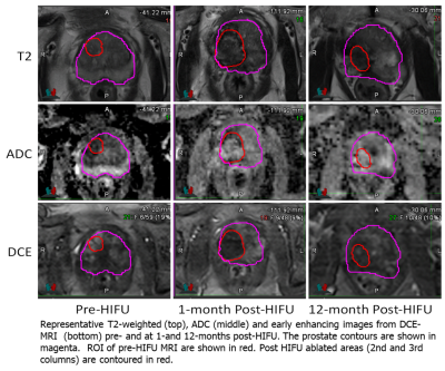 |
145 | Characterization of Longitudinal Anatomic and Functional Changes following HIFU Therapy and Implications for Prostate Cancer Surveillance
Adrian Breto1, Ahmad Algohary1, Michael Tradewell2, Leonardo Bittencourt3, Oleksandr Kryvenko4, Mark Gonzalgo2, Sanoj Punnen2, Dipen Parekh2, Bruno Nahar2, and Radka Stoyanova1
1Department of Radiation Oncology, University of Miami, Miami, FL, United States, 2Desai Seth Urology Institute, Miami, FL, United States, 3Department of Radiology, UH Cleveland Medical Center, Cleveland, OH, United States, 4Department of Pathology and Laboratory Medicine, University of Miami, Miami, FL, United States Keywords: Quantitative Imaging, Prostate, Focused Ultrasound Detecting prostate cancer recurrence after high intensity focused ultrasound (HIFU) therapy is a challenge for clinicians. We characterize longitudinal anatomical/functional changes of prostate in men undergoing HIFU therapy for prostate cancer. Nineteen men had mpMRI exams pre-HIFU, and at 1 and 12 months post-HIFU. Prostate volume decreased by 17% on the one year exam and in two-thirds of men changes in prostatic axis, up to 20 degrees in the direction of ablation. Functional mpMRI analysis shows a dramatic decrease in volume transfer constant (Ktrans) on dynamic contrast enhancing (DCE) imaging while there were no changes in apparent diffusion coefficient (ADC). |
|
1783.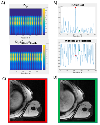 |
146 | Feasibility of T1/T2/T1ρ MR Multitasking of Arm Muscle for Imaging Amyotrophic Lateral Sclerosis
Matthew Dausch1, Sen Ma1, Xianglun Mao1, Hsu-Lei Lee1, Yibin Xie1, Debiao Li1, Frank Diaz2, and Anthony Christodoulou1
1Biomedical Imaging Research Institute, Cedaras Sinai Medical Center, Los Angeles, CA, United States, 2Neurology, Cedars-Sinai Medical Center, Los Angeles, CA, United States Keywords: Quantitative Imaging, Neurodegeneration, MR Multitasking, Amyotrophic Lateral Sclerosis Amyotrophic Lateral Sclerosis (ALS) is a neurodegenerative motor disorder that has a need for imaging methods for longitudinal studies. T1/T2/T1ρ MR multitasking is a multiparametric, quantitative, imaging method that shows promise in being a viable tool for this purpose. In this study, we tested the feasibility of a motion weighting modified T1/T2/T1ρ MR multitasking in the arm muscles, evaluating the accuracy and precision of quantitative measurements. |
|
1784.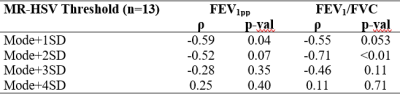 |
147 | Automatic Assessment of UTE MRI High Signal Volume in the Lung for Pediatric Patients with Cystic Fibrosis
Daniel Genkin1, Brandon Zanette2, Thomas Benkert3, Felix Ratjen2,4, Giles Santyr2,5, and Miranda Kirby6
1Department of Electrical, Computer, and Biomedical Engineering, Toronto Metropolitan University, Toronto, ON, Canada, 2Department of Translational Medicine, The Hospital for Sick Children, Toronto, ON, Canada, 3MR Application Predevelopment, Siemens Healthcare GmbH, Erlangen, Germany, 4Department of Pediatrics, University of Toronto, Toronto, ON, Canada, 5Department of Medical Biophysics, University of Toronto, Toronto, ON, Canada, 6Department of Physics, Toronto Metropolitan University, Toronto, ON, Canada Keywords: Quantitative Imaging, Lung, UTE, CF, Pediatric High signal volume (MR-HSV) obtained by Ultrashort Echo-Time (UTE) MRI has been shown to be associated with disease severity in adults with CF, but has yet to be investigated in pediatric CF patients. In this study, MR-HSV was automatically extracted from UTE MRI of pediatric CF (n=9) and healthy (n=4) participants. MR-HSV measurements were significantly increased in CF compared to health, and increased MR-HSV correlated with decreased lung function (ie. FEV1pp, FEV1/FVC). The findings in this work confirm that MR-HSV measurement is feasible in pediatric participants and may reflect disease severity associated with airway/parenchyma signal intensity abnormalities in CF. |
|
1785.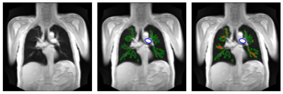 |
148 | Feasibility of pulse wave velocity measurement in pulmonary arteries from phase-resolved functional lung MRI
Marius Malte Wernz1,2, Andreas Voskrebenzev1,2, Robin Müller1,2, Maximilian Zubke1,2, Filip Klimeš1,2, Gesa Pöhler1,2, Frank Wacker1,2, and Jens Vogel-Claussen1,2
1Institute of Diagnostic and Interventional Radiology, Hannover Medical School, Hannover, Germany, 2Biomedical Research in Endstage and Obstructive Lung Disease Hannover (BREATH), German Center for Lung Research (DZL), Hannover, Germany Keywords: Quantitative Imaging, Velocity & Flow Phase-resolved functional lung (PREFUL) MRI-derived flow cycles are used to calculate time differences in blood flow and thus mean velocity between the truncus pulmonalis and voxels located in main pulmonary arteries. Two U-Net-based convolutional neural networks are used to identify voxels to include in the analysis. In 26 patients with chronic obstructive pulmonary disease (COPD) the resulting pulmonary artery pulse wave velocity (paPWV) values are reproducible after two weeks. The measured velocities in COPD patients and in 30 healthy subjects are in a physiologically plausible range compared to invasive measurements. |
|
1786.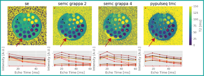 |
149 | Fast quantitative T2 mapping at ultra-high field using sparse sampling and dictionary-based reconstruction
Jochen Schmidt1,2, Dvir Radunsky3, Patrick Scheibe1, Noam Ben-Eliezer3,4,5, Robert Trampel1, and Nikolaus Weiskopf1,6
1Neurophysics, Max Planck Institute for Human Cognitive and Brain Sciences, Leipzig, Germany, 2International Max Planck Research School on Neuroscience of Communication: Function, Structure and Plasticity, Leipzig, Germany, 3Biomedical Engineering, Tel Aviv University, Tel Aviv, Israel, 4Sagol School of Neuroscience, Tel Aviv University, Tel Aviv, Israel, 5Center for Advanced Imaging Innovation and Research (CAI2R), New York University Langone Medical Center, New York, NY, United States, 6Felix Bloch Institute for Solid State Physics, Leipzig University, Leipzig, Germany Keywords: Quantitative Imaging, Sparse & Low-Rank Models Quantitative T2 mapping at ultra high-field via multi-echo spin-echo acquisitions suffers from bias introduced by pronounced transmit field inhomogeneities. Hence, accurate signal modeling is vital. Futhermore, high-resolution imaging results in longer scan times, which need to be compensated by k-space undersampling. Conventional undersampling schemes for multi-echo approaches skip identical phase encoding areas for all echo readouts. Image reconstruction quality might be improved by varying the sampling across echoes and using a low rank reconstruction approach. A multi-echo acquisition combining sparse undersampling with a dictionary modelling method was successfully used to acquire accurate T2 at 7T with a high acceleration. |
|
1787.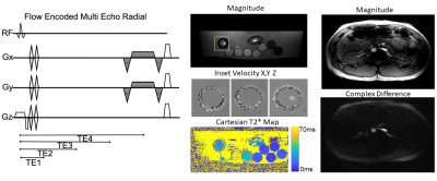 |
150 | Low Volume, Pneumatic Flow Phantom for Validation of Flowing T2* Estimation Using Projection Acquisition with Flow-Encoded Subtraction
Corina Margain1, Christopher Sandino2, Shreyas Vasanawala3, and Adam Bush1
1Department of Biomedical Engineering, University of Texas at Austin, Austin, TX, United States, 2Department of Electrical Engineering, Stanford University, Stanford, CA, United States, 3Department of Radiology, Stanford University, Stanford, CA, United States Keywords: Quantitative Imaging, Phantoms, non-cartesian Blood-oxygen level is a known biomarker for cardiovascular diseases which can be quantified by T2*-weighted MRI. Challenges with validation and motion have limited the clinical use of T2* oximetry methods. We present a PALMS sequence to measure flowing T2* which was validated using a pneumatically powered, low-volume, continuous flow phantom. Our results suggest that the presence of readout gradients influence T2* during flowing conditions while obliquity influences T2* in all settings conditions. |
|
1788.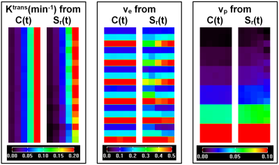 |
151 | Signal intensity form of extended Tofts model for quantitative analysis of dynamic contrast enhanced MRI data
Xiaobing Fan1, Aritrick Chatterjee1, Zhen Ren1, Aytekin Oto1, and Gregory S. Karczmar1
1Radiology, The University of Chicago, Chicago, IL, United States Keywords: Quantitative Imaging, Cancer The extended Tofts model (ETM) requires calculation of contrast agent concentration in tissue as function of time (C(t)) using a non-linear model that results in error propagation. Here, we introduce a signal intensity (S(t)) form of ETM (SI-ETM) without calculating C(t). QIBA DCE-MRI data was used to validate the SI-ETM, and then human prostate DCE-MRI data were analyzed to compare physiological parameters calculated from the ETM and the SI-ETM. The parameters calculated from S(t) were strongly correlated with the values calculated from C(t). Bland–Altman analysis showed good agreement between the parameters calculated from the ETM and the SI-ETM. |
|
1789.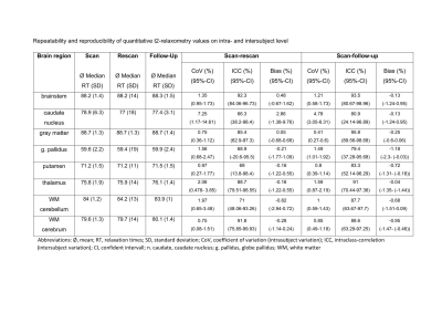 |
152 | Feasibility and clinical implementation of ultra-fast GRAPPATINI T2 mapping of the brain: A prospective study
Natascha Karolina Gruenebach1, Antoine Sanner1, Nils Friedrich Grauhan1, Mario Alberto Abello Mercado1, Andrea Kronfeld1, Vanessa Ines Schoeffling1, Tom Hilbert2,3,4, Marc A. Brockmann1, and Ahmed E. Othman1
1University medical center Mainz, Mainz, Germany, 2Advanced Clinical Imaging Technology, Siemens Healthineers International AG, Lausanne, Switzerland, 3Department of Radiology, Lausanne University Hospital and University of Lausanne, Lausanne, Switzerland, 4LTS5, École Polytechnique Fédérale de Lausanne (EPFL), Lausanne, Switzerland Keywords: Quantitative Imaging, Relaxometry, T2 Mapping We aimed to validate the clinical feasibility of the quantitative imaging MR-sequence GRAPPATINI. It offers T2 relaxometry values as well as synthetic T2-weighted images of the whole brain. 10 volunteers for the validation of repeatability and reproducebility on intra- and intersubject level and 52 patients for the morphological comparison between GRAPPATINI and standard T2-weighted sequence were included prospectively. T2 relaxation times of GRAPPATINI are robust and show only little variations among healthy subjects. Larger deviations only were found in caudate nucleus. Moreover its synthetic T2-weighted images achieve good interrater-reliability, although its subjective image quality is not equivalent to standard TSE. |
|
1790.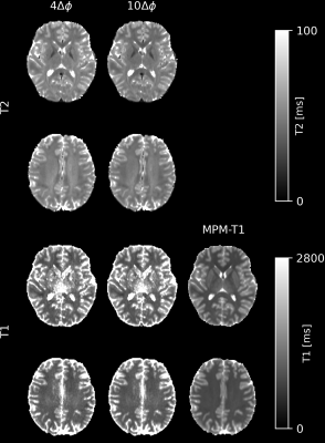 |
153 | Rapid gradient echo magnitude- and phase-based mapping of T2 and T1 using skipped-CAIPI 3D-EPI at 3T
Difei Wang1, Rüdiger Stirnberg1, and Tony Stöcker1,2
1MR Physics, DZNE, Bonn, Germany, 2Department of Physics and Astronomy, University of Bonn, Bonn, Germany Keywords: Quantitative Imaging, Modelling, Quantitative imaging, Relaxometry, Neuro A recent study demonstrated that phase-based T2 mapping with 3D-GRE is feasible. Only two phase images acquired with a small RF spoiling phase increment are needed to generate high-quality T2 maps. Here, we investigated simultaneous T2 and T1 mapping at 3T, by including the magnitude and more phase increments acquired rapidly with a skipped-CAIPI 3D-EPI sequence. This method is able to simultaneously provide T2 and T1 maps within 4-8 minutes. In addition to multiparametric mapping, a phase-based T2 map can be provided in less than 1 minute. |
|
1791.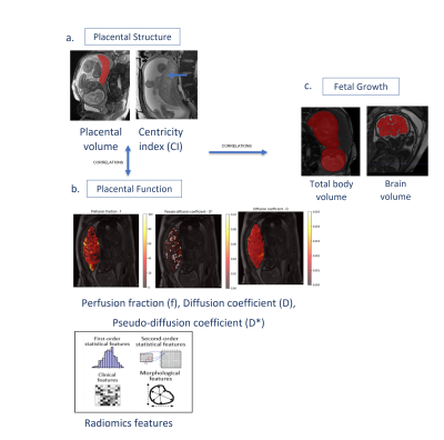 |
154 | Quantitative analysis of the placental structure and function and fetal growth using intravoxel incoherent motion MRI
Dana Schonberger1,2, Daphna Link-Sourani1, Netanell Avisdris1,3, Aviad Rabinowich1,4,5, Bella Specktopr Fadida1,3, Liat Ben-Sira4,6,7, Leo Joskowicz3, Liran Hiersch8, Ayala Zilberman8, and Dafna Ben Bashat1,4,7
1Sagol Brain Institute, Tel Aviv Sourasky Medical Center, Tel Aviv, Israel, 2The Department of Biomedical Engineering, Tel Aviv University, Tel Aviv, Israel, 3School of Computer Science and Engineering, The Hebrew University of Jerusalem, Jerusalem, Israel, 4Sackler Faculty of Medicine, Tel Aviv University, Tel Aviv, Israel, 5Department of Radiology, Tel Aviv Sourasky Medical Center, Israel, Tel Aviv, Israel, 6Division of Pediatric Radiology, Tel Aviv Sourasky Medical Center, Israel, Tel Aviv, Israel, 7Sagol School of Neuroscience, Tel Aviv University, Tel Aviv, Israel, 8Department of Maternal-Fetal Medicine and High Risk Pregnancy Outpatient clinic, Tel Aviv Sourasky Medical Center, Tel Aviv, Israel Keywords: Quantitative Imaging, Perfusion, Intravoxel incoherent motion, IVIM Placental structural and functional parameters are important for the assessment of placental insufficiency and fetal growth. Here we assessed quantitative measures of placental structural and functional parameters extracted from Intravoxel-incoherent motion (IVIM) with radiomics analysis, in fetuses with growth restriction (FGR) and appropriate for gestational age (AGA). Placental structure (volume and centricity index (CI, umbilical cord insertion location) were correlated with radiomics features of placental perfusion in AGA, and some were found to be significantly different between FGR and AGA, indicating higher functional heterogeneity in FGR. This multi-parametric quantitative placental assessment may improve identification of FGR fetuses. |
|
1792.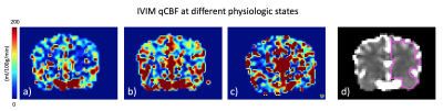 |
155 | Validation of a new method for Quantifying CBF with IVIM
Mira Liu1, Chisondi Simba Warioba1, Julian Bertini1, Niloufar Saadat2, Timothy Carroll1, and Gregory Christoforidis2
1University of Chicago, Chicago, IL, United States, 2Interventional Radiology, University of Chicago, Chicago, IL, United States Keywords: Quantitative Imaging, Perfusion Intravoxel Incoherent Motion (IVIM) is a non-contrast MR diffusion-based scan that uses a multitude of b-values to measure various speeds of molecular perfusion and diffusion, sidestepping problems of Gd-contrast or increased transit time in neurovascular disease. Quantification of IVIM CBF in ml/100g/min using pseudo-diffusion mean transit time led to strong Wilcoxon signed rank agreement, similar sensitivity, and a small offset compared to microsphere perfusion across three physiologic states. This suggests IVIM can be used to quantify CBF in ml/100g/min in a setting of cerebral steno-occlusive disease. |
|
1793.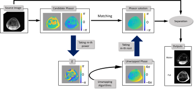 |
156 | Solving fat-water separation with arbitrary echo combination by phase unwrapping
Hao Peng1, Chuanli Cheng1, Xin Liu1, Hairong Zheng1, and Chao Zou1
1Shenzhen Institute of Advanced Technology,Chinese Academy of Sciences, Shenzhen, China Keywords: Quantitative Imaging, Quantitative Imaging The phase unwrapping technique is only applicable to the fat-water separation problems with IP/OP acquisition. This work developed a transition approach that enabled phase unwrapping technique applicable to fat-water separation with arbitrary TE combination. By establishing intermediate variables, the proposed method provided an effective way to diminish the traditional “ambiguity” problem. To validate the concept, datasets acquired with various field strength, anatomical areas and acquisition types (two-point and six-point acquisition) were applied, and no fat-water swaps were observed. |
|
1794.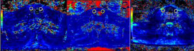 |
157 | T2* Mapping to Assess Hashimoto's Thyroiditis vs Healthy Thyroid at 3.0T: Feasibility
Nathan Ooms1, Humberto Monsevais1, and Ulrike Dydak1
1School of Health Sciences, Purdue University, West Lafayette, IN, United States Keywords: Quantitative Imaging, Body Hashimoto Thyroiditis is an autoimmune disease that largely effects women, and can cause fatigue, unexplained weight loss or gain, enlargement of the thyroid gland, and other symptoms. We are proposing utilizing T2* mapping to quantify the thyroid gland in the presence and absence of Hashimoto's Thyroiditis. Very little has been studied using T2* techniques in the thyroid gland, and what has been studies has focused on evaluating for nodules and cancer. We hope to broaden the capabilities to establish normal ranges and evaluate the effects of Hashimoto's Thyroiditis. |
|
1795.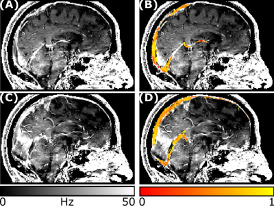 |
158 | Feasibility of R2*-based cerebral venous oxygenation mapping using a clinical susceptibility-weighted imaging protocol
Emma Biondetti1,2, Alessandro Villani1,2, Alessandra S. Caporale1,2, Antonio Maria Chiarelli1,2, Darien Calvo Garcia1,2, Massimo Caulo1,2,3, Valentina Tomassini1,2,4,5, and Richard G. Wise1,2,5
1Department of Neuroscience, Imaging and Clinical Sciences, 'G. d’Annunzio University' of Chieti-Pescara, Chieti, Italy, 2Institute for Advanced Biomedical Technologies (ITAB), 'G. d’Annunzio University' of Chieti-Pescara, Chieti, Italy, 3Department of Radiology, 'SS. Annunziata' University Hospital, Chieti, Italy, 4MS Centre, Department of Clinical Neurology, 'SS. Annunziata' University Hospital, Chieti, Italy, 5Cardiff University Brain Research Imaging Centre (CUBRIC), School of Psychology, Cardiff University, Cardiff, United Kingdom Keywords: Quantitative Imaging, Oxygenation The brain’s oxygen extraction fraction (OEF) is a key parameter in assessing cerebrovascular function and brain oxygen consumption. We investigated the feasibility of measuring cerebral venous oxygenation (SvO2), the key determinant of OEF, based on R2* estimated from a clinical susceptibility weighted imaging protocol and vessel segmentation aided by gadolinium administration, in a cohort of patients with Multiple Sclerosis. We were able to estimate venous oxygenation in the superior sagittal sinus, straight sinus, transverse sinuses and internal cerebral veins. The global median SvO2 ranged between 0.60 and 0.68, within the range of values expected in the human brain. |
|
1796.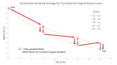 |
159 | Accelerated Sampling Strategy for High-resolution 3D T1rho Dispersion MRI with 4D Dynamic Radial Acquisitions
Gregory Peng1, Can Wu2, Jafari Ramin3, Yansong Zhao3, and Qi Peng1
1Department of Radiology, Albert Einstein College of Medicine and Montefiore Medical Center, Bronx, NY, United States, 2Department of Medical Physics, Memorial Sloan Kettering Cancer Center, New York, NY, United States, 3Philips Healthcare, Cambridge, MA, United States Keywords: Quantitative Imaging, Quantitative Imaging, T1rho dispersion imaging T1ρ dispersion imaging based on repeated T1ρ measurements at multiple spin-lock frequencies allows characterization of different dynamic processes of tissues. The clinical potential of these techniques is, however, limited by the total scan time needed to obtain reliable and consistent quantitative measurements. In this abstract, we propose the application of a 4D dynamic sequence with radial acquisitions in combination with an accelerated data-point sampling strategy to achieve efficient coverage of the high-dimensional data space in T1rho dispersion MRI, which leads to high-resolution 3D T1rho dispersion imaging acquired within clinically acceptable scan duration of ~5 minutes. |
|
1797. |
160 | New high-performance whole-body gradients to clarify Intravoxel incoherent motion DWI based multi-parametric MRI of prostate: Initial results
Roderic I Pettigrew1, Raja Muthupillai1, Erick Buko2,3, Michaela Martin4, and Andre Fischer4
1School of Engineering Medicine, Texas A&M University, Houston, TX, United States, 2Veterinary Clinical Sciences, University of Minnesota, Saint Paul, MN, United States, 3Center for Magnetic Resonance Research, University of Minnesota, Minneapolis, MN, United States, 4Siemens Healthcare, Erlangen, Germany Keywords: Quantitative Imaging, Prostate, multi parametric MRI We demonstrate the feasibility of obtaining high b-value (b > 2400 s/mm2) DWI images of the prostate obtained with short echo times (TE ~ 50 ms) on a prototype high-performance whole-body gradient system (Gmax = 200 mT/m, and SRmax = 200 T/m/s). Preliminary results from volunteers suggest that the apparent differences in the perfusion fraction metric of the IVIM model between the transition and peripheral zones of the prostate partly can be substantially diminished by choosing a shorter TE using high-performance gradients. |
|
 Back to Meeting Home
 Back to the Program-at-a-Glance
The International Society for Magnetic Resonance in Medicine is accredited by the Accreditation Council for Continuing Medical Education to provide continuing medical education for physicians.
 Back to Meeting Home
 Back to the Program-at-a-Glance
 View Presentation Video