Digital Poster
fMRI Acquisition & Analysis II
ISMRM & ISMRT Annual Meeting & Exhibition • 03-08 June 2023 • Toronto, ON, Canada

| Computer # | |||
|---|---|---|---|
2705.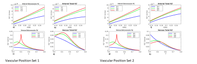 |
1 |
The dependence of the macrovascular transverse R2’ relaxation
and resultant BOLD fMRI signal on vascular position: A
simulation study
Xiaole Zhong1,2 and
J. Jean Chen1,2,3
1Department of Medical Biophysics, University of Toronto, Toronto, ON, Canada, 2Rotman Research Institute, Baycrest Hospital, Toronto, ON, Canada, 3Institute of Biomedical Engineering, University of Toronto, Toronto, ON, Canada Keywords: Signal Modeling, Simulations The R2’ effect is the foundation of BOLD fMRI contrast. R2’ is disproportionately sensitive to the presence of macrovasculature, which disrupts the homogeneity of the main magnetic field (B0), resulting in BOLD signal intensities that may heavily depend on macrovascular orientation and volume but also on vascular position. We simulate the BOLD signal strength in a voxel containing macrovasculature at various vascular positions within the voxel. This simulation highlights an additional source of variability in the macrovascular contributions to R2’-weighted MRI. |
|
2706.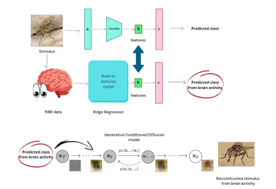 |
2 |
Brain Decoding and Reconstruction of concepts of visual stimuli
from fMRI through deep diffusion models
Matteo Ferrante1,
Tommaso Boccato2,
and Nicola Toschi3,4
1Biomedicine and prevention, University of Rome Tor Vergata, Roma, Italy, 2Biomedicine and prevention, University of Rome Tor Vergata, Rome, Italy, 3BioMedicine and prevention, University of Rome Tor Vergata, Rome, Italy, 4Department of Radiology,, Athinoula A. Martinos Center for Biomedical Imaging and Harvard Medical school, Boston, MA, USA, Boston, MA, United States Keywords: Machine Learning/Artificial Intelligence, Neuroscience, brain decoding, fMRI In vision, the brain is a feature extractor which works from images. We hypothesize that fMRI can mimic the latent space of a classifier, and employ deep diffusion models with BOLD data from the occipital cortex to generate images which are plausible and semantically close to the visual stimuli administered during fMRI. To this end, we mapped BOLD signals onto the latent space of a pretrained classifier and used its gradients to condition a generative model to reconstruct images. The semantic fidelity of our BOLD response to visual stimulus reconstruction model is superior to the state of the art. |
|
2707.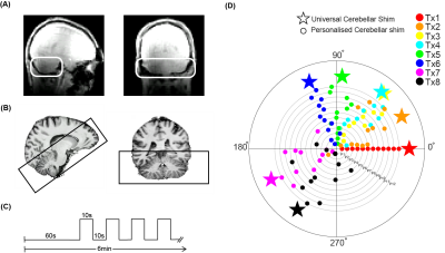 |
3 |
A universal B1 shim for the human cerebellum
Emma Brouwer1,2,
Wietske van der Zwaag1,2,
and Nikos Priovoulos1,2
1Spinoza Centre for Neuroimaging, Amsterdam, Netherlands, 2Computational Cognitive Neuroscience and Neuroimaging, Netherlands Institute for Neuroscience, Amsterdam, Netherlands Keywords: Data Acquisition, Shims, B1 shim The human cerebellum is underexplored in-vivo due to a lack of acquisition methods that effectively portray it. Its dense architecture necessitates high resolution, which benefits from 7T imaging. Unfortunately, at higher field strengths, images suffer from severely destructive B1-interference with typical coil geometries. Subject-optimized parallel-transmit approaches can mitigate B1 artifacts but increase scan-time. We designed a universal B1-shim specific to the cerebellum and immediately applicable across individuals for high-resolution 7T fMRI. We demonstrate improvements in tSNR and functional responses in cerebellar ROIs during a hand-motor task compared to a quadrature mode shim setting. |
|
2708.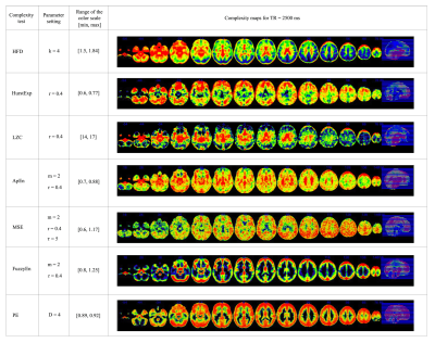 |
4 |
Statistical evaluation of complexity tests for fMRI timeseries
data
Dilmini Wijesinghe1,
Danny JJ Wang1,
and Kay Jann1
1USC Mark and Mary Stevens Neuroimaging and Informatics Institute, Keck School of Medicine at USC, Los Angeles, CA, United States Keywords: Software Tools, fMRI (resting state), Complexity, Higuchi Fractal Dimension, Hurst Exponent, Lempel-Ziv Complexity, Approximate Entropy, Multiscale Sample Entropy, Fuzzy Entropy, Permutation Entropy Complexity measures of rs-fMRI signals based on non-linear timeseries analyses have been proposed for quantifying the predictability of fMRI signals. There are multiple mathematical methods for evaluating complexity in timeseries signals. This study evaluates the optimal parameter settings in seven complexity tests by analyzing mean complexity measures of grey matter (GM) and CSF across multiple complexity tests with different parameter settings and different fMRI acquisition protocols. Furthermore, complexity evaluation was performed on surrogate timeseries data. Overall, Multiscale Sample Entropy, Fuzzy Entropy, and Hurst Exponent consistently showed an increase in mean complexity of GM compared to CSF in original data. |
|
2709.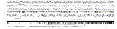 |
5 |
Using BOLD-fMRI to Compute Respiration Volume per Time (RVT) and
Respiration Variation (RV) with Convolutional Neural Network
(CNN) in Children
Abdoljalil Addeh1,2,
Fernando Vega1,2,
Rebecca J. Williams2,3,4,
Ali Golestani5,
G. Bruce Pike2,3,4,
and M. Ethan MacDonald 1,2,3,6
1Department of Biomedical Engineering, Schulich School of Engineering, University of Calgary, Calgary, AB, Canada, 2Hotchkiss Brain Institute, Cumming School of Medicine, University of Calgary, Calgary, AB, Canada, 3Department of Radiology, Cumming School of Medicine, University of Calgary, Calgary, AB, Canada, 4Department of Clinical Neurosciences, Cumming School of Medicine, University of Calgary, Calgary, AB, Canada, 5Department of Medical Physics, Alberta Heath Services, Calgary, AB, Canada, 6Department of Electrical & Software Engineering, Schulich School of Engineering, University of Calgary, Calgary, AB, Canada Keywords: Data Processing, fMRI, Physiological Noise In many fMRI studies, respiratory signals are unavailable or do not have acceptable quality. Consequently, the direct removal of low-frequency respiratory variations from BOLD signals is not possible. This study proposes a one-dimensional CNN model for reconstruction of two respiratory measures including RV and RVT. Results show that a CNN can capture informative features from the BOLD signals and reconstruct accurate RV and RVT timeseries. It is expected that application of the proposed method will lower the cost of fMRI studies, reduce complexity, and decrease the burden on participants because they will not be required to wear a respiratory bellows |
|
2710.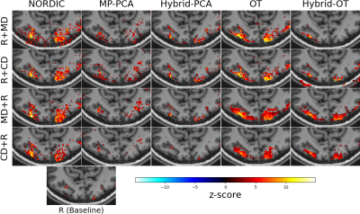 |
6 |
Benchmarking local low rank denoising methods for task-based
fMRI data analysis
Pierre-Antoine Comby1,
Zaineb Amor1,
Alexandre Vignaud1,
and Philippe Ciuciu1
1Neurospin (CEA), Gif-sur-Yvette, France Keywords: Data Processing, fMRI (task based), Denoising, Benchmak Local-low-rank denoising in task-based fMRI increases sensitivity to statistical detection of neural activity, without harming specificity. We compared 5 methods (NORDIC, MP-PCA, Hybrid-PCA, Optimal-Threshold, Hybrid-OT) in 4 preprocessing configuration (denoising on magnitude/complex data, before/after motion correction) and their effect on downstream analysis. For best performance, the denoising shoud be done prior to motion correction, and using complex-valued data is only valuable in some settings. In average (n=6), up to 8 times more activations can be detected (p < 0.05, controlling for FDR). We also provide open-source implementations for broader use of Local-Low-Rank denoising methods in fMRI. |
|
2711.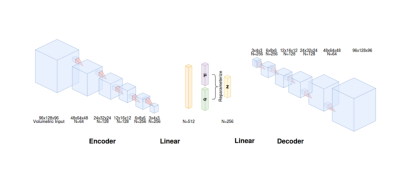 |
7 |
Representation learning of resting state functional MRI using a
volumetric variational autoencoder model (3D VAE)
Scott Peltier1,
Michelle Karker2,
Kuan Han2,
Doug Noll2,
and Zhongming M Liu2
1Functional MRI Laboratory, University of Michigan, Ann Arbor, MI, United States, 2Biomedical Engineering, University of Michigan, Ann Arbor, MI, United States Keywords: Machine Learning/Artificial Intelligence, Machine Learning/Artificial Intelligence In this work, we consider whether a VAE model trained with volumetric fMRI data (rather than a cortical subset of the data) is capable of encoding fMRI into low-dimensional representations, decoding these representations back into volumetric fMRI space, and also generating new fMRI patterns from the latent space. For 3D VAE model training, validation, and testing, volumetric resting-state fMRI data was used from the Human Connectome Project minimally preprocessed pipeline. We find the 3D VAE is able to accurately represent the spatial and temporal information in the data. In addition, it is able to synthesize realistic resting-state networks. |
|
2712.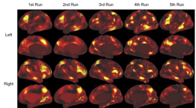 |
8 |
Using interpretable deep learning on task fMRI data to
understand brain regions related to working memory - a
repeatability study
Tianyun Zhao1,
Philip Tubiolo1,2,
Thomas Hagan1,
John C. Williams1,2,
Jared Van Snellenberg2,3,4,
and Chuan Huang1,4
1Biomedical Engineering, Stony Brook University, Stony Brook, NY, United States, 2Psychiatry and Behavioral Health, Renaissance School of Medicine at Stony Brook University, Stony Brook, NY, United States, 3Psychology, Stony Brook Univeristy, Stony Brook, NY, United States, 4Radiology, Renaissance School of Medicine at Stony Brook University, Stony Brook, NY, United States Keywords: Machine Learning/Artificial Intelligence, fMRI (task based) Deep learning, especially convolutional neural networks (CNN), has been shown to be able to identify the non-linear relation between functional magnetic resonance imaging (fMRI) and task performance. CNN can generate an interpretable result called saliency map highlighting regions that are important for task performance. It can uncover other neural processes that linear modeling cannot due to the high dimensionality nature of the fMRI. The CNN result can be presented as a saliency Previously, we developed a pipeline to produce the saliency map for working memory tasks. In this work, we further evaluated the repeatability of our pipeline. |
|
2713.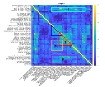 |
9 |
fMRI signal characteristics underlying human white-matter
functional connectivity
Nayana Menon1,
Jonathan Polimeni2,
and J. Jean Chen1,3,4
1Rotman Research Institute, Baycrest Health Sciences, Toronto, ON, Canada, 2Athinoula A. Martinos Center for Biomedical Imaging, Department of Radiology, Harvard Medical School, Massachusetts General Hospital, Boston, MA, United States, 3Department of Medical Biophysics, University of Toronto, Toronto, ON, Canada, 4Department of Biomedical Engineering, University of Toronto, Toronto, ON, Canada Keywords: Data Analysis, White Matter, fMRI, functional connectivity, Fourier analysis, Human Connectome Project In this study, we illustrate the BOLD fMRI signal characteristics underlying functional correlations in the white matter of healthy adults. We use a group-level white-matter only Pearson’s correlation matrix to identify several high-connectivity regions of interest. Our results show that the BOLD signal spectra of connected white-matter regions contain a low-frequency peak unseen in unconnected regions. |
|
2714.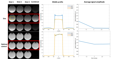 |
10 |
Joint Slice-Selective Pulse Design for Segmented FLEET-EPI using
a Differential Bloch Simulator and Conjugate Gradient-Optimal
Control
Imam Ahmed Shaik1,
Mukund Balasubramanian2,3,
Avery J.L. Berman4,5,
Jonathan R. Polimeni2,6,7,
and William Grissom1
1Institute of Imaging Science, Vanderbilt University, Nashville, TN, United States, 2Harvard Medical School, Boston, MA, United States, 3Boston Children's Hospital,, Boston, MA, United States, 4Carleton University, Ottawa, ON, Canada, 5University of Ottawa Institute of Mental Health Research, Ottawa, ON, Canada, 65Athinoula A. Martinos Center for Biomedical Imaging, Massachusetts General Hospital,, Charlestown, MA, United States, 7Harvard-MIT Division of Health Sciences and Technology,Massachusetts Institute of Technology, Cambridge, MA, United States Keywords: Data Acquisition, fMRI Multishot EPI-based fMRI can achieve high spatial resolutions to resolve functional activity at the level of layers and columns, but suffers sensitivity to motion and phase changes between shots. Reordering the shots in a multislice stack so that each slice’s shots are acquired sequentially (VFA-FLEET-EPI) reduces this sensitivity but requires specialized RF pulses to maintain consistent signal between shots as longitudinal magnetization evolves. In this work we show that designing these pulses jointly using an autodiff optimal control algorithm yields more consistent signal across shots which reduces ghosting compared to VFA sinc and recursively designed SLR pulses. |
|
2715.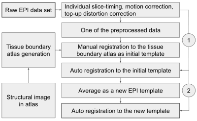 |
11 |
Manual registration and customized template for rodent fMRI data
spatial normalization
Wen-Ju Pan1,
Nmachi Anumba1,
Nan Xu1,
Lisa Meyer-Baese1,
and Shella Keilholz1
1Emory University/Georgia Institute of Technology, Atlanta, GA, United States Keywords: Data Analysis, Brain Rodent EPI image qualities may vary across coil types, coil positioning and different animals that challenge atlas registration. We proposed an accurate registration with study-group customized EPI template and initially manual registration with an assistance of the newly-introduced tissue-boundary atlas. Our studies demonstrated some visible mismatching in local anatomic structures by the standard registration methods for rodent data which were effectively corrected by the presented method. |
|
2716.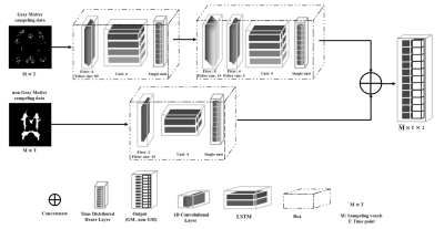 |
12 |
Functional MRI denoising Using Data-Driven Multi-Step Deep
Neural Network
Sina Ghaffarzadeh1,
Vahid Malekian2,
Faeze Makhsousi3,
and Seyyed Ali Seyyedsalehi3
1Biomedical Engineering, Amirkabir University of Technology (Tehran Polytechnic), Tehran, Iran (Islamic Republic of), 2University College London, London, United Kingdom, 3Amirkabir University of Technology (Tehran Polytechnic), Tehran, Iran (Islamic Republic of) Keywords: Data Processing, Brain In this study a novel method for sampling the active and noisy areas is proposed by using the purification of gray and non-gray matter areas of fMRI data. Also, a data-driven network is proposed in a parallel, multi-step and integrated manner for optimal noise reduction of t-fMRI data. Besides, the proposed method reduces substantially physiological noise without considering the specific noise source and only by using the ROI of noise and activity. Based on the results, the proposed method provides a more accurate and improved activity map than previous methods, which increases the power of activity analysis in fMRI data. |
|
2717.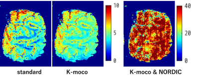 |
13 |
BISEPI high-resolution fMRI at 7T with NORDIC-PCA
Guoxiang Liu1,2,
Takashi Ueguchi1,2,
and Seiji Ogawa1,3
1NICT, Osaka, Japan, 2Graduate School of Frontier Biosciences, Osaka University, Osaka, Japan, 3Tohoku Fukushi University, Sendai, Japan Keywords: Image Reconstruction, fMRI (task based), Highe resolution Recently, a new noise reduction technique NOise Reduction with Distribution Corrected (NORDIC) PCA has been proposed for High resolution fMRI, and the improvements in keys including temporal-SNR, BOLD detectability have been demonstrated using this technique. In this work, we applied this technique to 0.4-mm isotropic human fMRI data acquired with our Block-interleaved segmented EPI, and compare the BOLD detectability of the data with and without NORDIC-performed. |
|
2718.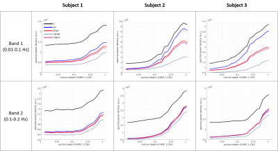 |
14 |
Depth-dependent effects of thermal and physiological noise
reduction in BOLD fMRI
Maria Guidi1,2,3,
Giovanni Giulietti4,5,
Harald E. Moeller2,
David G. Norris3,
and Federico Giove1,4
1MARBILab, Enrico Fermi Research Center, Rome, Italy, 2Max Planck Institute for Human Cognitive and Brain Sciences, Leipzig, Germany, 3Donders Centre for Cognitive Neuroimaging, Radboud University, Nijmegen, Netherlands, 4Fondazione Santa Lucia IRCCS, Rome, Italy, 5SAIMLAL Department, Sapienza University, Rome, Italy Keywords: Data Processing, Data Analysis, Denoising, Layers In this study, we evaluated the effect of common denoising steps (NORDIC, regression for motion parameters, RETROICOR and aCompCor) on a high-resolution resting-state BOLD fMRI dataset. We extracted the temporal standard deviation and the spectral power density at different cortical depths in the primary motor cortex and found that each denoising algorithm had a distinct signature on the profile shape. We further estimated the effect of denoising by calculating the temporal signal-to-noise ratio and delta variation signal (DVARS) for different tissue types and found that NORDIC and aCompCor had the largest impacts on the metrics considered. |
|
2719.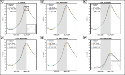 |
15 |
Validating NORDIC denoising on high-resolution fMRI data at 7 T
Viktor Pfaffenrot1 and
David Norris1,2
1Erwin L. Hahn Institute for Magnetic Resonance Imaging, University of Duisburg-Essen, Essen, Germany, 2Donders Institute for Brain, Cognition and Behavior, Radboud Iniversity Nijmegen, Nijmegen, Netherlands Keywords: Data Processing, fMRI, denoising, laminar fMRI, NORDIC PCA High-resolution fMRI is dominated by thermal noise. The NORDIC PCA filter is a promising method to remove thermal noise but its effect on the spatial fidelity of layer-dependent profiles is sparsely investigated. We evaluated the performance of NORDIC with several parameter setting for laminar fMRI at 7 T and sought to investigate the effect NORDIC has on the shape of the profiles. Our results suggest that NORDIC indeed affects high-resolution profiles but the overall effect can be seen to be small and can be further reduced with careful parameter settings. |
|
2720.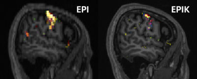 |
16 |
Where is each finger area in brain? Enhanced characterization of
the somatosensory area using high-resolution EPIK at 3T
Sung Suk Oh1,
Min Cheol Chang2,
Soyoung Kwak2,
Jong-ryul Choi3,
N. Jon Shah4,5,6,7,
and Seong Dae Yun4
1Medical Device Development Center, K-MEDI hub, Daegu, Korea, Republic of, 2Department of Rehabilitation Medicine, Yeungnam University, Daegu, Korea, Republic of, 3K-MEDI hub, Daegu, Korea, Republic of, 4Institute of Neuroscience and Medicine 4, INM-4, Forschungszentrum Juelich, Juelich, Germany, 5Institute of Neuroscience and Medicine 11, INM-11, Forschungszentrum Juelich, Juelich, Germany, 6JARA - BRAIN - Translational Medicine, Aachen, Germany, 7Department of Neurology, RWTH Aachen University, Aachen, Germany Keywords: Data Acquisition, fMRI (task based) The tactile stimulus of fingers in fMRI can effectively reveal the somatosensory functional region. However, this research requires a high-resolution fMRI technique, enabling a distinct delineation of functional areas for each finger. This work demonstrates the use of high-resolution EPIK (1.25 x 1.25 mm2) for enhanced characterization of somatosensory areas at 3T. EPIK provides improved spatial resolution than EPI (1.72 x 1.72 mm2) with increased brain coverage while keeping the same temporal resolution as EPI. The activation region from EPIK was shown to be more locally specific, thereby enabling a clearer recognition of functional areas for each finger. |
|
2721.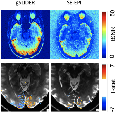 |
17 |
Spin-echo-based generalized Slice Dithered Enhanced Resolution
(gSLIDER) for mesoscale fMRI at 3 Tesla
Salvatore John Torrisi1,2,3,
Congyu Liao4,
Jennifer Townsend1,3,
and An (Joseph) Vu1,3
1Radiology, SF VA Medical Center, San Francisco, CA, United States, 2Northern California Institute of Research and Education, San Francisco, CA, United States, 3Radiology, University of California, San Francisco, San Francisco, CA, United States, 4Division of Radiological Sciences Laboratory, Stanford University, Palo Alto, CA, United States Keywords: Pulse Sequence Design, Contrast Mechanisms, Neuro We demonstrate sub-millimeter generalized Slice Dithered Enhanced Resolution (gSLIDER) for high-resolution spin-echo (SE) fMRI at 3T. Activations were significantly greater than standard SE fMRI, demonstrating the suitability of this method for high-resolution, mesoscale fMRI. |
|
2722.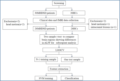 |
18 |
The ALFF alterations in maintenance hemodialysis patients with
sleep disorder:a rs-fMRI study combined with machine learning
analysis
Menghan Feng1,
Yue Zhang2,
Zeying Wen3,4,
Yuchi Wu2,
Chengwei Fu3,
Kan Deng5,
Qizhan Lin2,
and Bo Liu2
1Xin-Huangpu Joint Innovation Institute of Chinese Medicine in Guangdong Province, Guangzhou, China, 2The Second Affiliated Hospital of Guangzhou University of Chinese Medicine, Guangzhou, China, 3Guangzhou University of Chinese Medicine, Guangzhou, China, 4The First Affiliated Hospital of Henan University of Chinese Medicine, Zhengzhou, China, 5Philips Healthcare, Guangzhou, China Keywords: fMRI (resting state), Kidney The prevalence of SD is high in chronic kidney disorder patients who suffering maintenance hemodialysis whereas little is known about the neuropathologic mechanism. We investigated the ALFF alterations between MHDSD patients and HCs and those meaningful features were used for constructing discriminating model based the SVM algorithm to classify the MHDSD patients. We found the aberrant spontaneous activities in the DMN, AN, CEN, and VIN in MHDSD patients. Additionally, the classifier indicated the discriminative ALFF features in the above regions demonstrated good performance. This will contribute to well understanding the neuropathological mechanism and seeking biomarkers for discrimination in MHDSD patients. |
|
2723.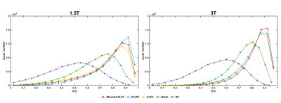 |
19 |
Reliability and sensitivity of fMRI at 3T and 1.5 T: metrics
toward fMRI guided individualized precise TMS treatment
Qiu Ge1,2,3,
MinLiang Yao4,
Juan Yue1,2,3,4,
Xue Yang1,2,3,
YueJiao Ding1,2,3,
Yong Zhang5,
and Yufeng Zang1,2,3,4
1Center for Cognition and Brain Disorders, The Affiliated Hospital of Hangzhou Normal University, Hangzhou, China, 2Zhejiang Key Laboratory for Research in Assessment of Cognitive Impairments, HangZhou, China, 3Institute of Psychological Sciences, Hangzhou Normal University, HangZhou, China, 4Hangzhou Normal University Affiliated Deqing Hospital, HangZhou, China, 5Yong Zhang, GE Healthcare, Shanghai, China Keywords: Data Acquisition, fMRI FDA cleared a TMS treatment protocol with fMRI-guided individualized targeting for major depressive disorder. This protocol was based on 3T scanner. However, 1.5 T MRI is more widely available in hospitals. We comprehensively tested the reliability and sensitivity of fMRI metrics related to TMS treatment on 1.5T and 3T scanners. In general, the sensitivity of 3T is larger than 1.5T. However, the reproducibility of peak FC location and the activation location, the reliability of RS-fMRI local metrics, are similar for 1.5T and 3T. In conclusion, 1.5 fMRI meets the needs for guiding individualized precise rTMS treatment. |
|
2724.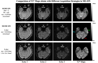 |
20 |
When BOLD Optimized ME-EPI Echo Combination Isn't BOLD Optimized
R. Allen Waggoner1,
Chisato Suzuki1,
Ken-ichi Ueno1,
and Keiji Tanaka1
1RIKEN Center for Brain Science, Wako-shi, Saitama, Japan Keywords: Artifacts, fMRI, Multi-Echo, Multi-Shot In regions suffering from magnetic susceptibility effects, the use of BOLD optimized echo combination for averaging Single-Shot/Multi-Echo EPI data across echos, leads to voxels with improves signal but reduced BOLD sensitivity. The use of Multi-shot/Multi-Echo EPI can suppress the susceptibility effects allowing BOLD optimized echo averaging that retains BOLD sensitivity. |
|
 Back to Meeting Home
 Back to the Program-at-a-Glance
The International Society for Magnetic Resonance in Medicine is accredited by the Accreditation Council for Continuing Medical Education to provide continuing medical education for physicians.
 Back to Meeting Home
 Back to the Program-at-a-Glance
 View Presentation Video