Digital Poster
Quantitative Imaging Beyond Relaxometry I
ISMRM & ISMRT Annual Meeting & Exhibition • 03-08 June 2023 • Toronto, ON, Canada

Digital Poster
Quantitative Imaging Beyond Relaxometry I
| Computer # | |||
|---|---|---|---|
3391.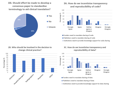 |
161 |
Current UK perspectives on the challenges for clinical
translation of quantitative MR imaging biomarkers.
Julia Markus1,
Po-Wah So2,
Harpreet Hyare3,
and Penny Cristinacce4
1Centre for Medical Imaging, UCL, London, United Kingdom, 2Department of Neuroimaging,Institute of Psychiatry, King's College London, London, United Kingdom, 3Radiology Department, University College London, London, United Kingdom, 4Division of Cancer Sciences, The University of Manchester, Manchester, United Kingdom Keywords: Quantitative Imaging, Challenges, Consensus A web-based survey was developed from the “Steps on the Path to Clinical Translation” workshop, at the British & Irish Chapter-ISMRM conference held on 7th September 2022. The survey explored the UK MRI community’s perspective on the clinical translation of quantitative MR imaging biomarkers. Three main themes emerged from the results: the need for 1) consensus; continued development of resources that unite existing work and improve shared lexicon; 2) context dependency; defining the steps to clinical translation, in ways that appreciate the uniqueness of the imaging biomarker and the clinical question; 3) a clearer definition of imaging biomarker expectation or product profile. |
|
3392.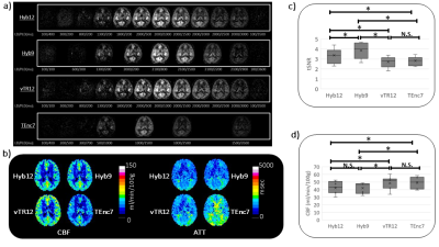 |
162 |
Comparison of a hybrid multi-delay pseudo-continuous arterial
spin labelling scheme with time-encoded and variable-TR schemes
Makoto Obara1,
Osamu Togao2,
Lena Vaclavu3,
Ryoji Mikayama4,
Tatsuhiro Wada4,
Shota Ishida5,
Hiroshi Hamano1,
Matthias J.P. van Osch3,
Kim van de Ven6,
Yu Ueda1,
Jihun Kwon1,
Masami Yoneyama1,
and Marc Van Cauteren7
1Philips Japan, Tokyo, Japan, 2Department of Molecular Imaging & Diagnosis, Graduate School of Medical Sciences, Kyushu University, Fukuoka, Japan, 3C.J. Gorter MRI Center, Department of Radiology, Leiden University Medical Center, Leiden, Netherlands, 4Division of Radiology, Department of Medical Technology, Kyushu University, Fukuoka, Japan, 5Department of Radiological Technology, Faculty of medical sciences, Kyoto College of Medical Science, Kyoto, Japan, 6Philips Healthcare, Best, Netherlands, 7Philips Healthcare, Tokyo, Japan Keywords: Quantitative Imaging, Perfusion, ASL To accurately calculate cerebral blood flow (CBF) and arterial transit time (ATT), there are two kinds of time-efficient approaches in use for multi-delay pseudo-continuous arterial spin labelling: time-encoded and sequential variable-TR. Hybrid schemes have recently been proposed that exploit the strengths of these approaches, but they have not been evaluated head-to-head, and the purpose of this study is therefore to investigate the clinical validity of hybrid schemes. The SNR, CBF and ATT were measured and compared in seven healthy subjects. A higher SNR was obtained in the hybrid scheme with high correlation coefficients with other ATT schemes, suggesting quantitative ability. |
|
3393.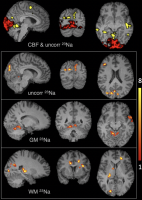 |
163 |
Mapping changes in tissue 23Na concentration and cerebral blood
flow during a visual task using ASL and functional Sodium
Imaging (fNaI)
Iris Asllani1,2,
Balázs Örzsik1,
Samira Bouyagoub1,
Joseph Woods3,
Itamar Ronen1,
Guillaume Madelin4,
and Mara Cercignani5
1University of Sussex, Brighton, United Kingdom, 2Rochester Institute of Technology, Rochester, NY, United States, 3University of Oxford, Oxford, United Kingdom, 4New York University, New York, NY, United States, 5Cardiff University, Cardiff, United Kingdom Keywords: Quantitative Imaging, Neuro, Sodium MRI, fMRI, Arterial Spin Labeling, Partial Volume Correction Sodium MRI was combined with arterial spin labeling (ASL) perfusion MRI to detect changes in total sodium concentration (TSC) and cerebral blood flow (CBF) associated with visual tast activation on 6 healthy volunteers. There was no overlap between the areas in the visual cortex where there was a significant change in CBF (~38%) and the regions where a change (~11%) in TSC was detected. Results from partial volume correction indicate that most of the changes in TSC originated in the white matter. |
|
3394.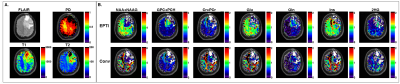 |
164 |
Integrated Fast High-Resolution MR Fingerprinting and
Spectroscopic Imaging for Absolute Quantification of Metabolic
Imaging
Mehran Baboli1,2,
Fuyixue Wang1,2,
Zijing Dong1,2,
Jorg Dietrich2,3,
Erik Uhlmann2,4,
Tracy Batchelor2,5,6,
Daniel Cahill2,7,
and Ovidiu C. Andronesi1,2
1Radiology, A. A. Martinos Center for Biomedical Imaging, Charlestown, MA, United States, 2Radiology, Harvard Medical School, Boston, MA, United States, 3Neurology, Division of Neuro-Oncology, Boston, MA, United States, 4Neurology, Beth Israel Deaconess Medical Center, Boston, MA, United States, 5Neurology, Brigham’s and Women Hospital, Boston, MA, United States, 6Neuro-Oncology, Dana Farber Cancer Institute, Boston, MA, United States, 7Neurosurgery, Massachusetts General Hospital, Boston, MA, United States Keywords: Tumors, Spectroscopy Quantification of metabolite concentration is the primary concern in clinical MR Spectroscopic Imaging which is valuable to assess disease pathology. Absolute metabolite quantification requires correction of MRSI signal for T1/T2 relaxation and proton density, which due to time limitations are not measured in the subject of interest but assumed to be constant across all voxels based on assumed literature values. Here, we integrated 3D-Echo-Planar Time-resolved Imaging (3D-EPTI) that allows fast MR-fingerprinting of T1, T2 and PD with fast MRSI metabolic imaging in each subject. The metabolite quantification based on voxel-based MRF was compared to literature based relaxations and PD values. |
|
3395.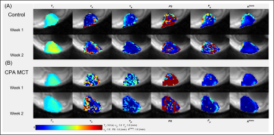 |
165 |
Monitoring metronomic chemotherapy response in orthotopic breast
cancer mouse model using DCE-MRI
Sawwal Qayyum1,
Jin Zhang1,
and Gene Kim1
1Radiology, Weill Cornell Medical, New York City, NY, United States Keywords: Quantitative Imaging, DSC & DCE Perfusion, Cancer, Orthotopic, 4T1, Metronomic Metronomic chemotherapy (MCT) is a cost-effective combinatorial treatment that can be used to normalize tumor vasculature for further treatment. Current monitoring schemes of MCT response are either invasive or are partially sampled. Dynamic contrast enhanced (DCE)-MRI can be utilized to measure the spatial heterogeneity of the tumor microenvironment which can serve as a noninvasive biomarker for treatment response. Using an MCT dose scheme of cyclophosphamide (70mg/kgx3 IP/week), the preliminary result shows that the DCE-MRI can detect the tumor vascular normalization in the treated group. |
|
3396.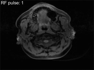 |
166 |
The effect of black blood and fat suppression prepulses on
signal modelling in T1-mapping and dynamic contrast enhanced MRI
Wilhelm Stehling1,
Myrte Wennen1,2,
Eric Schrauben1,
Pim van Ooij1,
Kak Khee Yeung3,
Aart Nederveen1,
and Oliver Gurney-Champion1,4
1Radiology and Nuclear Medicine, Amsterdam University Medical Centers, Amsterdam, Netherlands, 2Department of Intensive Care, Erasmus Medical Centre, Rotterdam, Netherlands, 3Vascular Surgery, Amsterdam University Medical Centers, Amsterdam, Netherlands, 4Cancer Center Amsterdam, Imaging and Biomarkers, Amsterdam, Netherlands Keywords: System Imperfections: Measurement & Correction, Simulations In this work, we derived an equation describing the signal evolution during an SGRE sequence with preparation pulses. The signal changes especially during the earlier RF pulses and differs from the values for later RF pulses, where it approaches the signal from an SPGR sequence without prepulses. Using the derived equation might enable more accurate DCE examinations. |
|
3397.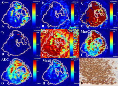 |
167 |
Differentiation of Triple-negative and HER2 Positive Breast
Cancer Using DISCO CE-MRI
Guo Haodong1,
Dmytro Pylypenko2,
Li Haige1,
Zhu Jianguo1,
Yuan Xiaofan1,
and Zhang Ziyan1
1the Second Affiliated Hospital of Nanjing Medical University, Nanjing, China, 2GE Healthcare, China, Beijing, Beijing, China Keywords: Quantitative Imaging, Breast, DISCO CE-MR This study aims to investigate the feasibility triple-negative and HER2 positive breast cancer using DISCO CE-MRI. A total of 96 patients were recruited in the study. Quantitative and semi-quantitative parameters were used. kep and TTP had diagnostic value for triple-negative breast cancer. The AUC of kep and TTP were 0.870 and 0.928. kep had diagnostic value for HER2 positive breast cancer.The AUC of kep was 0.832. Our findings suggest that DISCO CE-MRI parameters might be reliable quantitative indicators for the differential diagnosis of triple-negative breast cancer and HER2 positive breast cancer. |
|
3398.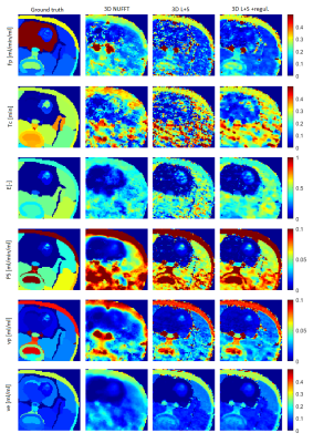 |
168 |
Advanced-Model 3D DCE-MRI of Small Animals with Spatially
Regularized Processing Steps
Radovan Jirik1,
Ondrej Macicek1,
Michal Bartos2,
Marie Mangova3,
Pavel Rajmic3,
Denisa Hyvlova1,
and Zenon Starcuk, jr.1
1Czech Academy of Sciences, Institute of Scientific Instruments, Brno, Czech Republic, 2Czech Academy of Sciences, Institute of Information Theory and Automation, Praha, Czech Republic, 3Department of Telecommunications, Brno University of Technology, Faculty of Electrical Engineering and Communication, Brno, Czech Republic Keywords: Quantitative Imaging, DSC & DCE Perfusion In DCE-MRI, advanced pharmacokinetic models, such as the 2CX, TH and ATH models, provide a more complete set of pharmacokinetic parameters than the commonly used simpler models, such as the Tofts and Patlak models. However, their use requires higher temporal resolution and signal-to-noise ratio. Hence, advanced-model DCE-MRI allows for imaging of only a few slices. We present an approach making 3D advanced-model DCE-MRI possible and evaluate it under the low-signal-to-noise-ratio conditions of small-animal MRI. Our methodology is based on compressed sensing and spatially regularized fitting of the pharmacokinetic model. The approach is evaluated on simulated and real datasets. |
|
3399.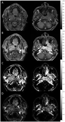 |
169 |
Comparing L1 and L2 Regularizations for Quantitative Transport
Mapping of Tumor: an Image Quality Analysis
Dominick Romano1,2,
Qihao Zhang1,2,
Ilhami Kovanlikaya2,
Pascal Spincemaille2,
and Yi Wang2,3
1Biomedical Engineering, Cornell University, New York, NY, United States, 2Radiology, Weill Cornell Medical College, New York, NY, United States, 3Biomedical Engineering, Cornell University, Ithaca, NY, United States Keywords: Quantitative Imaging, Perfusion, Regularization This study compared L1 and L2 regularized Quantitative Transport Mapping (QTM1-3) of dynamic contrast enhanced (DCE) MRI in breast and neck tumor using image quality scoring. Improved consistent soft tissue and lesion characterization was observed when using the L1 norm. |
|
3400.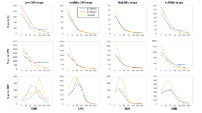 |
170 |
Quantitative BOLD with Variational Bayesian inference: model
comparisons with Monte Carlo simulations and in an elderly
cohort
Linh N. N. Le1,
Gregory J. Wheeler1,
Nicholas P. Blockley2,
and Audrey P. Fan1,3
1Department of Biomedical Engineering, University of California, Davis, Davis, CA, United States, 2School of Medicine & Health Sciences, University of Nottingham, Nottingham, United Kingdom, 3Department of Neurology, University of California, Davis, Davis, CA, United States Keywords: Quantitative Imaging, Oxygenation This study uses Monte Carlo simulations to understand the behavior of quantitative BOLD in different physiological conditions, using a Variational Bayesian inference framework with prior information and data from Asymmetric Spin Echo (ASE) scans. The performance of the three models at 7 SNR levels (from 5 to 500) showed that one-compartment and two-compartment models estimated oxygen extraction fraction (OEF) more accurately than linear model across a full range of deoxygenated blood volume (DBV) (p<0.05 using two-way ANOVA with pairwise comparisons). In vivo data showed that Bayesian inference approach effectively enables smoother and quantitatively different parameter maps of OEF and DBV. |
|
3401.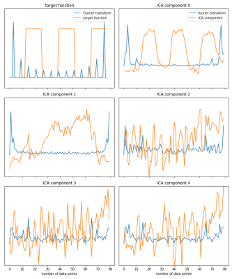 |
171 |
Automating Component Selection in Independent Component Analysis
(ICA) in dynamic Oxygen-Enhanced MRI (dOE-MRI)
Annika Hofmann1,2,
Jennifer H.E. Baker3,
Firas Moosvi4,
and Stefan A Reinsberg2
1Department of Physics, TU Dortmund University, Dortmund, Germany, 2Department of Physics & Astronomy, University of British Columbia, Vancouver, BC, Canada, 3Radiation Biology Unit, British Columbia Cancer Research Centre, Vancouver, BC, Canada, 4Department of Computer Science, Mathematics, Physics and Statistics, University of British Columbia, Kelowna, BC, Canada Keywords: Quantitative Imaging, Cancer, Independent Component Analysis Using Independent Component Analysis (ICA) in dynamic Oxygen-Enhanced MRI has been shown to improve the sensitivity of this technique. However, the ICA component has to be identified manually by an observer. In this work we propose an optimization process that automatically determines the best number of components and extracts the component in best accordance to the target function of the breathing challenge. |
|
3402.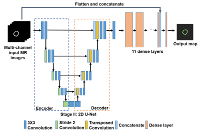 |
172 |
Deep learning assisted quantification of myocardial oxygen
extraction fraction
Ran Li1,
Cihat Eldeniz1,
Thomas Schindler1,
Linda Peterson1,
Pamela Woodard1,
and Jie Zheng1
1Washington University in St. Louis, St. Louis, MO, United States Keywords: Quantitative Imaging, Myocardium A previously developed MRI method for quantitative myocardial oxygen extraction mapping showed promising results, but image quality suffered from distortion and inhomogeneity artifacts. A new deep learning-based approach was developed and tested in healthy subjects. This preliminary study showed excellent reproducibility and consistent myocardial oxygen extraction values with other reported data using positron emission tomography methods. |
|
3403.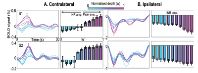 |
173 |
Spatial variation of the negative BOLD response in human primary
visual cortex
Artemy Vinogradov1,
Nooshin J. Fesharaki1,
Amanda Taylor2,
David Ress2,
and Jung Hwan Kim1
1University of Texas Health Center at Houston, Houston, TX, United States, 2Baylor College of Medicine, Houston, TX, United States Keywords: Quantitative Imaging, fMRI (task based), Depth-dependent BOLD, neurovascular coupling, hemodynamic response function and ultra-high-field mri The negative hemodynamic response functions (nHRF), temporal dynamics of the negative BOLD response (NBR) evoked by a brief stimulus, have been reported as an inverted version of the corresponding positive HRF (pHRF). With a careful delineation of the nHRF from the stimulus representation (SR), we found that the nHRF adjacent to the SR was not the inverted version of the corresponding pHRF. We also characterized the nHRF with respect to the depth and found inverse proportion of the nHRF amplitudes between contralateral and ipsilateral sides, which can indicate different underlying mechanisms of the nHRF. |
|
3404.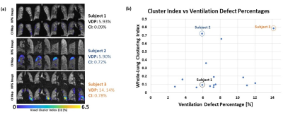 |
174 |
Quantification of Spatial Ventilation Defect Distribution in
Hyperpolarized Gas MRI of Lungs Using A 3D Clustering Algorithm
Gabriela Maria Garcia Delgado1,
Ummul Afia Shammi1,
Talissa Ann Altes2,
John P Mugler III3,4,
and Robert Paul Thomen1,2
1Biomedical, Biological and Chemical Engineering, University of Missouri, Columbia, MO, United States, 2Radiology, School of Medicine, University of Missouri, 65201, MO, United States, 3Radiology and Medical Imaging, School of Medicine, University of Virginia, Charlottesville, VA, United States, 4Biomedical Engineering, University of Virginia, Charlottesville, VA, United States Keywords: Quantitative Imaging, Hyperpolarized MR (Gas), Body, Lung, Contrast Mechanisms, Data Analysis Hyperpolarized gas (HPG) MR imaging allows for quantification of a patient’s lung function. The spatial distribution of ventilation defect patterns is often overlooked in quantitative analyses but may be important for further understanding the nature of lung disease. Here we present a method for quantifying the extent to which defect voxels tend to be sparsely distributed or clustered. This technique involves spherical region-growing for each defect voxel to assess the fraction of neighbors which are also part of a defect. A ‘clustering index’ is reported which quantifies the extent to which defect voxels are spatially congregated or scattered. |
|
3405.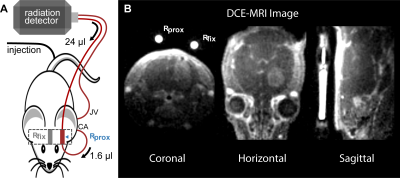 |
175 |
Stepwise validation of Perfusion MRI Kinetic Modeling using an
Extracorporeal Circulation for AIF determination in Mice
Florian Gierse1,
Florian Büther2,
Juela Cufe2,
Bastian Maus3,
Michael Claesener2,
Sven Hermann1,
Klaus Peter Schäfers1,
Katharina Kronenberg4,
Uwe Karst4,
Michael Andreas Schäfers1,
Cornelius Faber3,
and Philipp Backhaus2
1European Institute for Molecular Imaging, University of Münster, Münster, Germany, 2Department for Nuclear Medicine, University Hospital Münster, Münster, Germany, 3Translational Research Imaging Center, Clinic of Radiology, University of Münster, Münster, Germany, 4Institute of Inorganic and Analytical Chemistry, University of Münster, Münster, Germany Keywords: Quantitative Imaging, Perfusion Calculation of perfusion parameters from DCE-MRI using pharmacokinetic modeling depends on accurate determination of the dynamic tissue concentration and the dynamic arterial blood concentration (AIF). However, precise AIFs are difficult to measure in small animals. We introduce a novel extracorporeal circulation approach from the external carotid artery for feasible AIF determination. DCE derived AIF and tissue contrast agent concentrations corresponded well with radioactive analogs and mass spectrometry. Perfusion parameters derived by different pharmacokinetic models demonstrated variable agreement with simultaneous radiotracer derived calculations of modeling parameters. Overall, we introduce an accurate and precise framework for quantitative perfusion MRI in small animals. |
|
3406.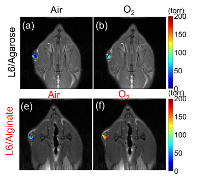 |
176 |
Quantification of oxygenation levels within biocompatible
hydrogels subcutaneously implanted in rats
Yuka Sugamura1,
Vikram Kodibagkar1,
Amy Emerson1,
and Jessica Weaver1
1Arizona State University, Tempe, AZ, United States Keywords: Quantitative Imaging, Oxygenation, pO2 mapping, T1 mapping, hydrogels Cell therapy has the potential to repair irreversible pathologies including infarction and diabetes. High survival rate of the delivered cells is essential for effective treatment, where cell oxygenation is a critical factor. Biocompatible hydrogels are often used as cell delivery vehicles. Here, we propose an approach to quantify the oxygen availability that the cells encapsulated in biocompatible hydrogels experience after cell transplantation. In this study, we demonstrate the feasibility of assessing the pO2 within the hydrogels implanted in rats by combining the hydrogel with a previously developed magnetic resonance imaging (MRI) -based tissue oximetry technique. |
|
3407.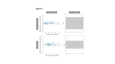 |
177 |
Influence of different confounders on measurements results of 2D
Flow MRI Examinations in Healthy Travelling Volunteers
Ralf Felix Trauzeddel1,2,
Thomas Grandy3,
Elias Daud1,2,
Maximilian Müller1,2,
Darian Viezzer1,2,
Thomas Hadler1,2,
Ning Jin4,
Daniel Giese5,6,
and Jeanette Schulz-Menger1,2,3
1Working Group on Cardiovascular Magnetic Resonance, Experimental and Clinical Research Center, a joint cooperation between the Charité Universitätsmedizin Berlin and the Max-Delbrück-Center for Molecular Medicine, Berlin, Germany, Charité - Universitätsmedizin Berlin, Berlin, Germany, 2Partner Site Berlin, DZHK (German Centre for Cardiovascular Research), Berlin, Germany, 3Department of Cardiology and Nephrology, Helios Hospital Berlin-Buch, Berlin, Germany, 4Cardiovascular MR R&D, Siemens Medical Solutions USA, Inc., Cleveland, Ohio, USA, Cleveland, OH, United States, 5Magnetic Resonance, Siemens Healthcare GmbH, Erlangen, Germany, 6Institute of Radiology, University Hospital Erlangen, Friedrich-Alexander-Universität Erlangen-Nürnberg (FAU), Erlangen, Germany, Erlangen, Germany Keywords: Quantitative Imaging, Heart, 2D Flow Various confounders can have an influence on the results of cardiovascular MRI (CMR) examinations. Data about systematic investigations of such confounders are sparse. 2D Flow CMR using segmented and realtime measurements was performed in 20 healthy travelling volunteers to examine the influence of beat-to-beat variability between different heart cycles, sequence types, field strengths and scanner configurations as well as examiners on forward flow volume and peak velocity and compared to 95% tolerance intervals established by intraobserver analysis to test for comparability and precision of measurements which revealed good to very good comparability regarding various physiological, technical and post- processing confounders. |
|
3408.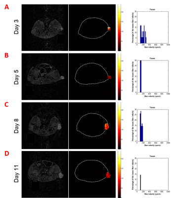 |
178 |
Magnetic Ferritin Nanoprobe Imaging Analysis with MRI Slow
Velocity Mapping Measurement
FangFei He1,2,
Wentao Liu2,
Tongwei Zhang3,
Fan Yang4,
Yi Hou1,
Zhihua Gan1,
and Dong Han2,5
1College of Life Science and Technology, Beijing University of Chemical Technology, Beijing, China, 2CAS Center for Excellence in Nanoscience, National Center for Nanoscience and Technology, Beijing, China, 3Institute of Geology and Geophysics, Chinese Academy of Sciences, Beijing, China, 4Beijing Intelligent Brain Cloud Inc, Beijing, China, 5University of Chinese Academy of Sciences, Beijing, China Keywords: Quantitative Imaging, Velocity & Flow, Tumor Interstitial Fluid For effectively penetrating into tumors, drug diffusion speed should be greater than the outward tumor interstitial fluid (TIF) convection speed. Studying TIF is helpful to deeply understand the fluid behavior of tumor fluid flow and its impact on the delivery of nano drugs. In this experiment, an improved PC-MRI slow flow measurement sequence was used to detect the velocity of interstitial fluid in different tumor growth periods. Combined with clinical standard sequence, the relationship between the distribution of the magnetic ferritin (M-HFn) contrast agent in tumor and the velocity of TIF was observed and analyzed. |
|
 Back to Meeting Home
 Back to the Program-at-a-Glance
The International Society for Magnetic Resonance in Medicine is accredited by the Accreditation Council for Continuing Medical Education to provide continuing medical education for physicians.
 Back to Meeting Home
 Back to the Program-at-a-Glance
 View Presentation Video