Digital Poster
Quantitative Imaging Beyond Relaxometry II
ISMRM & ISMRT Annual Meeting & Exhibition • 03-08 June 2023 • Toronto, ON, Canada

Digital Poster
Quantitative Imaging Beyond Relaxometry II
| Computer # | |||
|---|---|---|---|
3565.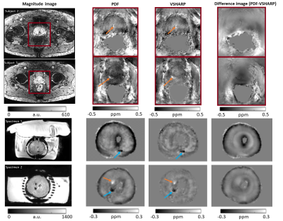 |
161 |
An Optimized High Resolution Acquisition and Processing Pipeline
for QSM in the Prostate
Laxmi Muralidharan1,
Manju Mathew2,
Joey Clemente2,
Lucy Caselton2,
Sumandeep Kaur2,
Mrishta Brizmohun2,
Shonit Punwani2,
and Karin Shmueli1
1Dept of Medical Physics and Bioengineering, University College London, London, United Kingdom, 2Centre for Medical Imaging, University College London, London, United Kingdom Keywords: Quantitative Imaging, Susceptibility, Background field removal We aimed to optimize MRI acquisition with 1mm isotropic resolution and Quantitative Susceptibility Mapping (QSM) reconstruction for prostate clinical research. Acquisition parameters optimized in two subjects included fat-water phase artifact removal, parallel imaging acceleration factor, resolution and number of echoes. QSM masking, background field removal and susceptibility calculation were optimized in six subjects and three prostatectomy specimens. In-phase acquisition removed more fat-water phase artifacts than post-processing. VSHARP and excluding rectal gas reduced residual background fields and Iterative Tikhonov regularization reduced noise. This optimized (8.5 minute) protocol and pipeline will allow incorporation of prostate QSM in clinical research studies. |
|
3566.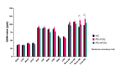 |
162 |
Quantitative evaluation of deep gray matter in
striatal-cerebellar-brainstem circuits in Parkinson’s disease
patients with freezing of gait
Youmin Zhang1,
Naying He1,
Zhijia Jin 1,
Yu Liu1,
Xinhui Wang1,
E. Mark Haacke1,2,
and Fuhua Yan1
1Department of Radiology, Ruijin Hospital, Shanghai Jiao Tong University School of Medicine, Shanghai, China, 2Department of Radiology, Wayne State University, Detroit, MI, United States Keywords: Quantitative Imaging, Quantitative Susceptibility mapping This study evaluated the structural changes of the deep gray matter in the cortico-basal ganglia and cerebellar motor circuit in Parkinson’s disease patients with freezing of gait. Using region-of-interest-based quantitative susceptibility mapping analysis, we found pronounced striatum atrophy combined with caudal substantia nigra abnormal iron accumulation in Parkinson’s disease patients with freezing of gait. This result offered new insight into future research investigating the pathophysiology of the freezing of gait. |
|
3567. |
163 |
Magnetic Susceptibility Separation Reveals Longitudinal Lesion
Changes in Multiple Sclerosis
Ziyan Zhu1,
Javad Hamidi Esfahani1,
Nashwan Naji1,
Peter Seres1,
Derek Emery2,
Gregg Blevins3,
Penelope Smyth3,
and Alan Wilman1,2
1Department of Biomedical Engineering, University of Alberta, Edmonton, AB, Canada, 2Department of Radiology and Diagnostic Imaging, University of Alberta, Edmonton, AB, Canada, 3Division of Neurology, Department of Medicine, University of Alberta, Edmonton, AB, Canada Keywords: Multiple Sclerosis, Multiple Sclerosis, Iron Change, Myelin Change Susceptibility separation may distinguish iron and myelin in multiple sclerosis (MS) lesions, but little work has been done on longitudinal changes. Twenty MS subjects were followed for 14 months to investigate para and diamagnetic changes independently using susceptibility separation in comparison to transverse relaxation and susceptibility mapping. Susceptibility separation provided distinction between myelin and iron events, offering more insight into MS lesion evolution. |
|
3568.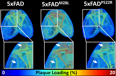 |
164 |
Detecting whole mouse brain beta-amyloid plaques in 5xFAD mice
using high-resolution quantitative susceptibility mapping
Nian Wang1,
Chengqian Zhou2,
Surendra Maharjan1,
and Abigail Wallace1
1Radiology and Imaging Sciences, Indiana University, Indianapolis, IN, United States, 2Boston University, Boston, MA, United States Keywords: Alzheimer's Disease, Alzheimer's Disease, beta-amyloid Alzheimer’s disease (AD) is an age-associated neurodegenerative disease that is reaching epidemic proportions as a result of the aging of the world’s population. It is likely that the most effective treatment for AD will need to be administrated before cognitive symptoms occur, necessitating a biomarker for the early stages of AD. Novel high-resolution MRI technologies with the capability to characterize the individual beta-amyloid plaque through the whole brain is highly desirable. |
|
3569.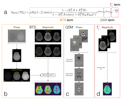 |
165 |
BTX: Simultaneous 3D Quantitative Magnetization Transfer Imaging
and Susceptibility Mapping
Albert Jang1,2,
Hyungseok Jang3,
and Fang Liu1,2
1Athinoula A. Martinos Center for Biomedical Imaging, Harvard Medical School, Charlestown, MA, United States, 2Radiology, Massachusetts General Hospital, Boston, MA, United States, 3Radiology, University of California, San Diego, San Diego, CA, United States Keywords: Quantitative Imaging, Multi-Contrast, Magnetization Transfer, Quantitative Susceptibility Mapping We propose a novel sequence that enables simultaneous quantitative magnetization transfer (qMT) imaging and susceptibility mapping (QSM) for assessing tissue composition, microstructure, and microenvironment. We extend our BTS sequence to incorporate a multi-echo acquisition scheme for probing tissue susceptibility information. The acquisition allows quantification of B1+, bias-corrected T1, qMT parameters (macromolecule bound proton fraction and proton exchange rate), tissue susceptibility and T2*. The method’s feasibility is demonstrated using an in-vivo brain scan, where 3D concurrent qMT and QSM were obtained for the whole brain at feasible scan time. |
|
3570.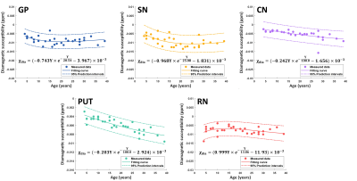 |
166 |
Imaging the myelin in brain deep gray matters during brain
development using sub-voxel QSM
Zhenghao Li1,
Ruimin Feng1,
and Hongjiang Wei1
1Shanghai Jiao Tong University, Shanghai, China Keywords: Quantitative Imaging, Quantitative Susceptibility mapping Myelin is a non-neglectable susceptibility source in deep gray matter (DGM) nuclei. Myelination and demyelination processes are essential in DGMs during brain development and aging. However, they have not been investigated by MRI yet. We used a sub-voxel QSM method to independently image and quantify the myelin concentration in brain DGM. We recruited 32 healthy subjects with ages from 4 to 39 years old to describe the DGM myelination and demyelination trajectories. The results suggest that different DGMs display various development trajectories during brain development. |
|
3571.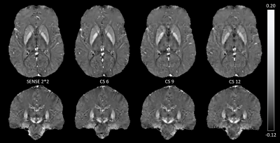 |
167 |
Using Compressed Sensing techniques for QSM on multiple
sclerosis patients, to what extent can we accelerate?
Émilie Poirion1,
Julien Savatovsky1,
Jessica Guillaume2,
and Mathieu Santin3,4,5
1Imaging department, Rothschild Foundation Hospital, Paris, France, 2Research clinical department, Rothschild Foundation Hospital, Paris, France, 3Institut du Cerveau (ICM) - Paris Brain Institute, Inserm U 1127, CNRS UMR 7225, Sorbonne Université, Paris, France, 4Center for NeuroImaging Research (CENIR), Paris, France, 5MDS Research International, La Rochette, France Keywords: Quantitative Imaging, Quantitative Susceptibility mapping Despite the characterization of in-vivo biomarkers of MS pathophysiology, there is still a gap between histological knowledges and morphological MRI as it is performed in clinical practice. New MRI advanced techniques, such as quantitative susceptibility mapping (QSM) might help to better describe the disease, but long acquisition times are limiting their uses. Adding acceleration techniques can reduce this time. However, there is no current evidence about their impact on QSM values. Thus, we added four QSM sequences to our clinical protocol of MS follow-up, varying the acceleration factor from classical SENSE (2*2) to high compressed-sense (6, 9, and 12). |
|
3572. |
168 |
QSM and R2* relaxometry analysis for mapping of white matter
hyptension and cognition in patients with lacunar Infarction
HONGWEI LI 1,
XUCHEN YU1,
MIN HE2,
WEIBO CHEN3,
Jing Ding 2,
and He Wang1
1Institute of Science and Technology for Brain-Inspired Intelligence, Fudan University, Shanghai, China, 2Department of Neurology, The Affiliated Zhongshan Hospital of Fudan University, Shanghai, China, 3Philips Healthcare, Shanghai, China Keywords: Vessels, Perfusion Due to the lacunar infarcts are small, they are asymptomatic in most cases. However, the location and accumulation of multiple lacunar infarcts might lead to severe physical and cognitive impairment. This lesion, caused by the involvement of cerebral small vessel in the brain, is thought to be associated with an increased risk of Vessel dementia (VaD). Therefore, exploring the perfusion alterations in patients with early lacunar infarction might help with interventions and preventive measures, thereby reducing the social and healthcare burden. |
|
3573.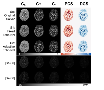 |
169 |
DeepDECOMPOSE: A Deep Learning based framework for solving
DECOMPOSE QSM
Jingjia Chen1,
Alfredo De Goyeneche1,
and Chunlei Liu1,2
1Electrical Engineering and Computer Sciences, University of California, Berkeley, Berkeley, CA, United States, 2Helen Wills Neuroscience Institute, University of California, Berkeley, Berkeley, CA, United States Keywords: Quantitative Imaging, Quantitative Susceptibility mapping We propose a deep learning (DL) approach to accelerate and improve the accuracy of the parameter fitting problem of DECOMPOSE-QSM. The approach allows a triple complex exponential model to be fitted in <1s on CPUs and <20ms on a GPU for a 256x256 image, vs. 5+ min for the original solver. The DL solver can be implemented with either fixed echo times or adaptive to a range of echo times and number of echoes. Trained with various additive noise levels, the DL-solver performs more robustly compared to the conventional optimization-based solver when the signal has a very low SNR. |
|
3574.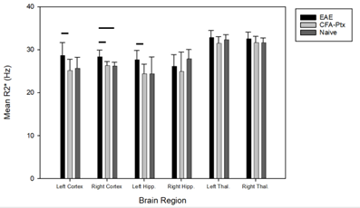 |
170 |
Magnetic Susceptibility vs Relaxometry in the Characterization
of an Animal Model of Multiple Sclerosis
Rehman Ali Tariq1,
Rania Muhammed1,
Ying Wu1,
Qandeel Shafqat1,
Hongfu Sun2,
and Jeff F. Dunn1
1Radiology, University of Calgary, Calgary, AB, Canada, 2School of Information Techology and Electrical Engineering, University of Queensland, Brisbane, Australia Keywords: Quantitative Imaging, Susceptibility, Multiple Sclerosis, Animal Models There is evidence for changes in magnetic susceptibility in the CNS of people with MS. MRI techniques utilizing relaxometry and magnetic tissue properties – R2* and quantitative susceptibility mapping, respectively – can be used to detect these changes. Both techniques were applied to study pathological changes in EAE mice compared to controls. Significant differences were found in the cortex between EAE mice and controls using R2*, while no differences in magnetic susceptibility were found between the two groups. Although this indicates that R2* may be better at detecting differences, perhaps it can be utilized as a complement to QSM analysis. |
|
3575. |
171 |
Realistic Abdominal QSM phantom
Javier Ignacio Silva1,2,3,
Mathias Lambert1,2,3,
Carlos Milovic3,4,
Sergio Uribe2,3,5,
and Cristian Tejos1,2,3
1Department of Electrical Engineering, Pontificia Universidad Catolica de Chile, Santiago, Chile, 2Biomedical Imaging Center, Pontificia Universidad Catolica de Chile, Santiago, Chile, 3Millennium Institute for Intelligent Healthcare Engineering (iHEALTH), Santiago, Chile, 4School of Electrical Engineering, Pontificia Universidad Catolica de Valparaiso, Valparaiso, Chile, 5Department of Radiology, Pontificia Universidad Catolica de Chile, Santiago, Chile Keywords: Signal Modeling, Susceptibility Abdominal Quantitative Susceptibility Mapping (QSM) involves a series of additional challenges compared with brain QSM, mainly because of the presence of undesired contributions related to fat and gasses. Previously, we proposed a QSM phantom with fat contributions, which emulated different susceptibility scenarios in the abdominal region. In this work, we present an improved version which now considers additional features including a variable multi-peak fat model, variable R2* and background field features. Simulation experiments show how these new components produce of different effects, like increased signal decay in specific tissues, 1st and 2nd kind chemical shift artifacts and water/fat swaps in graph cuts reconstructions. |
|
3576.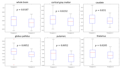 |
172 |
Brain iron content increase and oxygen extraction fraction
reduction after iron repletion in blood donation induced iron
deficiency
Hangwei Zhuang1,2,
Alexey Dimov3,
Gary Brittenham4,
Pascal Spincemaille3,
Thanh Nyugen3,
Yi Wang3,
and Eldad Hod4
1Biomedical Engineering, Cornell University, Ithaca, NY, United States, 2Department of Radiology, Weill Medical College of Cornell University, New York, NY, United States, 3Weill Medical College of Cornell University, New York, NY, United States, 4Columbia University Medical Center, New York, NY, United States Keywords: Hematologic, Neuro We examined brain iron content using susceptibility source separation and oxygen extraction fraction (OEF) in adult healthy blood donors with blood donation-induced iron deficiency treated with intravenous iron repletion at the first donation and a second donation after approximately 5 months and compared it with a control group who was not treated after the first donation. Iron content increases in the parietal cortex, the caudate and the putamen were observed in the treatment group. OEF reduction was observed in the whole brain and multiple ROIs, suggesting iron repletion treatment helps reduce oxidative stress. |
|
3577.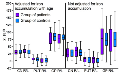 |
173 |
Brain iron status in children and adolescents with
beta-thalassemia.
Daniil Kirdyashkin1,
Vera Lopatina1,
Petr Bulanov1,2,
Natalia Kriventsova1,
Galina Tereshchenko1,
and Petr Menshchikov1,2
1Dmitry Rogachev National Research Center of Pediatric Hematology, Oncology and Immunology, Moscow, Russian Federation, 2Philips, Moscow, Russian Federation Keywords: Quantitative Imaging, Quantitative Susceptibility mapping, Iron accumulation Background TDT patients are susceptible to secondary iron overload. Determining brain iron content may be crucial to prevent brain iron poisoning. TDT Patient and control groups consisted of 23 and 50 children and adolescents respectively. 3D, T1-weighted and susceptibility-weighted images were obtained and QSM maps were reconstructed. Magnetic susceptibility values in brain structures were adjusted for age-related iron accumulation. Statistical analysis, carried out with and without correction for age, did not reveal significant differences in brain iron content between the two groups. Brain iron overload does not occur in TDT patients, whether it be children or adults. |
|
3578.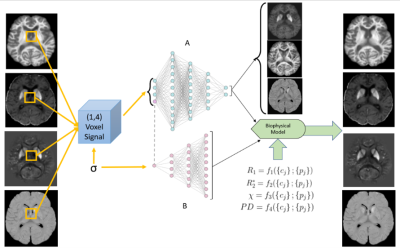 |
174 |
Myelin, Iron, and Free Water Content Quantification Through
BIOPHYSICSS Deep Learning
Ilyes Benslimane1,
Günther Grabner2,
Simon Hametner3,
Thomas Jochmann1,4,
Robert Zivadinov1,5,
and Ferdinand Schweser1
1Department of Neurology, Buffalo Neuroimaging Analysis Center, Buffalo, NY, United States, 2Department of Medical Engineering, Carinthia University of Applied Sciences, Klagenfurt, Austria, 3Department of Neuropathology and Neurochemistry, Medical University of Vienna, Vienna, Austria, 4Department of Computer Science and Automation, Technische Universität Ilmenau, Ilmenau, Germany, 5Department of Computer Science and Automation, Center for Biomedical Imaging, Clinical and Translational Science Institute at the University at Buffalo, Buffalo, NY, United States Keywords: Quantitative Imaging, Machine Learning/Artificial Intelligence Clinical translation of quantitative MRI (qMRI) is challenged by the multiple dependencies MR signal has on different tissue compartments. We previously introduced a neural network designed for single subject analysis (BIOPHYSICSS-DL) that overcomes the multiple tissue dependencies of qMRI and produces maps of source content. BIOPHYSICSS-DL has previously focused to myelin and iron quantification in the brain but in this work we expand that model to include a free water compartment. The network also used exclusively quantitative MRI metrics but the expansion of this model was accomplished with weighted MRI data which is another critical step to eventual clinical adoption. |
|
3579.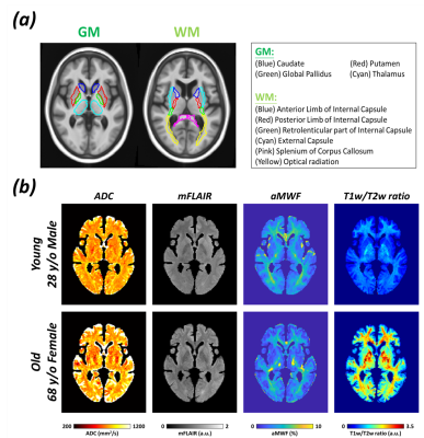 |
175 |
Age-dependent changes of myelin-related feature analysis in gray
matter and white matter
Nan-Hao Chen1,2,
Li-Ping Chen3,
Chia-Wei Hsu3,
Chin-Hua Yang1,3,4,
and Hsu-Hsia Peng1
1Department of Biomedical Engineering and Environmental Sciences, National Tsing Hua University, Hsinchu, Taiwan, 2Institute of Biomedical Engineering and Nanomedicine, National Health Research Institutes, Miaoli, Taiwan, 3Department of Medical Imaging, National Taiwan University Hospital Hsinchu Branch, Hsinchu, Taiwan, 4Department of Radiology, Taoyuan General Hospital, Taoyuan, Taiwan Keywords: Quantitative Imaging, Aging, Myelin-related images The regional characteristics extracted from myelin-related images have been considered the markers of brain degradation. However, a systematic investigation of age-dependent changes in myelin-related images remains deficient. We aimed to investigate the associations between regional features and age in myelin-related images. Our results revealed that ADC, mFLAIR, and T1w/T2w ratio were associated with age while aMWF showed no age-dependency. The increased heterogeneity and decreased uniformity in certain brain regions were observed in the elderly, indicating changes of microstructural tissue integrity in ADC, mFLAIR, and T1w/T2w ratio and the clinical usefulness of aMWF without a confounding factor of age in diagnosis. |
|
3580. |
176 |
Mapping brain development : An ex vivo multi-contrast MRI study
on the ferret
Laura Mouton1,
Anthony Ruze1,
Romain Valabrègue1,
Jean-Baptiste Pérot1,
Lucas Soustelle2,
Vaibhav Sahu3,
Katja Heuer3,
Stéphane Lehéricy1,
Roberto Toro3,
and Mathieu D. Santin1
1Institut du Cerveau (ICM) - Paris Brain Institute, Inserm U 1127, CNRS UMR 7225, Sorbonne Université, Center for Neuroimaging research (CENIR), Paris, France, 2Aix Marseille Univ, CNRS, CRMBM, Marseille, France, 3Institut Pasteur, Université de Paris, Département de neuroscience, Paris, France Keywords: Quantitative Imaging, Ex-Vivo Applications, Multi-Contrast Brain development during the first weeks after birth in ferrets is similar to the one in the last trimester pregnancy in humans. Studying the ferret brain could thus provide insights about brain development and the underlying processes. We used a multi-contrast MRI approach combining diffusion-weighted and quantitative MRI at 11.7T to investigate normal brain development in the ex vivo ferret brain. We were able to reconstruct fiber tracts even at a very early stage and assess their myelination level based on macromolecular fraction values. Combining diffusion-weighted with quantitative MRI is thus an interesting imaging approach to study normal brain development. |
|
3581.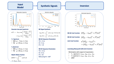 |
177 |
Joint Inversion of Multi-Echo Gradient-Echo and Spin-Echo Data
for the Estimation of Myelin Water Fraction
Ségolène Dega1,
Ravi Dadsena2,
Hendrik Paasche1,
and Tony Stöcker2,3
1Helmholtz Centre for Environmental Research (UFZ), Leipzig, Germany, 2German Center for Neurodegenerative Diseases (DZNE), Bonn, Germany, 3Department of Physics and Astronomy, University of Bonn, Bonn, Germany Keywords: Quantitative Imaging, Multi-Contrast, Myelin Myelin water fraction (MWF) mapping using MRI has enabled researchers to directly examine myelination and demyelination in both developing and diseased brains. T2-, and T2*-weighted multi-echo data have been proposed to estimate MWF in the human brain. Even for the simple two pool signal models of myelin and non-myelin associated water, number of dimensions of the parameter space for obtaining MWF estimates remains high, making parameter estimation challenging. The aim of this research is to improve the accuracy of brain myelin mapping using a novel joint inversion imaging method that combines data from multiple contrasts in a single optimization process. |
|
3582.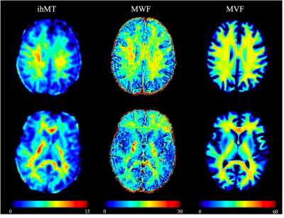 |
178 |
Myelin Measurement: Comparison Between Simultaneous Relaxometry,
Inhomogeneous Magnetization Transfer Imaging, and Myelin Water
Fraction
Moto Nakaya1,2,
Akifumi Hagiwara1,
Masanori Ozaki3,
Hiroshi Kusahara3,
Yasunobu Hoshino4,
Yuji Tomizawa4,
Kazumasa Yokoyama4,
Shohei Fujita1,2,
Christina Andica1,
Koji Kamagata1,
Masaaki Hori1,5,
Akihiko Wada1,
Osamu Abe2,
and Shigeki Aoki1
1Department of Radiology, Juntendo University, Tokyo, Japan, 2Department of Radiology, The University of Tokyo, Tokyo, Japan, 3Canon Medical Systems Corporation, Kawasaki, Japan, 4Department of Neurology, Juntendo University, Tokyo, Japan, 5Department of Radiology, Toho University Omori Medical Center, Tokyo, Japan Keywords: Multiple Sclerosis, Validation In 16 healthy adults, we examined the correlation between three MR myelin measurement methods, including myelin volume fraction (MVF) measured with simultaneous tissue relaxometry of R1 and R2 relaxation rates and proton density, inhomogeneous magnetization transfer imaging (ihMT), and myelin water fraction (MWF). A strong correlation was obtained between ihMT and MVF, and MWF was less correlated with the others. We should be mindful that these metrics are measuring different aspects of “myelin”. |
|
3583.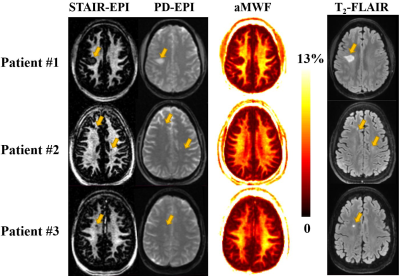 |
179 |
Quantitative Myelin Water Imaging Using Short TR Adiabatic
Inversion Recovery Prepared FSE (STAIR-FSE) and EPI (STAIR-EPI)
Sequences
Dina Moazamian1,
Sam Sedaghat1,
Jiyo S Athertya1,
Bhavsimran Singh Malhi1,
Soo Hyun Shin1,
James Lo1,2,
Hyungseok Jang1,
Eric Y Chang1,3,
Jiang Du1,2,3,
Graeme M Bydder1,
and Yajun Ma1
1Radiology, UC San Diego, San Diego, CA, United States, 2Bioengineering, UC San Diego, San Diego, CA, United States, 3Radiology Service, Veterans Affairs San Diego Healthcare System, San Diego, CA, United States Keywords: Multiple Sclerosis, Multiple Sclerosis, Myelin water Myelin water imaging (MWI) techniques have shown great promise for the early detection of demyelination and monitoring the effectiveness of neuroprotective therapies in remyelination. The purpose of this study is to develop new clinical transitional MWI techniques, which employ a short TR adiabatic inversion recovery (STAIR) preparation in combination with clinical FSE and EPI acquisitions. Quantified apparent myelin water fractions (aMWFs) show myelin loss in lesions and normal-appearing white matter (NAWM) in patients with multiple sclerosis compared to normal white matter (NWM) in healthy volunteers, demonstrating their potential in clinical practice. |
|
3584.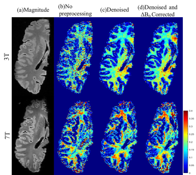 |
180 |
Comparison of myelin water fraction mapping on ex vivo human
brain at 3T and 7T
Guojun Xu1,
Zhiyong Zhao1,
Qinfeng Zhu1,
Zuozhen Cao1,
Keqing Zhu2,3,
Jing Zhang2,3,
and Dan Wu1
1Department of Biomedical Engineering, Zhejiang University, Hangzhou, China, 2China Brain Bank and Department of Neurology in Second Affiliated Hospital, Zhejiang University School of Medicine, Hangzhou, China, 3Department of Pathology, Zhejiang University, Hangzhou, China Keywords: Quantitative Imaging, White Matter Multi-echo gradient-echo (mGRE) is an important method to quantify myelin water fraction (MWF) of the human brain, but the results may depend on field strength given the difference in T2*. This study performed mGRE-based MWF on ex vivo human brain at high resolution at both 3T and 7T. We found MWF-derived from 7T showed higher measurements with lager standard deviations compared to those 3T, and the 3T and 7T results showed moderate agreement. These findings indicated the MWF mapping result was field-strength dependent and further validations were needed to support their reliability. |
|
 Back to Meeting Home
 Back to the Program-at-a-Glance
The International Society for Magnetic Resonance in Medicine is accredited by the Accreditation Council for Continuing Medical Education to provide continuing medical education for physicians.
 Back to Meeting Home
 Back to the Program-at-a-Glance
 View Presentation Video