Digital Poster
Relaxometry & Diffusion II
ISMRM & ISMRT Annual Meeting & Exhibition • 03-08 June 2023 • Toronto, ON, Canada

| Computer # | |||
|---|---|---|---|
1798.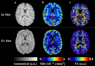 |
161 | Diffusion Tensor Imaging Parameters in ALS Post Mortem In Situ versus Ex Situ MR Acquisitions
Dominique Neuhaus1,2, Maria Janina Wendebourg3,4, Eva Scheurer1,2, Celine Berger1,2, Melanie Bauer1,2, Tanja Haas5, Regina Schlaeger3,4, and Claudia Lenz1,2
1Institute of Forensic Medicine, Dept. of Biomedical Engineering, University of Basel, Basel, Switzerland, 2Institute of Forensic Medicine, Health Department Basel-Stadt, Basel, Switzerland, 3Neurology Clinic and Polyclinic, Dept. of Clinical Research, University Hospital Basel, University of Basel, Basel, Switzerland, 4Translational Imaging in Neurology (ThINk), Dept. of Biomedical Engineering, University of Basel, Basel, Switzerland, 5Division of Radiological Physics, Dept. of Radiology and Nuclear Medicine, University Hospital Basel, Basel, Switzerland Keywords: Quantitative Imaging, Diffusion Tensor Imaging The development of magnetic resonance imaging (MRI) biomarkers in neurodegenerative diseases such as amyotrophic lateral sclerosis (ALS) might alleviate current challenges. Generally, post mortem (PM) ex situ quantitative imaging is used for the validation of potential biomarkers, which shows several limitations. In this study, PM MRI brain scans of three deceased ALS patients have been conducted. The objective was to identify differences between in situ and ex situ scans regarding the diffusion tensor imaging (DTI) measures fractional anisotropy (FA) and mean diffusivity (MD). A significant difference was found for the FA values in white matter. |
|
1799. |
162 | Application of six diffusion-weighted MRI models in predicting lymph node metastasis for resectable gastric cancer: A pilot study
Jing Li1, Shao-yu Wang2, and Xue-jun Chen1
1Radiology, the Affiliated Cancer Hospital of Zhengzhou University (Henan Cancer Hospital), Zhengzhou, China, 2MR Scientific Marketing, Siemens Healthneers, Shanghai, Shanghai, China Keywords: Quantitative Imaging, Tumor This study firstly explored the potential of six diffusion-weighted MRI models for preoperative prediction of lymph node metastasis (LNM) in resectable gastric cancer (GC). The DKI_K, CTRW_D, DKI_D, FROC_D, IVIM_D*, IVIM_f, Mono_ADC, as well as morphologic indicators of tumor thickness, clinical T staging on MRI (cT), MRI reported LN status were significantly different between LNM positive and LNM negative groups. These parameters were statistically correlated with LNM. The cT, MRI reported LN, and CTRW_D were independent risk factors of LNM, combining these three parameters yielded the highest predictive efficacy. |
|
1800.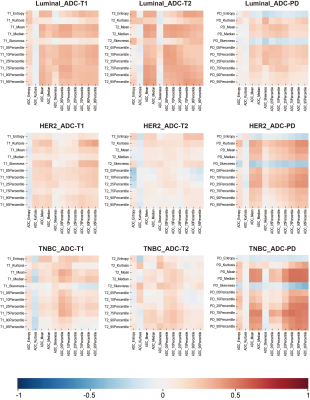 |
163 | Correlation of T1/T2/PD map and apparent diffusion coefficient for neoadjuvant therapy response evaluation in three breast cancer subtypes
Wenhong Jiang1, Siyao DU1, Lizhi Xie2, Min Zhao2, Zichuan Xie3, and Lina Zhang1
1Department of Radiology, The First Hospital of China Medical University, shenyang, China, 2GE Healthcare, Beijing, China, 3Guangzhou institute of technology, Xidian University, Guangzhou, China Keywords: MR Fingerprinting/Synthetic MR, Breast In this study, we evaluated and analyzed the potential correlation of first-order feature pairs from T1/T2/PD and ADC maps within different treatment response groups in locally advanced breast cancer patients undergoing neoadjuvant chemotherapy (NAC). In TNBC subtype, multiple features on ADC and PD maps strongly correlated only in pCR group, which may indicate PD map as complements to ADC for monitoring NAC response. |
|
1801. |
164 | Does simultaneous multi-slice technique affect quantitative measurements of diffusion-weighted MR imaging in hepatocellular carcinoma?
Zheng Ye1, Shan Yao1, Ting Yang1, Qing Li2, Robert Grimm3, Zhenlin Li1, and Bin Song1
1Department of Radiology, West China Hospital, Sichuan University, Chengdu, China, 2MR collaborations, Siemens Healthcare Ltd., Shanghai, China, 3MR Application Predevelopment, Siemens Healthcare GmbH, Erlangen, Germany Keywords: Quantitative Imaging, Diffusion/other diffusion imaging techniques Although diffusion weighted MR imaging is of great importance in the diagnosis and evaluation of hepatocellular carcinoma (HCC), it is limited by long acquisition times. Simultaneous multi-slice (SMS) technique can reduce scan time and has been proved to be feasible in liver imaging. Our study included twenty patients with HCC, and acquired conventional and SMS-accelerated diffusion data with different models (DWI, DKI and IVIM). By analyzing quantitative parametric maps, we found SMS technique did not affect the quantitative measurements of HCC. However, the difference of mean kurtosis between conventional and SMS-accelerated DKI in liver parenchyma should be noted. |
|
1802.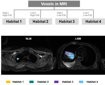 |
165 | Multiparametric MRI Based Habitat Imaging for Differentiating Lymph Node Metastasis in Lung Adenocarcinoma
Gaofeng Shi1, Qi Wang1, Hui Feng1, Hui Liu1, Mengyu Song1, Xinyue Liang2, and Yongming Dai2
1Department of Radiology, Fourth Hospital of Hebei Medical University, Shijiazhuang, China, 2Central Research Institute, United Imaging Healthcare, Shanghai, China Keywords: Quantitative Imaging, Cancer Analysis of lymph node metastasis (LNM) in lung cancer is vital for disease detection and treatment planning optimization. Multi-parametric MRI was widely used not only to characterize tumor size and anatomy, but also to assess the tissue metabolism and physiology. Conventionally, these are evaluated independently and/or are combined into an average parameter for the entire tumor, while the spatial information within the lesions was discarded. Habitat imaging allows to capture these subtle differences in tumors. In this context, a multi-parametric MRI based habitat analysis was approved to predict the LNM status. |
|
1803.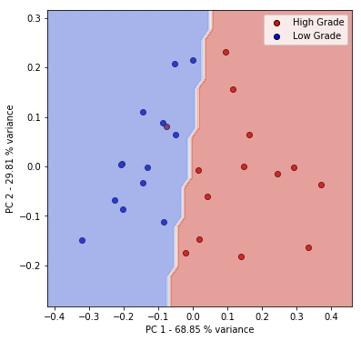 |
166 | A Machine Learning based Preoperatively Grading Rectal Method with Continue Time Random Walk DWI Model.
Zhijun Geng1, Shaolei Li2, Yunfei Zhang2, Yongming Dai2, and Chuanmiao Xie1
1Sun Yat-sen University Cancer Center, Guangzhou, China, 2MR Collaboration, Central Research Institute, United Imaging Healthcare, Shanghai, China Keywords: Quantitative Imaging, Machine Learning/Artificial Intelligence Machine learning offers a principled approach for developing automatic algorithms for analysis of high-dimensional biomedical data. The continuous-time random-walk model (CTRW) is novel non-Gaussian diffusion model that provides promising evidence indicating a possible link between voxel-level spatiotemporal diffusion heterogeneity and microscopic intravoxel tissue heterogeneity giving the model advantages in diagnosing many diseases1. In this study, we apply a machine-learning algorithm, the principal component analysis (PCA), for quantitative automatic diagnosis to grade rectal cancer using parameters from CTRW model. Our study shows that PCA has the potential to grade rectal cancer with higher accuracy than the original parameters. |
|
1804.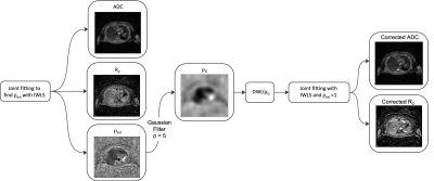 |
167 | Joint fitting of ADC and R2 with an estimate of the signal offset improves noise characteristics of whole-body R2 maps.
Annemarie Knill1,2, Jessica Winfield1,2, Hannah Barnsley2, Benjamin Malawo2, Georgina Hopkinson2, Dow-Mu Koh1,2, Christina Messiou1,2, and Matthew Blackledge1
1The Institute of Cancer Research, London, United Kingdom, 2The Royal Marsden NHS Foundation Trust, London, United Kingdom Keywords: Quantitative Imaging, Modelling Joint fitting of ADC and R2 improves the noise characteristics of whole-body R2 maps when using an iterative weighted least squares (IWLS) estimation that accounts for the signal offset, ρ, between diffusion-weighted and multi-echo-time imaging sequences. This is demonstrated using simulation experiments using relevant values of R2, ADC, and signal-to-noise ratio (SNR), where we find an improvement in R2 precision using the joint-fitting approach. Initial validation was further performed in clinical imaging examples to demonstrate the improvement in R2 map detail. |
|
1805.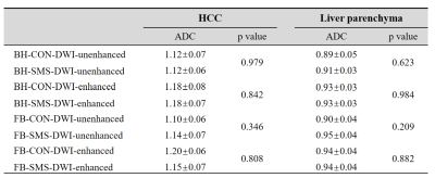 |
168 | Quantitative DWI in patients with hepatocellular carcinoma: effects of simultaneous multi-slice acquisition and gadoxetic acid injection
Ting Yang1, Zheng Ye1, Shan Yao1, Qing Li2, and Bin Song1
1West China Hospital, Chengdu, China, 2MR Collaborations, Siemens Healthineers Ltd., Shanghai, China Keywords: Quantitative Imaging, Liver, simultaneous multi-slice diffusion weighted imaging Diffusion weighted imaging (DWI) and gadoxetic acid-enhanced magnetic resonance imaging (MRI) are important in diagnosing hepatocellular carcinoma (HCC). Previous studies have validated that gadoxetic acid administration during enhanced MRI improves signal-to-noise ratio without influencing apparent diffusion coefficient (ADC), while the impact of simultaneous multi-slice (SMS) technique on the measurement of ADC is still unknown. Our study assessed the influence of gadoxetic acid administration on ADC values of liver with and without SMS acceleration. We found that SMS technique and gadoxetic acid administration did not affect the ADC values of liver parenchyma and HCC. |
|
1806.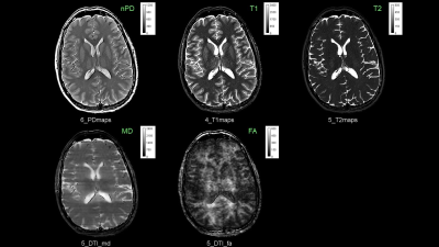 |
169 | Combined Multiparameter Quantitative MRI of PD, T1, T2 and the Diffusion Tensor with MP6-qMRI-Turbo Spin Echo with SPLICE
Ning Hua1, Andrew Ellison1, Yansong Zhao2, and Hernan Jara1
1Radiology, Boston University, Boston, MA, United States, 2Philips Healthcare, Boston, MA, United States Keywords: Quantitative Imaging, Diffusion Tensor Imaging, qMRI, DTI Purpose: To augment the qMRI multi-parametricity of the Triple TSE to additionally include primary diffusion tensor imaging (DTI) functionality and with seamless geometric compatibility with the nPD, T1, and T2 maps of the Triple-TSE framework. Methods: qMRI maps were generated with algorithms based on Bloch and Bloch-Torrey equations. Results: histogram analyses for the phantom and volunteer reveal well behaved qMRI data for nPD, T1, T2, MD, and FA. Conclusion: Creating a unified clinical MP-qMRI protocol for PD, T1, T2 and diffusion tensor seems within reach. |
|
1807.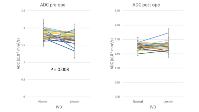 |
170 | Apparent diffusion coefficient of the lumbar intervertebral disc as a quantitative indicator of disease severity and treatment effectiveness
Daiki Sakamoto1, Xinnan Li1, Hiroyuki Hamaguchi1, Katsuhisa Yamada2, Hideki sudo2, and Khin Khin Tha1,3
1Laboratory for Biomarker Imaging Science, Hokkaido University Graduate School of Biomedical Science and Engineering, Hokkaido, Japan, 2Department of Orthopedic Surgery, Hokkaido University Faculty of Medicine, Hokkaido, Japan, 3Grobal Center for Biomedical Science and Engineering, Hokkaido University Faculty of Medicine, Hokkaido, Japan Keywords: Quantitative Imaging, Degenerative, Intervertebral disc degeneration, ADC, T1rho, T2*, Lumbar This prospective study aimed to evaluate the role of T1ρ, T2*, and ADC – the quantitative MRI indices, in determining the severity of intervertebral disc degeneration and treatment effectiveness. All three indices decreased with increasing Pfirmman grade, which determines the severity, suggestive of their potential as indicators of disease severity. The mean ADC of the degenerated disc increased significantly after surgery, suggesting its potential as a treatment effectiveness indicator. |
|
1808.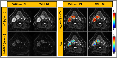 |
171 | Application of Deep Learning-based Reconstruction for Diffusion Kurtosis Imaging in Head and Neck Cancer
Amaresha Shridhar Konar1, Jaemin Shin2, Ramesh Paudyal1, Akash Deelip Shah3, Abhay Dave4, Maggie Fung2, Eve LoCastro1, Suchandrima Banerjee5, Nancy Lee6, and Amita Shukla-Dave1,3
1Medical Physics, Memorial Sloan Kettering Cancer Center, New York City, NY, United States, 2GE Healthcare, New York City, NY, United States, 3Radiology, Memorial Sloan Kettering Cancer Center, New York City, NY, United States, 4Touro College of Osteopathic Medicine, New York City, NY, United States, 5GE Healthcare, Menlo Park, CA, United States, 6Radiation Oncology, Memorial Sloan Kettering Cancer Center, New York City, NY, United States Keywords: Quantitative Imaging, Machine Learning/Artificial Intelligence A Deep learning (DL)-based reconstruction is a promising method to achieve higher resolution for diffusion-weighted Kurtosis imaging (DKI) without increasing signal averaging. The DKI phantom and patient results demonstrated improved image quality and reduced Gibbs (ringing) artifact, aiding in the robust estimation of Dapp and Kapp. In all phantom and patient data, the standard deviation of Dapp and Kapp measured in images reconstructed without DL was higher than in images reconstructed using DL. The NEX=1 significantly reduced the multi-b-value data acquisition time, and the DL-based reconstruction can produce images comparable to the standard NEX=2 or 4, depending on the b-value. |
|
1809.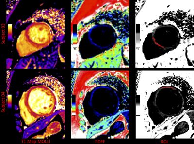 |
172 | A fat quantification approach based on longitudinal relaxation time
Yanglei Wu1, Xiaoyue Zhou2, Ke Xu3, and Huayan Xu3
1MR Collaboration, SIEMENS Healthineers Ltd., Beijing, China, 2MR Collaboration, SIEMENS Healthineers Ltd., Shanghai, China, 3Department of radiology, West China Second University Hospital, Chengdu, China Keywords: Quantitative Imaging, Fat The quantification of fat in humans is plays an important role in clinical diagnosis and scientific research. Especially in some cardiomyopathy characterized by fat deposition like arrhythmogenic right ventricular cardiomyopathy, and duchenne muscular dystrophy-associated cardiomyopathy. Current magnetic resonance imaging-based fat quantification methods have difficulty to characterizing cardiac etc. fat fractions. But the application of cardiac T1 mapping has been very extensive, and our proposed approach to build a partial volume model to utilize the T1 value of voxel obtained by T1 mapping can complete the measurement of cardiac fat content. |
|
1810.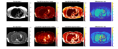 |
173 | PDFF and R2* Reconstruction Error Mapping using Cramér-Rao Lower Bounds for Quality Assessment In-Vivo
Alexandre Triay Bagur1,2, Paul Aljabar2, Matthew Robson2, Michael Brady2, and Daniel P Bulte1
1University of Oxford, Oxford, United Kingdom, 2Perspectum Ltd, Oxford, United Kingdom Keywords: Quantitative Imaging, Relaxometry Image analysts would welcome a measure of proton density fat fraction (PDFF) and R2* mapping error for image quality assessment in-vivo. This work adopts prior methodology using Cramér-Rao lower bound formulation and applies it to PDFF and R2* precision mapping in-vivo. The method is demonstrated in a software phantom and on one subject with fatty liver. The calculated maps agree with the ground truth and are informative in regions where the reconstruction signal model holds. The method should be extended to calculation of PDFF error maps. |
|
1811.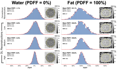 |
174 | Addressing Bias and Precision in Low SNR Chemical Shift Encoded MRI Proton Density Fat Fraction Estimation using a Deep Learning Reconstruction
Nathan T Roberts, PhD1, Nikolaos Panagiotopoulos, MD2, Ty Cashen, MD, PhD1, Daiki Tamada, PhD2, Diego Hernando, PhD2,3,4,5, Arnaud Guidon, PhD1, and Scott B Reeder, MD, PhD2,3,4,6,7
1GE HealthCare, Waukesha, WI, United States, 2Radiology, University of Wisconsin - Madison, Madison, WI, United States, 3Medical Physics, University of Wisconsin - Madison, Madison, WI, United States, 4Biomedical Engineering, University of Wisconsin - Madison, Madison, WI, United States, 5Electrical and Computer Engineering, University of Wisconsin - Madison, Madison, WI, United States, 6Medicine, University of Wisconsin - Madison, Madison, WI, United States, 7Emergency Medicine, University of Wisconsin - Madison, Madison, WI, United States Keywords: Quantitative Imaging, Machine Learning/Artificial Intelligence Low bias and high precision are important for accurate diagnosis, staging, and treatment monitoring of chronic liver disease using chemical shift-encoded (CSE)-MRI. However, CSE-MRI proton density fat fraction (PDFF) measurements are often biased by an asymmetric noise distribution present in PDFF maps acquired with low/moderate signal-to-noise ratio (SNR). This work investigates the use of deep learning de-noising to mitigate this bias in phantoms and in vivo. Results demonstrate that deep learning reconstruction removes or reduces noise-related PDFF estimation bias while maintaining the expected noise distribution characteristic of PDFF. |
|
1812.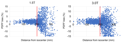 |
175 | Dependence of PDFF Measurement Accuracy on Location, Resolution, and Field Strength – a Phantom Experiment
Nikolaos Panagiotopoulos1,2, David Rutkowski3, Alexandra Anagnostopoulos1, Thekla H Oechtering1,2, Diego Hernando1,3,4,5,6, Jean Brittain3, and Scott B Reeder1,3,4,5,7,8
1Department of Radiology, University of Wisconsin-Madison, Madison, WI, United States, 2Department of Radiology and Nuclear Medicine, Universität zu Lübeck, Lübeck, Germany, 3Calimetrix, Madison, WI, United States, 4Department of Medical Physics, University of Wisconsin-Madison, Madison, WI, United States, 5Department of Biomedical Engineering, University of Wisconsin-Madison, Madison, WI, United States, 6Department of Departments of Electrical & Computer Engineering, University of Wisconsin-Madison, Madison, WI, United States, 7Department of Medicine, University of Wisconsin-Madison, Madison, WI, United States, 8Department of Emergency Medicine, University of Wisconsin-Madison, Madison, WI, United States Keywords: Quantitative Imaging, Phantoms The purpose of this work was to determine the spatial dependence of PDFF measurements at both 1.5T and 3.0T in a phantom setting. We showed that bias in CSE-MRI-based PDFF measurements is low when performed in areas not more than 20 cm from the isocenter in any direction. These results have immediate practical relevance for clinical routine, e.g., in prescribing MRI acquisitions for clinical PDFF measurements and for the correct placement of pocket phantoms in the MRI bore when used as quality assurance tools. |
|
1813.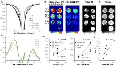 |
176 | T1-compesated quantification of NOE and amide for brain tumor patients using a QUEST-like 10-point fitting
Chu Wang1, Chuyu Liu1, Benqi Zhao2, Zhuozhao Zheng2, and Xiaolei Song1
1center for biomedical imaging research, school of medicine, Tsinghua Univisity, Beijing, China, 2Beijing Tsinghua Changgung Hospital, Beijing, China Keywords: Quantitative Imaging, CEST & MT CEST MRI is a promising molecular imaging technique, but the in vivo quantification inherently faces challenges because of multiple types of signal contaminations. Herein we introduced a QUEST-like 10-point fitting method for simultaneous NOE and amide quantification at 3T with reduced T1 contamination. The quantification method was evaluated by simulations and phantom experiments. Furthermore, for glioblastoma patients, the highlighted regions on fitted amide images are consistent with the hyperintensities on Gd-T1W maps, which is a smaller enhanced area than those on conventional APTw images. The initial results on tumor patients indicate its potential for clinical use. |
|
1814.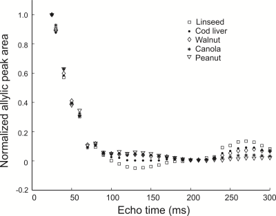 |
177 | Effect of Omega-3 Fatty Acid Content on Allylic Proton T2 at 9.4 T
Adam Carscadden1, Nathaniel Bly1, Anthony G. Tessier1,2, Catherine J. Field3, and Atiyah Yahya1,2
1Department of Oncology, University of Alberta, Edmonton, AB, Canada, 2Department of Medical Physics, Cross Cancer Institute, Edmonton, AB, Canada, 3Department of Agricultural, Food and Nutritional Science, University of Alberta, Edmonton, AB, Canada Keywords: Quantitative Imaging, Spectroscopy, fat quantification In this work at 9.4 T, it is demonstrated that the apparent T2 (includes losses due to J-coupling) relaxation times of the allylic fat protons (≈ 2.1 ppm) is correlated with higher ω-3 content. J-coupling evolution appears to refocus with a PRESS TE of 270 ms rendering it a suitable TE for obtaining T2 estimates. Experiments were performed on oils of varying ω-3 content and preliminary in-vivo experiments were performed on two mice, each fed a different fat diet. |
|
1815.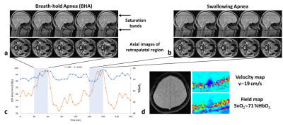 |
178 | Concurrent mapping of brain metabolism and airway architecture during respiration and volitional apneas
Michael C Langham1, Jing Xu1, and Felix Wehrli1
1Radiology, University of Pennsylvania, Philadelphia, PA, United States Keywords: Quantitative Imaging, Quantitative Imaging, Obstructive sleep apnea Obstructive sleep apnea is associated with changes in upper airway morphology and sleep disturbances from transient nocturnal hypoxemia and hypoxia. The latter adversely affects brain metabolism and neurologic function. MRI is the only non-invasive modality cable of concurrently visualize airway architecture and quantify neurometabolism during sleep. We present an interleaved pulse sequence and demonstrate its ability to detect metabolic response and airway anatomic alteration from a swallowing apnea (SA). Although airway anatomy during SA is distinctively different from a breath-hold apnea both result in similar physiological responses in terms of cerebral blood flow and venous blood oxygenation. |
|
1816. |
179 | Amide proton transfer-weighted imaging for hepatocellular carcinoma: Correlation with Ki-67 labeling index
Jingcheng Huang1, Qingqing Wen2, Weiqiang Dou2, and Xianfu Luo1
1Clinical Medical School of Yangzhou University, Northern Jiangsu People’s Hospital, Yangzhou, China, Yangzhou City, China, 2GE Healthcare, MR Research China, Beijing, P.R. China, Beijing City, China Keywords: Quantitative Imaging, CEST & MT, APTw Conventional MRI methods are difficult to reveal histologic and molecular characteristic heterogeneity of HCC. In this study, we aimed to investigate the relationship between APTw and Ki-67 labeling index (LI) in hepatocellular carcinoma (HCC) to evaluate whether APTw can reflect the pathological information of HCC at the molecular level. It was found that APTw values were significantly different between low and high Ki-67 LI in HCC, and the AUC to distinguish high and low Ki-67 LI was 0.794, indicating APTw imaging might be a potential molecular technique for predicting the malignancy grade of HCC |
|
1817. |
180 | Quantification of APT and NOE(-3.5) using chemical exchange saturation transfer MRI with double saturation powers (DSP-CEST)
Yu Zhao1 and Zhongliang Zu1
1Vanderbilt University Medical Center, Nashville, TN, United States Keywords: Quantitative Imaging, CEST & MT In this study, we proposed a new data-postprocessing method to specifically quantify the APT and NOE effects based on two canonical CEST MRI acquisitions with double saturation powers (DSP). First, Numerical simulations based on Bloch equations is used to demonstrate that the proposed method can detect the APT and NOE effects with a removal of background signals from the direct water saturation (DS) and the semi-solid magnetization transfer (MT) and the CEST signals that originate from the fast-exchange pools. Then, an in vivo validation of the proposed method is conducted using an animal tumor model at a 4.7-T scanner. |
|
 Back to Meeting Home
 Back to the Program-at-a-Glance
The International Society for Magnetic Resonance in Medicine is accredited by the Accreditation Council for Continuing Medical Education to provide continuing medical education for physicians.
 Back to Meeting Home
 Back to the Program-at-a-Glance
 View Presentation Video