Digital Poster
The Bench: Preclinical Studies Throughout the Body
ISMRM & ISMRT Annual Meeting & Exhibition • 03-08 June 2023 • Toronto, ON, Canada

Digital Poster
The Bench: Preclinical Studies Throughout the Body
| Computer # | |||
|---|---|---|---|
3740.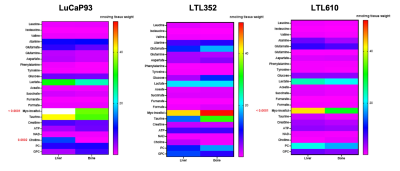 |
161 |
Metabolic similarity of prostate cancer patient-derived
xenografts propagated in the bone versus liver
Deepti Upadhyay1,
Shubhangi Agarwal1,
Jinny Sun1,
Robert A Bok1,
Rahul Aggarwal2,
Donna M Peehl1,
John Kurhanewicz1,
and Renuka Sriram1
1Radiology and Biomedicla Imaging, University of California, San francisco, San Francisco, CA, United States, 2University of California, San francisco, San Francisco, CA, United States Keywords: Cancer, Metabolism, Models, metastasis, tumor, microenvironment Metabolic plasticity due to cell intrinsic properties, its inherent dependence on organ of origin as well as its interaction with microenvironmental factors are considered key to establishment of metastases and is believed to be heterogenous. This in turn has a significant impact and differential response to the treatment of choice making this a lethal disease. Here, we investigate using magnetic resonance imaging and spectroscopy the biochemical profile of the same patient derived xenografts of small cell neuroendocrine prostate cancer, an aggressive phenotype, implanted in two different sites, liver and bone that are associated with reduced survival. |
|
 |
3741.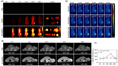 |
162 |
manganese-based nanosystem for dual-modality imaging guided
STING pathway with immunotherapy against triple-negative breast
cancer
Xiuhong Guan1,
Yue Zhao1,
Xin Huang2,
Zhiyong Wang3,
and Ruimeng Yang4
1Department of Radiology, First Affiliated Hospital of Jinan University, Guangzhou, China, 2Guangzhou Medical University, Guangzhou, China, 3School of Materials Science and Engineering, Sun Yat-sen University, Guangzhou, China, 4Department of Radiology, Guangzhou First People’s Hospital, Guangzhou, China Keywords: Molecular Imaging, Cancer, tumor, dual-modality imaging, STING pathway, Immunotherapy Due to the high incidence and mortality rate, breast cancer has become the major cause of cancer death among females. This study reports a kind of functional nanocomposite, which was designed to comprises Pluronic F127 to self-assembly manganese carbonyl (MnCO) and IR780 dyes into one system, exhibiting NIR-II and MR dual-modal imaging guided STING pathway and anti-tumor immunity against triple-negative breast cancer. |
3742.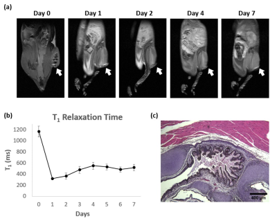 |
163 |
A Novel MRI Platform for Long-term Stem Cell Tracking In Vivo
Keyu Zhuang1,2,
Rocco Romagnuolo3,
Daniel A. Szulc1,2,
Hai-Ying Mary Cheng4,5,
Michael A. Laflamme3,6,7,
and Hai-Ling Margaret Cheng1,2,8
1Institute of Biomedical Engineering, University of Toronto, Toronto, ON, Canada, 2Translational Biology and Engineering Program, Ted Rogers Centre for Heart Research, Toronto, ON, Canada, 3McEwen Stem Cell Institute, University Health Network, Toronto, ON, Canada, 4Department of Biology, University of Toronto Mississauga, Mississauga, ON, Canada, 5Department of Cell and Systems Biology, University of Toronto, Toronto, ON, Canada, 6Peter Munk Cardiac Centre, University Health Network, Toronto, ON, Canada, 7Department of Laboratory Medicine and Pathobiology, University of Toronto, Toronto, ON, Canada, 8The Edward S. Rogers Sr. Department of Electrical and Computer Engineering, University of Toronto, Toronto, ON, Canada Keywords: Molecular Imaging, Cell Tracking & Reporter Genes, stem cell
To extend our previously reported bright-ferritin cell
tracking platform to monitoring stem cell therapy, we
investigated tracking human embryonic stem cells
transplanted intramuscularly in immune-compromised mice.
With this technology, the stem cells impart a bright,
T1-induced contrast. In-vitro assays of viability and |
|
3743.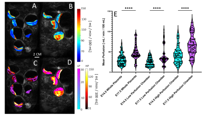 |
164 |
Modeling Mouse Placental Perfusion Dynamics and Contractions via
Dynamic Contrast Enhancement MRI
Devin Raine Everalo Cortes1,2,3,4,
Margaret C. Stapleton2,4,
Samuel Wyman2,4,
Kristina E. Schwab5,6,
Noah W. Coulson2,7,
Dalton R West2,4,
Thomas Becker-Szurszewski5,6,
Sean Hartwick5,6,
Anthony G. Christodoulou8,
and Yijen L Wu2,3,4,9
1Department of Bioengineering, University of Pittsburgh, Pittsburgh, PA, United States, 2Department of Developmental Biology, School of Medicine, University of Pittsburgh, Pittsburgh, PA, United States, 3Department of Bioengineering, Swanson School of Engineering, University of Pittsburgh, Pittsburgh, PA, United States, 4Rangos Research Center Small Animal Imaging Core, Children's Hospital of Pittsburgh of UPMC, Pittsburgh, PA, United States, 5Rangos Research Center Animal Imaging Core, Children's Hospital of Pittsburgh of UPMC, Pittsburgh, PA, United States, 6Department of Pediatrics, University of Pittsburgh, School of Medicine, Pittsburgh, PA, United States, 7Rangos Research Center Small Animal Imaging Core, University of Pittsburgh, Pittsburgh, PA, United States, 8Biomedical Imaging Research Institue, Cedars-Sinai Medical Center, Los Angeles, CA, United States, 9Department of Pediatrics, School of Medicine, University of Pittsburgh, Pittsburgh, PA, United States Keywords: Contrast Agent, Placenta, Perfusion, PCA, FASD, DCE Placental development is a dynamic process, and the timing of placental development events is crucial for appropriate fetal development. However, the placenta itself remains critically understudied in the context of fetal development disorders. We propose novel models for correlating DCE-MRI perfusion estimation with biologically relevant compartments and extracting dynamic biomarkers for placental development. We validate this model by demonstrating that placental perfusion is disrupted in a murine model of peri-conceptual alcohol exposure, and that this disruption occurs differently across various compartments from E14.5 to E17.5. |
|
3744.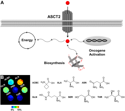 |
165 |
ALAw-CEST MRI of the ASCT2 transporter in a mouse model of
prostate cancer
Behnaz Ghaemi1,
Yuguo Li2,
Martin Pomper1,
Jeff Bulte1,
Peter van Zijl2,
and Aline Thomas1
1Johns Hopkins University, Baltimore, MD, United States, 2Kennedy Krieger Institute, Baltimore, MD, United States Keywords: Cancer, Preclinical, Prostate Cancer Increased utilization of glutamine is characteristic of many advanced, aggressive cancers. Established glutamine imaging strategies require the use of labeled probes (positron emission tomography, hyperpolarized spectroscopy) that can limit their accessibility. Here, we investigated the utility of chemical exchange saturation transfer (CEST) MRI upon intravenous injection of unlabeled alanine to monitor differences in cellular glutamine uptake as a potentially complementary imaging biomarker for profiling cancers and monitoring their progression. |
|
3745.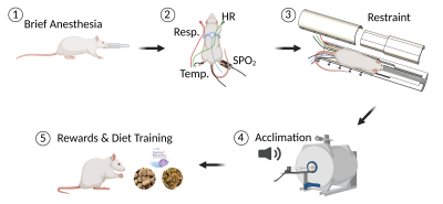 |
166 |
Gastrointestinal MRI with Conscious Rats
Xiaokai Wang1,
Angela Yee1,
Jiayue Cao1,
Ashley Cornett1,
Fatimah Alkaabi1,
Yushi She1,
Ulrich Scheven1,
and Zhongming Liu1
1University of Michigan, Ann Arbor, MI, United States Keywords: Data Acquisition, Animals Anesthesia has been required for preclinical gastrointestinal MRI but may confound gastric physiology. This study demonstrates, for the first time, the feasibility of gastrointestinal MRI with awake rats, and reports the effects of anesthesia on gastric motor function. Results suggest that awake rats show greater rates of gastric emptying and intestinal filling, and stronger gastric and intestinal motility relative to those anesthetized. Anesthesia with 2.5% isoflurane can entirely stop muscle contraction. The methods and findings reported herein lay the foundation for using awake GI-MRI in preclinical studies of gastrointestinal function or dysfunction. |
|
3746.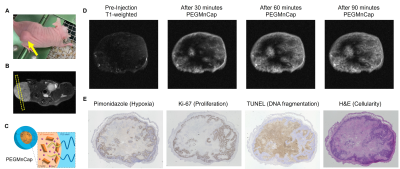 |
167 |
Distinct contrast patterns in patient-derived tumor xenografts
(PDX) mouse model revealed by pH-sensitive MR contrast agent
Akira Sumiyoshi1,
Takahiro Nomoto2,
Megumi Iiyama1,
Kensuke Osada1,
Rumiana Bakalova1,
Nobuhiro Nishiyama2,
and Ichio Aoki1
1National Institutes for Quantum Science and Technology, Chiba, Japan, 2Tokyo Institute of Technology, Yokohama, Japan Keywords: Molecular Imaging, Cancer Here we performed a molecular MR imaging study on patient-derived tumor xenografts (PDX) mouse model. In vivo molecular MR imaging demonstrated that proliferative vs. non-proliferative regions exist in PDX model and highly specific contrast agent such as PEGMnCap (pH-responsive nanoparticle based on Mn) can delineate active and hypoxic regions while small-molecular size contrast agent such as Gd-DOTA (gadoterate meglumine) can delineate non-active tumor regions. These MR image separations were not clearly obtained with a cancer cell line, suggesting more complex microenvironment of PDX model and may provide valid translational feedbacks for clinical practice. |
|
3747.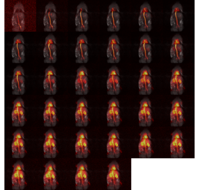 |
168 |
Rat angiography from far away: design and performance of a small
bath cryostat with NMR capability for transport of
hyperpolarized samples.
Andrea Capozzi1,2,
Yupeng Zhao3,
Magnus Karlsson3,
Esben Søvsø Szocska Hansen4,
Lotte Bonde Bertelsen4,
Christoffer Laustsen4,
Mathilde Hauge Lerche3,
and Jan Henrik Ardenkjær-Larsen3
1IPHYS, EPFL, Lausanne, Switzerland, 2Health Tech, Technical University of Denamrk, Kgs. Lyngby, Denmark, 3Health Tech, Technical University of Denmark, Kgs. Lyngby, Denmark, 4Aarhus University Hospital, Aarhus, Denmark Keywords: New Devices, Hyperpolarized MR (Non-Gas), transportable hyperpolarization Hyperpolarization via dissolution Dynamic Nuclear Polarization has the potential to revolutionize diagnostic radiology. Nevertheless, the methodology struggles to enter everyday clinical practice. One of the reasons why broad consensus among clinicians is still missing lies in the technical complexity that characterize hyperpolarization via dDNP. Differently from PET, hyperpolarized MR contrast agent cannot be transported and have to be prepared on-site. We developed a robust methodology to change this paradigm. We herein present our latest updates on transportable hyperpolarization technology. |
|
3748.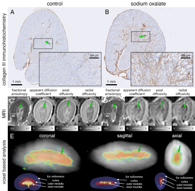 |
169 |
Voxel-based analysis of renal fibrosis with diffusion MRI in
mouse models
Rohan S Virgincar1,
Shimrit Avraham2,
Kai H Barck1,
Joshua Webster3,
Jeffrey Hung3,
Aaron K Wong4,
Hans D Brightbill4,
Patrick Caplazi3,
Bill Forrest5,
Laura Bell6,
Man Kin Choy1,
Andrey Shaw2,
Alex J de Crespigny6,
and Luke Xie1
1Translational Imaging, Genentech, South San Francisco, CA, United States, 2Research Biology, Genentech, South San Francisco, CA, United States, 3Research Pathology, Genentech, South San Francisco, CA, United States, 4Translational Immunology, Genentech, South San Francisco, CA, United States, 5Bioinformatics, Genentech, South San Francisco, CA, United States, 6Clinical Imaging Group, Genentech, South San Francisco, CA, United States Keywords: Kidney, Diffusion Tensor Imaging Voxel-based analysis (VBA) can enable voxel-wise statistical analysis of control and disease images without the need for user-defined regions-of-interest (ROI), but has not been widely used beyond neuroimaging. We demonstrate that VBA can be successfully applied to mouse models of renal fibrosis. Diffusion MRI parametric maps of control and disease groups were registered non-linearly to a common coordinate space and VBA successfully identified distinct regions of fibrosis in two different models, which strongly corresponded to whole-kidney histology. VBA therefore has potential in the clinic to identify regions of fibrosis, where whole-kidney histology cannot be obtained. |
|
3749.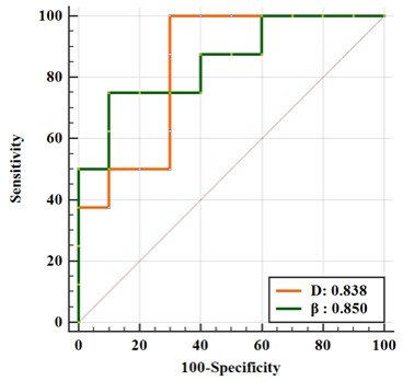 |
170 |
Fractional order calculus model in liver cancer with
immunohistochemical indexes CK19 and Hep par-1: a preliminary
animal study
Jinhuan Xie1,
Liling Long1,
Chenhui Li1,
and Huiting Zhang2
1The First Affiliated Hospital of Guangxi Medical University, Nanning, China, 2Siemens Healthineers, Wuhan, China Keywords: Liver, Diffusion/other diffusion imaging techniques The purpose of this study was to explore the feasibility of preoperative non-invasive evaluation of immunohistochemical indexes of hepatocellular carcinoma, CK19 and Hep par-1 positive, with Fractional Order Calculus (FROC) model using nude mice model. Results showed that the diffusion coefficient D and fractional order derivative in space β had statistically significant differences and good diagnostic performances between the CK19 and Hep par-1 positive groups. These finding showed that FROC is useful diffusion model and D and β are useful biomarkers in liver cancer with expression of the immunohistochemical indexes CK19 and Hep par-1. |
|
3750.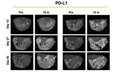 |
171 |
MR Molecular Imaging of Pancreatic Cancer Reveals Differences in
Tumor Microenvironment during Therapeutic Monitoring with
Immunotherapy
Victoria Laney1 and
Zheng-Rong Lu1
1Case Western Reserve University, Cleveland, OH, United States Keywords: Molecular Imaging, Cancer PDAC is a highly aggressive malignant cancer and the 3rd leading cause of cancer related deaths in the US. The aggressive nature of PDAC is in part due to the desmoplastic tumor microenvironment, which promotes rapid tumor growth and impedes therapeutic efficacy of conventional therapies. Immune checkpoint inhibitors have showed specific efficacy in PDAC promotes extracellular remodeling for effective treatment of aggressive tumors. In this study we investigated the ability of an MR molecular imaging technology for image guidance and therapeutic monitoring of PD-L1 treated PDAC tumors. |
|
3751.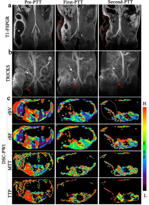 |
172 |
Dynamic MR Imaging-guided Photothermal Therapy of Solid Tumor
and Monitoring of Vascular Invasion
Rong Zhang1 and
Zhang Hua1
1Department of Radiology, Affiliated Hospital of Qingdao University, Qingdao, China Keywords: Molecular Imaging, DSC & DCE Perfusion, Magnetic resonance angiography Timely evaluating the abundance of vessels and infiltration by noninvasive ways together with synchronous photothermal therapy (PTT) still faces great challenges. A multifunctional and biocompatible nanoprobes (NaBiF4: Gd@PDA@PEG) with high longitudinal relaxation rate and photothermal conversion efficiency was designed as promising photosensitizer for tumor PTT and contrast agent for MR imaging. The anatomic decrease and thinning of tumor blood vessels could be observed in real-time using noninvasive MR angiography ( TRICKS and DSC-PWI) during PTT. We demonstrated that the vascular invasion status of tumor during PTT can be monitored by utilizing our multifunctional nanoprobes, which provide possibility for clinical transformation. |
|
3752.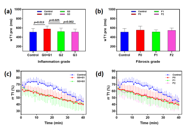 |
173 |
Can dynamic gadoxetic acid-enhanced MRI combined with water
specific T1 mapping predict the early-stage of Nonalcoholic
Steatohepatitis in rat?
Qian Wan1,
Hao Peng1,
Feng Liu2,
Chuanli Cheng1,
Xiaoyi Liu2,
Changjun Tie1,
Xin Liu1,
Yi Wang2,
and Chao Zou1
1Shenzhen Institute of Advanced Technology, Chinese Academy of Sciences, Shenzhen, China, 2Peking University People's Hospital, Beijing, China Keywords: Contrast Agent, Liver The early detection of the inflammation and fibrosis grade are important to Nonalcoholic steatohepatitis (NASH) patients. Gadoxetic acid with MRI T1 quantitation is one of promising noninvasive method. However, intracellular hepatocyte lipid is a confounding factor for the accurate T1 quantitation in NASH case. Here, we used a water specific T1 (wT1) mapping with dynamic gadoxetic acid enhanced MRI in a rat NASH model induced by MCD diet. The results demonstrated that gadoxetic acid was not helpful to differentiate the inflammation and fibrosis severity in the early stage of NASH in rats. |
|
3753.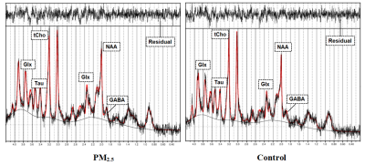 |
174 |
1H-MRS reveals changes of metabolites in the olfactory bulb of
PM2.5-exposed mice
Ren Li1,
Yong Zhang2,
and Xiao-Yong Zhang1
1Institute of Science and Technology for Brain-Inspired Intelligence, Fudan University, 200433, Shanghai, China, 2GE. Healthcare, shanghai, China Keywords: Spectroscopy, Quantitative Imaging, PM2.5 PM2.5 has been shown to be harmful to the central nervous system of mammals. Long term exposure to PM2.5 may cause changes in brain neural metabolites. However, there were few magnetic resonance spectroscopy (MRS) studies in rodents under PM2.5 exposure. In this work, we developed an air enrichment system to concentrate PM2.5. Using 1H-MRS, we successfully observed the brain metabolic profile of olfactory bulb in PM2.5-exposed mice, and found the concentration changes of some key metabolites in the brain, indicating that PM2.5 has a non-negligible impact on brain metabolism. |
|
3754.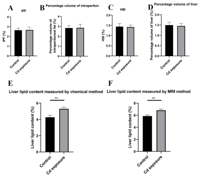 |
175 |
Three-dimensional CUBE MR imaging accurately quantified lipid
accumulation in reflection of longitudinal fish health
Dong Liu1,
Lei Pan2,
Weiyin Vivian Liu3,
and Wenzhen Zhu1
1Department of Radiology, Tongji Hosptial of Tongji Medical College of Huazhong University of Science and Technology, Wuhan, China, 2Faculty of Resources and Environmental Science, Hubei University, Wuhan, China, 3MR Research, GE Healthcare, Wuhan, China Keywords: Liver, Animals, assess fish health Quantitative methods of fish lipid accumulation have attracted a lot of attention in ecotoxicology. Traditionally, invasive approaches were time-consuming and generally required animal sacrifice, drastically restricting applicability in ecotoxicology. In this study, a noninvasive MRI approach was proposed to quantify lipid accumulation in two represented fish species with IDEAL-IQ and Cube-Flex-T2 sequences. We demonstrated the accuracy of our novel MRI approach in quantifying fish lipid accumulation. In addition, we conducted cadmium exposure experiment and observed no significant differences in lipid deposition between MRI and traditional methods. Thus, this MRI method could have a wide range of applications in ecotoxicological research. |
|
3755.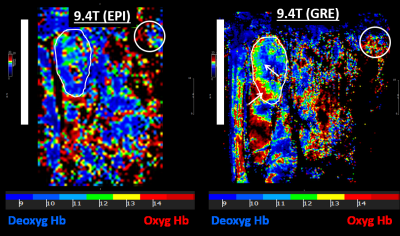 |
176 |
Optimized metabolic imaging of rat’s kidney on clinical MRI
scanners. Comparison to small-animal high-field imaging
El-Sayed H. Ibrahim1,
Abdul Parchur1,
Srividya Kidambi1,
Mingyu Liang1,
and Allen Cowley1
1Medical College of Wisconsin, Milwaukee, WI, United States Keywords: Kidney, Hypertension T2*-based blood oxygenation level-dependent (BOLD) MRI has been used to investigate renal metabolism in the rat’s kidney, which is typically conducted on high-field small-animal MRI scanners. However, T2* is very small at high-field strength, e.g., 9.4T, which results in borderline signal-to-noise ratio and questions the measurements accuracy. In this study, we investigated the capability of BOLD imaging of the rat’s kidney on a clinical 3T MRI scanner, where T2* is much higher, and compared the results to those acquired on 9.4T small-animal scanner. The results showed the capability for distinguishing different oxygenation levels in the kidney on clinical 3T scanners. |
|
3756.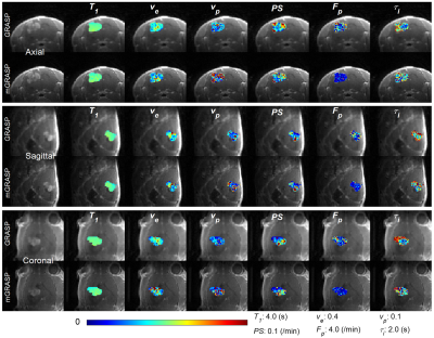 |
177 |
Contrast Kinetic Model-based GRASP Reconstruction for Small
Animal Imaging at 7T
Jin Zhang1,
Sawwal Qayyum1,
Jonghyun Bae1,
Eddy Solomon1,
and Gene Kim1
1Weill Cornell Medical College, New York, NY, United States Keywords: Image Reconstruction, DSC & DCE Perfusion In this study, we propose a contrast kinetic model-based GRASP (mGRASP) method which can improve the spatial image quality as well as temporal characteristics of DCE-MRI. The proposed mGRASP method was assessed with 9L rat glioma model and GL261 mouse glioma model in an Active Contrast Encoding (ACE)-MRI experiment which simultaneously measures the pre-contrast T1 and contrast kinetic model parameters including intracellular water lifetime. The results show that mGRASP provides improved image quality and temporal patterns compared to GRASP. |
|
3757.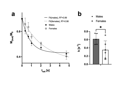 |
178 |
Assessment of mouse brain creatine kinase rate using 31P-MRS at
7 Tesla
Antoine Cherix1,
Mohamed Tachount1,
and Jason Lerch1,2
1Nuffield Department of Clinical Neurosciences, University of Oxford, Oxford, United Kingdom, 2Mouse Imaging Centre (MICe), Hospital for Sick Children, Toronto, ON, Canada Keywords: Spectroscopy, Animals 31P-MRS experiments in mouse brain are scarce and often limited to ultra-high field scanners. We have recently reported that 31P-MRS is feasible in mouse brain at 7T as well. We now tested the potential of using saturation transfer experiments in the same setting to measure the creatine kinase rate in mouse brain. |
|
3758.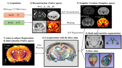 |
179 |
In vivo T1-mapping template of the mouse brain at 4.7T and 7T
with a MP2RAGE sequence
Elise Cosenza1,2,
Aurélien Trotier1,
Gauthier Massone1,
Emeline J Ribot1,
Laurent Petit2,
Sylvain Miraux1,
and Valery Ozenne1
1CNRS, CRMSB, Bordeaux, France, 2CNRS, GIN IMN, Bordeaux, France Keywords: Data Analysis, Animals, brain parcellation 3D T1 templates of the mouse brain at 7T and 4.7T were created with a MP2RAGE pulse sequence. The template was next coregistered with the DORR atlas. The resulting parcellation allows volume and T1 measurements for each labels. The values obtained can be compared according to the magnetic field, intra and inter individual. Volumes were found in agreement with the literature. The T1 measurements are reproducible between individuals and significantly different at 4.7T and 7T. |
|
3759.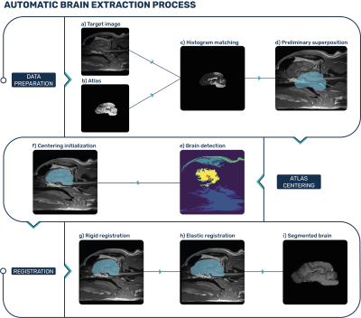 |
180 |
Automatic brain extraction on multi-contrast dogs and cats MRI
images
Jamil Nour Eddin1,
Hugo Dorez1,
and Valentina Curcio1
1HawkCell, Marcy-l'Étoile, France Keywords: Segmentation, Animals Brain extraction in an MRI image is the first pre-processing step of a neuroimaging quantification pipeline. Once the brain is extracted, further post-processing becomes faster, more specific, and easier to implement and interpret. Existing brain extraction tools are mostly adapted to work on the human anatomy, or on specific animal species. This gives poor results when applied to animal brain images. We developed an atlas-based Veterinary Images Brain Extraction (VIBE) algorithm that is fully automatized and was successfully tested on different cats and dogs breeds, 3D and 2D MRI images, different contrasts (T1-weighted, T2-weighted and T2 FLAIR) and acquisition planes. |
|
 Back to Meeting Home
 Back to the Program-at-a-Glance
The International Society for Magnetic Resonance in Medicine is accredited by the Accreditation Council for Continuing Medical Education to provide continuing medical education for physicians.
 Back to Meeting Home
 Back to the Program-at-a-Glance
 View Presentation Video