Digital Poster
The Bench: Preclinical Studies Within the Brain
ISMRM & ISMRT Annual Meeting & Exhibition • 03-08 June 2023 • Toronto, ON, Canada

Digital Poster
The Bench: Preclinical Studies Within the Brain
| Computer # | |||
|---|---|---|---|
 |
3914.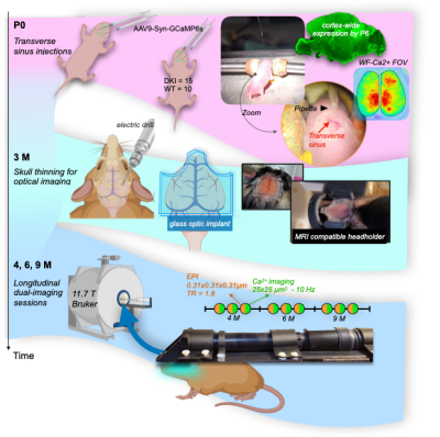 |
161 |
Longitudinal simultaneous fMRI and mesoscale calcium imaging in
a mouse model of Alzheimer’s disease
Francesca Mandino1,
Xilin Shen2,
Gabriel Desrosiers-Gregoire3,
David O'Connor4,
Bandhan Mukherjee5,
Kristin DeLuca5,
Ali Hamodi4,
Ashley Owens5,
Yonghyun Ha1,
An Qu1,
John Onofrey4,
Xenophon Papademetris4,
Mallar Chakravarty3,
Michael C. Crair4,
Stephen M Strittmatter5,
and Evelyn Lake1
1Radiology, Yale University, New Haven, CT, United States, 2Radiology and bioimaging sciences, Yale University, New Haven, CT, United States, 3Douglas Mental Health University Institute, McGill, Montreal, QC, Canada, 4Yale University, New Haven, CT, United States, 5Neurology and Neuroscience, Yale University, New Haven, CT, United States Keywords: Alzheimer's Disease, Brain Multimodal neuroimaging plays an active role in understanding clinically accessible biomarkers of health and disease. Using simultaneous mesoscopic calcium imaging and BOLD-fMRI, we examine spontaneous activity in a GCaMP-positive mouse model of Alzheimer’s disease (AD)—longitudinally—at 4, 6, and 9 months. We find that both imaging modalities show early wide-spread changes in connectivity in AD compared to controls, with diverging trends at 6months. Intriguingly, these neuroimaging changes precede typical behaviour deficits. Further, preliminary evidence of cross-modal uncoupling hints at a complex relationship between excitatory neural activity (calcium imaging data), and the BOLD signal that is affected by AD-related progression. |
 |
3915.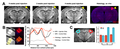 |
162 |
MRI detection of gene expression in the mammalian brain using
the Manganese transporter Zip14
Harikrishna Rallapalli1,
Eleanor C McCall1,
Nadia Bouraoud1,
and Alan P Koretsky1
1Laboratory of Functional and Molecular Imaging, NIH/NINDS, Bethesda, MD, United States Keywords: Molecular Imaging, Cell Tracking & Reporter Genes, Brain, Neuroscience, Cell-type Imaging, Brain connectivity We report the use of Zip14, a Manganese transporter, as an MRI-visible gene expression reporter system. Mice were intracranially injected into various brain areas with AAVs carrying a plasmid construct of Synapsin promoter for neuronal-specific expression, Zip14, and histological marker. MRI was performed immediately after, 2 weeks after, and 4 weeks after injection. Significant, large magnitude hyperintensity was observed 2 and 4 weeks after injection, but not immediately after, at the injection site and areas projecting away or towards the injection site. This gene expression reporter system is viable without the use of additional contrast agents or non-standard imaging protocols. |
|
|
3916.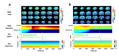 |
163 |
Identifying spreading depression during coma induction with
multi-modal fMRI
Yuanyuan Jiang1,
Zeping Xie1,
Wenchao Yang1,
Bei Zhang1,
David Hike1,
Andy Liu1,2,
Brian L. Edlow1,3,
and Xin Yu1
1Martinos Center for Biomedical Imaging, Massachusetts General Hospital, Charlestown, MA, United States, 2Department of Neuroscience, Boston University, Boston, MA, United States, 3Center for Neurotechnology and Neurorecovery, Department of Neurology, Massachusetts General Hospital, Boston, MA, United States Keywords: Stroke, Ischemia, coma, spreading depression We have demonstrated spreading depression as a key event contributing to coma induction following the ET-1 injection to the brainstem of rats. Multi-modal fMRI enables the whole brain functional mapping to present the spatial dynamic changes during coma induction, in particular, paired with the incidence of SD events. |
3917. |
164 |
Therapeutic response assessment to IDH inhibitor using
hyperpolarized [5-12C-1-13C]α-ketoglutarate MRSI in rat models
of low-grade glioma
Donghyun Hong1,
Yaewon Kim1,
Chandrasekhar Mushti2,
Noriaki Minami1,
Jing Wu2,
Murali Krishna Cherukuri2,
Rolf E. Swenson3,
Daniel B Vigneron1,
and Sabrina Ronen1,4
1Department of Radiology and Biomedical Imaging, University of California, San Francisco, San Francisco, CA, United States, 2National Cancer Institute, NIH, Bethesda, MD, United States, 3National Heart, Lung, and Blood Institute, NIH, Bethesda, MD, United States, 4Brain Tumor Research Center, University of California, San Francisco, San Francisco, CA, United States Keywords: Hyperpolarized MR (Non-Gas), Treatment, Tumor, Preclinical, Non-proton, Spectroscopy Hyperpolarized [1-13C]α-ketoglutarate is useful for monitoring 2HG and glutamate production but has limited SNR and the [5-13C]α-ketoglutarate natural abundance peak is within 0.1ppm of 2HG. To address these challenges, this study investigated the utility of HP [5-12C-1-13C]α-ketoglutarate to monitor 2HG and glutamate production as biomarkers of response to treatment with the mutant IDH inhibitor BAY-1436032 in an in vivo rat glioma model using an optimized acquisition strategy. We were able to clearly monitor the dynamic localized production of both 2HG and glutamate in vivo in real-time, and early metabolic changes predicted enhanced survival. |
|
3918.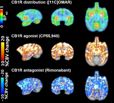 |
165 |
Agonism/antagonism on cannabinoid 1 receptor reveal opposite
cerebral blood volume change: a simultaneous PET/fMRI study on
primates
Chi-Hyeon Yoo1 and
Hsiao-Ying Wey1
1Athinoula A. Martinos Center for Biomedical Imaging, Boston, MA, United States Keywords: Molecular Imaging, Drug Development The goal of this study was to detect the changes of cerebral blood volume (CBV) in the non-human primate brain induced by administration of agonist and antagonist of cannabinoid receptor 1 (CB1R) using simultaneous positron emission tomography (PET) and function magnetic resonance imaging (fMRI). After the antagonist injection, a bi-phasic response, fast increase and prolonged decrease of CBV, were identified throughout the brain, while CB1R agonist administration evoked elevation of CBV. Future studies integrating PET-derived receptor occupancy information with the CBV changes will provide deeper understanding on CB1R and brain response. |
|
3919. |
166 |
First neuroimaging experience at 11.7 Tesla on the anaesthetized
non-human primate.
Fawzi Boumezbeur1,
Alexis Amadon1,
Marion Gay1,
Franck Mauconduit1,
Vincent Gras1,
Maxime Roustan1,
Frederic Lepretre1,
Aurélien Massire2,
Cécile Rabrait-Lerman1,
Sebastien Mériaux1,
Alexandre Vignaud1,
and Nicolas Boulant1
1NeuroSpin, CEA, Gif-sur-Yvette, France, 2Siemens Healthcare SAS, Saint-Denis, France Keywords: Bioeffects & Magnetic Fields, High-Field MRI After more than 20 years of research and development, the 11.7T Iseult MRI scanner is in the last stage of its commissioning. However, authorization of Human MRI examination at this unprecedented magnetic field strength is still pending. In this context, MRI examinations of healthy anaesthetized non-human primates are being carried out to check on the absence of acute physiological effects following exposure and gain some insights into the opportunities and challenges ahead. |
|
3920. |
167 |
Development and application of dynamic MRSI of an HP
neuroprotective agent in an MCAO mouse model of ischemic stroke
at 14.1 T
Thanh Phong Lê1,2,
Lara Buscemi3,
Mario Lepore4,
Andrea Capozzi1,5,
Lorenz Hirt3,
Mor Mishkovsky1,
and Jean-Noël Hyacinthe2,6
1Laboratory of Functional and Metabolic Imaging, École polytechnique fédérale de Lausanne (EPFL), Lausanne, Switzerland, 2Geneva School of Health Sciences, HES-SO University of Applied Sciences and Arts Western Switzerland, Geneva, Switzerland, 3Department of Clinical Neurosciences, Lausanne University Hospital (CHUV), Lausanne, Switzerland, 4CIBM Center for Biomedical Imaging, École polytechnique fédérale de Lausanne (EPFL), Lausanne, Switzerland, 5Department of Health Technology, Center for Hyperpolarization in Magnetic Resonance, Technical University of Denmark, Kgs Lyngby, Denmark, 6Image Guided Intervention Laboratory, University of Geneva (UNIGE), Geneva, Switzerland Keywords: Hyperpolarized MR (Non-Gas), Preclinical, Molecular Imaging Advanced acquisition hardware and sequences boosting the spatiotemporal resolution overcome the existing limitations of HP metabolic MRI in animal disease models, providing new contrasts and opening up new perspectives. Herein, we designed a 1H/13C volume/surface-combined head coil providing full mouse brain coverage with high sensitivity, as well as a high-efficiency acquisition scheme to achieve superior spatial and temporal resolution images. This setup was employed for time-resolved imaging of the cerebral metabolic response to a neuroprotective bolus of hyperpolarized pyruvate in a transient hypoxia/ischemia mouse model. The experiments suggest a distinct metabolism between the ischemic and healthy tissues. |
|
3921.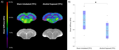 |
168 |
Exercise and Environmental Complexity Superintervention Improves
Mechanical Property Recovery in FASD Rodents Measured via MRE
L. Tyler Williams1,
Ian F. Smith2,
Katrina A. Milbocker2,
Diego A. Caban-Rivera1,
Samuel Kurtz3,4,
Matthew D. J. McGarry5,
Elijah E. W. Van Houten4,
Anna Y. Klintsova2,
and Curtis L. Johnson1,2
1Dept. of Biomedical Engineering, University of Delaware, Newark, DE, United States, 2Dept. of Psychological & Brain Sciences, University of Delaware, Newark, DE, United States, 3Laboratorie de Mécanique et Génie Civil, CNRS, Université de Montpellier, Montpellier, France, 4Département de Génie Mécanique, Université de Sherbrooke, Sherbrooke, QC, Canada, 5Thayer School of Engineering, Dartmouth College, Hanover, NH, United States Keywords: Elastography, Preclinical In this longitudinal study, we investigate the effects of fetal alcohol exposure on mechanical properties in the developing rodent brain using magnetic resonance elastography (MRE). Additionally, we test whether behavioral “superintervention,” consisting of combined exposure to wheel running followed by environmental complexity, can mitigate changes to property measures. We found that alcohol exposed rats exhibit lower stiffness than sham intubated rats at baseline but recover stiffness to near baseline levels after superintervention. Furthermore, we confirmed MRE is sensitive to small property variations in rodents which may reflect myelination and microstructural integrity. |
|
3922.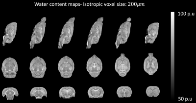 |
169 |
In vivo measurement of rat brain water content at 9.4T using
super-resolution reconstruction
Dennis C. Thomas1,2,
Ana-Maria Oros-Peusquens*1,
Michael Schöneck1,
Antje Willuweit1,
Zaheer Abbas1,
Markus Zimmermann1,
Jörg Felder1,
Avdo Celik1,
and N. Jon Shah1,3,4,5
1Institute of Neuroscience and Medicine-4, Forschungszentrum, Jülich, Germany, 2Institute of Neuroradiology, University Hospital Frankfurt, Frankfurt, Germany, 3Institute of Neuroscience and Medicine 11, INM-11, JARA, Forschungszentrum Jülich,, Jülich, Germany, 4JARA - BRAIN - Translational Medicine, Aachen, Germany, Aachen, Germany, 5RWTH Aachen University, Aachen, Germany Keywords: Quantitative Imaging, Neurofluids, Super-resolution reconstruction, High-field MRI, ex vivo Animal models have an indisputable role in the investigation of brain pathology. Investigating water content in vivo could lead to a better understanding of pathogenesis and hence, a robust technique to measure water content using MRI would be very beneficial. Here, we adapt a super-resolution-based technique, previously developed for humans, to the rat brain and report in vivo high-resolution (200μm isotropic) water content maps, obtained using a 9.4T MRI scanner. High resolution, isotropic water content maps of the rat brain are demonstrated. The water content values obtained using the proposed MR technique are compared with ex vivo wet/dry methods. |
|
3923.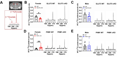 |
170 |
Hyperpolarized [1-13C]pyruvate detects neuronal metabolic
impairment in glucose transporter and pyruvate kinase deficient
mice
Caroline Guglielmetti1,2,
Huihui Li3,
Yoshitaka Sei3,
Lydia Le Page1,2,
Lauren Schields3,4,
Brice Tiret1,2,
Xiao Gao1,2,5,
Iris Lo3,
Talya Dayton6,
Jeffrey Rathmell7,
Matthew Vander Heiden6,8,
Ken Nakamura3,4,9,10,
and Myriam Chaumeil1,2,4,5
1Department of Physical Therapy and Rehabilitation Science, University of California San Francisco, San Francisco, CA, United States, 2Department of Radiology and Biomedical Imaging, University of California San Francisco, San Francisco, CA, United States, 3Gladstone Institute of Neurological Disease, Gladstone Institutes, San Francisco, CA, United States, 4Graduate Program in Biomedical Sciences, University of California San Francisco, San Francisco, CA, United States, 5UCSF/UCB Graduate Program in Bioengineering, University of California San Francisco, San Francisco, CA, United States, 6Koch Institute for Integrative Cancer Research and the Department of Biology, Massachusetts Institute of Technology, Boston, MA, United States, 7Department of Pathology, Microbiology, and Immunology, Vanderbilt Center for Immunobiology, Nashville, TN, United States, 8Dana-Farber Cancer Institute, Boston, MA, United States, 9Department of Neurology, University of California San Francisco, San Francisco, CA, United States, 10Graduate Program in Neuroscience, University of California San Francisco, San Francisco, CA, United States Keywords: Hyperpolarized MR (Non-Gas), Neuro We generated mice with deletion of the glucose transporter 3 (GLUT3cKO) or pyruvate kinase 1 (PKM1cKO) in CA1 hippocampal neurons. GLUT3cKO and PKM1cKO mice showed memory impairment. Hyperpolarized (HP) 13C magnetic resonance spectroscopic imaging showed that female, but not male, PKM1cKO mice had increased HP [1-13C]pyruvate-to-lactate conversion, while female GLUT3cKO mice had decreased conversion and brain volume, evaluated by T2-MRI. Fluorine-18-fluorodeoxyglucose ([18F]-FDG) positron emission tomography imaging did not detect changes, highlighting HP [1-13C]pyruvate’s potential to detect downstream alterations in brain glucose metabolism. Altogether, our findings demonstrated that neurons metabolize glucose through glycolysis in vivo, and require glycolysis for normal function. |
|
3924.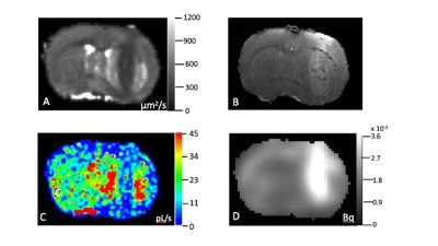 |
171 |
Metabolic Activity Diffusion Imaging [MADI] of Rat Brain Glioma
Joshua Schlegel1,
Eric Baker1,
Samantha Holland2,
Jared Stoller3,
William Packwood4,
Xin Li1,
Ramon Barajas3,
Charles Springer1,
and Martin Pike1
1AIRC, OHSU, Portland, OR, United States, 2Neurology, OHSU, Portland, OR, United States, 3Diagnostic Radiology, OHSU, Portland, OR, United States, 4Research Cores and Shared Resources, OHSU, Portland, OR, United States Keywords: Molecular Imaging, Metabolism, Cancer We compared the novel DWI MRI-based metabolic activity [MA] imaging approach [MADI] to 18FDG-PET, employing an in vivo rat glioma model. The MADI-derived parameter, kio*V product, is proportional to Na+/K+ ATPase [NKA] activity, the primary cellular energy consumer. The kioV decreased within the tumor vs. contralateral while 18FDG uptake increased. This is consistent with the Warburg effect in glioma, which activates glucose uptake but also switches energy production from mitochondrial oxidative phosphorylation to glycolysis, thus reducing overall ATP production and NKA activity. MADI expands DWI MRI utility, providing a novel, noninvasive MA imaging method, with greater resolution than PET.
|
|
3925.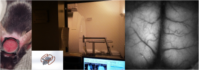 |
172 |
Feasibility of simultaneous fMRI and optical imaging for
head-fixed freely-behaving mice
Wen-Ju Pan1,
Harrison Watters1,
Lisa Meyer-Baese1,
Annabelle Singer1,
and Shella Keilholz1
1Emory University/Georgia Institute of Technology, Atlanta, GA, United States Keywords: Multimodal, Brain For simultaneous optical imaging and fMRI with freely-behaving mice, head-fixed without body restriction in minimal stress awake state, optimizations were conducted on multiple aspects, including multi-wavelength optical imaging system for both optical intrinsic signals and extrinsic fluorescence studies of genetically encoded calcium indicators (GECI) and voltage indicators (GEVI) mice with tube lens design instead of imaging fiber bundle, transparent cranial window development, integrated quadrature coil in head holder, and the designed belt treadmill/ virtual reality (VR) system to fit a customized rodent cradle in the commonly used 12cm ID gradient for the small animal MRI system. |
|
3926.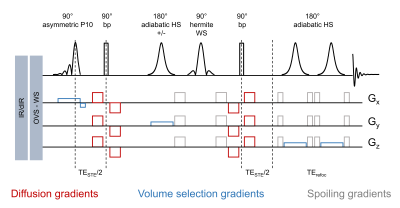 |
173 |
Improved glutamine detection using diffusion-weighted SPECIAL at
14.1T
Jessie Mosso1,2,3,
Dunja Simicic2,3,
Bernard Lanz2,3,
Rolf Gruetter1,
and Cristina Cudalbu2,3
1LIFMET, EPFL, Lausanne, Switzerland, 2CIBM Center for Biomedical Imaging, Lausanne, Switzerland, 3Animal Imaging and Technology (AIT), EPFL, Lausanne, Switzerland Keywords: Spectroscopy, Diffusion/other diffusion imaging techniques, brain, preclinical, sequence design Brain glutamine (Gln) is a key biomarker of hepatic encephalopathy (HE). The estimation of its diffusion properties with diffusion-weighted MRS is of high interest in the field but remains challenging due to its low concentration. We propose a new diffusion-weighted MRS sequence, DW-SPECIAL, which enables to reach shorter echo times and is thus beneficial for strongly J-coupled metabolites such as Gln. DW-SPECIAL reduces the uncertainty in Gln diffusion decays and the standard deviation across animals for Gln diffusivity in a homogenous control population and paves the way for an in-depth study of Gln diffusion properties in HE. |
|
3927.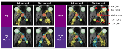 |
174 |
9 crossings per 1 bend: Tractography of contra- and ipsilateral
connections in mouse optic pathways improved with
ODF-Fingerprinting
Patryk Filipiak1,
Thajunnisa A. Sajitha2,
Timothy Shepherd1,
Kamri Clarke1,
Dimitris G. Placantonakis3,
Jiangyang Zhang1,
Kevin C. Chan2,
Fernando E. Boada4,
and Steven H. Baete1
1Center for Advanced Imaging Innovation and Research (CAI2R), Department of Radiology, NYU Langone Health, New York, NY, United States, 2NeuroImaging and Visual Science Laboratory, Departments of Ophthalmology and Radiology, NYU Langone Health, New York, NY, United States, 3Department of Neurosurgery, Perlmutter Cancer Center, Neuroscience Institute, Kimmel Center for Stem Cell Biology, NYU Langone Health, New York, NY, United States, 4Radiological Sciences Laboratory and Molecular Imaging Program at Stanford, Department of Radiology, Stanford University, Stanford, CA, United States Keywords: Image Reconstruction, White Matter, tractography, optic pathways, optic tract, optic chiasm, crossing fibers Reconstruction of rodent optic pathways is particularly challenging for dMRI tractography due to the relatively small size of the crossing area in the optic chiasm and the unbalanced proportion between the contra- and ipsilateral axonal links. In this study, we replace the commonly used Orientation Distribution Function peak finding approach with our dictionary-based technique called ODF-Fingerprinting to improve the reconstruction of crossing fibers and thus correct the proportion of tracts exiting the optic chiasm. Our results from in vivo diffusion-weighted images of 18 mice show significant improvement (p<0.05) in many cases, helping decrease the discrepancies between reconstruction and gold-standard histology. |
|
3928.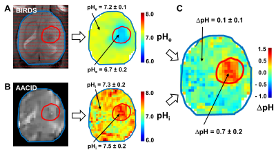 |
175 |
Imaging the transmembrane pH gradient in gliomas
Sandeep Kumar Mishra1,
Jelena Mihailović1,
Fahmeed Hyder1,2,
and Daniel Coman1
1Radiology & Biomedical Imaging, Yale University, New Haven, CT, United States, 2Biomedical Engineering, Yale University, New Haven, CT, United States Keywords: Molecular Imaging, Cancer, transmembrane pH Extracellular acidosis in relation to intracellular milieu is a unique feature of the tumor microenvironment. The difference between intracellular pH (pHi) and extracellular pH (pHe) is much larger in tumors than normal tissue. Measuring the transmembrane pH gradient (ΔpH=pHi–pHe) could provide a tool for assessing tumor aggressiveness, monitoring treatment efficacy, guiding localized drug delivery, and understanding tumor responsiveness. This work establishes transmembrane pH gradient imaging in brain tumors. We observed a significantly higher transmembrane pH gradient in RG2 tumors compared to normal brain. Decreasing transmembrane pH gradient may serve as a functional biomarker for positive therapeutic outcome. |
|
3929.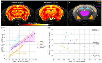 |
176 |
Mapping the effect of oxytocin on the mouse brain using Q2TIPS
arterial spin labelling and BOLD fMRI at 9.4T
Eugene Kim1,
Davide di Censo1,
Zhuoni Li2,
Eilidh MacNicol1,
Alexandra Hertz2,
Michael Craig2,
Diana Cash1,
and Marija M Petrinovic2
1Department of Neuroimaging, King's College London, London, United Kingdom, 2Department of Forensic and Neurodevelopmental Sciences, King's College London, London, United Kingdom Keywords: Brain Connectivity, Arterial spin labelling, pharmacological MRI Currently, standard Bruker protocols for arterial spin labelling (ASL) in mice are limited to single-slice methods. Here, we implemented multi-slice Q2TIPS ASL on a Bruker 9.4T scanner for whole-brain mapping of cerebral blood flow (CBF) in mice. There was good agreement between Q2TIPS and the standard Bruker FAIR ASL. Pharmacological MRI using Q2TIPS ASL and BOLD fMRI revealed that systemic administration of a behaviorally relevant dose of oxytocin caused a global decrease in CBF and increase in functional connectivity in healthy, wildtype mice. These opposing effects corroborate a recent concept of mutual exclusivity between neuronal activity and functional connectivity. |
|
3930. |
177 |
A water-exchange dynamic contrast-enhanced MRI protocol
optimized for AQP4 measurement in glioma
Guangxu Han1,
Yinhang Jia1,
Zejun Wang1,
Yi-Cheng Hsu2,
and Ruiliang Bai3,4
1Key Laboratory of Biomedical Engineering of Ministry of Education, College of Biomedical Engineering and Instrument Science, Zhejiang University, Hangzhou, China, 2MR Collaboration, Siemens Healthcare, Shanghai, China, 3Zhejiang University School of Medicine, Hangzhou, China, 4College of Biomedical Engineering and Instrument Science, Zhejiang University, Hangzhou, China Keywords: Contrast Agent, Tumor A dynamic contrast-enhanced MRI (DCE-MRI) protocol was optimized to measure transmembrane water exchange rate (kio), which was correlated kio with aquaporin-4 (AQP4) immunohistochemistry results in a rodent subcutaneous glioma model. From the acquired results, a significant correlation was demonstrated between the optimized kio and AQP4, indicating that kio measured by DCE-MRI could reveal the expression of AQP4.
|
|
3931.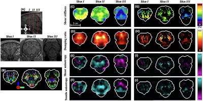 |
178 |
Mechanical Stiffness and Anisotropy Measured by MR Elastography
during Brain Development in the Minipig
Shuaihu Wang1,
Charlotte A Guertler1,
Ruth J Okamoto1,
Curtis L Johnson2,
Matthew DJ McGarry3,
and Philip V Bayly1
1Washington University in St. Louis, St. Louis, MO, United States, 2University of Delaware, Newark, DE, United States, 3Dartmouth College, Hanover, NH, United States Keywords: Elastography, Tissue Characterization The relationship between brain development and mechanical properties of brain tissue is important but remains incompletely understood. Here we use data from magnetic resonance elastography (MRE) and diffusion tensor imaging (DTI) to estimate anisotropic mechanical properties in six female Yucatan minipigs at ages from 3 to 6 months with a transversely- isotropic nonlinear inversion (TI-NLI) algorithm. Our results show that white matter is more dissipative and anisotropic than gray matter, and reveal effects of brain development on brain stiffness and structural anisotropy. Changes in brain mechanical properties may be a fundamental biophysical signature of brain development. |
|
3932.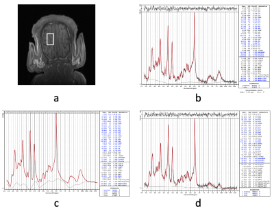 |
179 |
Bias-Reduced LCModel Quantification of 1H Brain MRS with
Baseline-and-MM Removal: Application in a Circulatory Arrest
Study using Neonatal Pigs
Meng Gu1,
Ralph Hurd1,
and Daniel Spielman1
1Radiology, Stanford University, Stanford, CA, United States Keywords: Data Processing, Spectroscopy, Accurate Quantification Quantification of in-vivo short-TE MRS using LCModel suffers from biases due to varying baseline and macro-molecule components, especially for metabolites with coupled resonances. This problem exacerbates at low SNR as the baseline estimated becomes less accurate. To improve quantification, baseline and macro-molecule components were estimated from a high SNR line-broadened spectrum and then subtracted from the original spectrum. Using the baseline and macro-molecule removed spectrum, significantly reduced biases for Gln/tCr and GABA/tCr were achieved at different SNRs. Using this method, LCModel quantification revealed a more prominent trend of increasing Gln/tCr and increased GABA/tCr during circulatory arrest in neonatal pig brain. |
|
3933.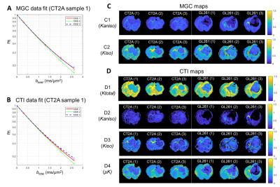 |
180 |
Correlation Tensor Imaging Enhances Histological Differences
Between Mouse Glioblastoma Subtypes
Rafael Neto Henriques1,
Rui V Simões1,2,
Sara Monteiro1,3,
Andrada Ianuş1,
Tânia Carvalho1,
Sune Jespersen4,5,
and Noam Shemesh1
1Champalimaud Research, Champalimaud Foundation, Lisbon, Portugal, 2Institute for Research & Innovation in Health (i3S), University of Porto, Porto, Portugal, 3Institute for Systems and Robotics, Department of Bioengineering, Instituto Superior Técnico, Universidade de Lisboa, Lisboa, Portugal, 4Center of Functionally Integrative Neuroscience (CFIN) and MINDLab, Department of Clinical Medicine, Aarhus University, Aarhus, Denmark, 5Department of Physics and Astronomy, Aarhus University, Aarhus, Denmark Keywords: Diffusion/other diffusion imaging techniques, Diffusion/other diffusion imaging techniques Correlation Tensor Imaging (CTI) has been recently proposed as a novel methodology for resolving kurtosis sources without relying on strong multi-gaussian component assumptions. Here, CTI is harnessed to decipher kurtosis sources in two different mouse glioblastoma subtypes. Our results reveal that CTI’s kurtosis source separation resolves the underlying microstructural differences between the glioblastoma subtypes and faithfully represents their specific histopathological features. In comparison to previous diffusion MRI approaches, CTI kurtosis maps present enhanced sensitivity towards tumor differences. These first results show the potential of CTI in providing more sensitive and specific future biomarkers for monitoring tumor progression and therapy outcome. |
|
 Back to Meeting Home
 Back to the Program-at-a-Glance
The International Society for Magnetic Resonance in Medicine is accredited by the Accreditation Council for Continuing Medical Education to provide continuing medical education for physicians.
 Back to Meeting Home
 Back to the Program-at-a-Glance
 View Presentation Video