Digital Poster
Deep Learning Image Reconstruction II
ISMRM & ISMRT Annual Meeting & Exhibition • 03-08 June 2023 • Toronto, ON, Canada

Digital Poster
Deep Learning Image Reconstruction II
| Computer # | |||
|---|---|---|---|
3096.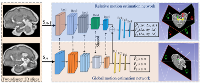 |
41 |
Geometric Constrained Deep Learning for Motion Correction of
Fetal Brain MR Images
Laifa Ma1,2,
Liangjun Chen1,
Fenqiang Zhao1,
Zhengwang Wu1,
Li Wang1,
Weili Lin1,
He Zhang3,
Kenli Lin2,
and Gang Li1
1University of North Carolina at Chapel Hill, Chapel Hill, NC, United States, 2Hunan University, Changsha, China, 3Fudan University, Shanghai, China Keywords: Image Reconstruction, Brain Robust motion correction of fetal brain MRI slices is crucial for fetal brain volume reconstruction. However, conventional methods can only handle a limited range of motion. Hence, a deep learning model based on prior geometric constraints is proposed to predict the motion of 2D slices. It consists of a global and a relative motion estimation network. Sharing features between two networks make the model to learn more unique feature representations for global motion correction. Moreover, we present a control point-based approach to simulate complex fetal motion trajectories. The experimental results demonstrate that the proposed method is effective and efficient. |
|
3097.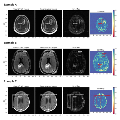 |
42 |
Effect of pathology on quantitative metrics of image
reconstruction using a deep learning-based brain MRI
reconstruction model
Shengjia Chen1,
Patricia Johnson1,
and Yvonne W. Lui1
1Department of Radiology, New York University Langone Health, New York, NY, United States Keywords: Image Reconstruction, Brain We evaluate image quality in brain MR images with pathology, reconstructed by a deep learning-based image reconstruction algorithm. We have two main contributions: 1) a procedure for evaluating the image reconstruction quality of images, both globally and in patches with labelled pathology, and 2) report quantitative differences between two groups of reconstructed images (abnormal vs. normal). The pathology evaluation results find pathology regions have more losses and lower structural similarity when compared to normal patches and entire normal brains. |
|
3098.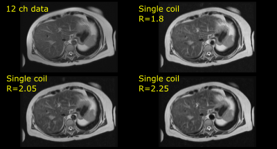 |
43 |
Accelerated acquisition and DL reconstruction to enable single
shot fast spin echo (SSFSE) imaging of diagnostic quality using
single coil
Sudhanya Chatterjee1,
Florintina C1,
Rohan Patil1,
Sajith Rajamani1,
Rajagopalan Sundaresan1,
Uday Patil1,
Preetham Shankapal1,
Suresh Emmanuel Joel1,
Ramesh Venkatesan1,
and Harsh Agarwal1
1GE Healthcare, Bangalore, India Keywords: Image Reconstruction, Image Reconstruction, artificial intelligence, abdomen, SSFSE Single shot fast spin echo (SSFSE) is a popular imaging approach for acquisition of high-resolution MR images in motion sensitive areas such as abdomen. In certain clinical settings, use of multiple coils setup for acquisition is not feasible (such as abdominal scans for obese subjects in non-wide bore MRI scanners). SSFSE imaging with single coil using the popular partial Fourier approach only presents risk of excessive blurring in the images. In this study we present a method to enable SSFSE T2 imaging using single coil. Proposed method is evaluated on prospectively accelerated data. |
|
3099.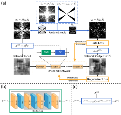 |
44 |
SSMo-QSM: A self-supervised learning method for model-based
quantitative susceptibility mapping reconstruction
Jie Feng1,
Ming Zhang1,
Ruimin Feng1,
and Hongjiang Wei1
1School of Biomedical Engineering, Shanghai Jiao Tong University, Shanghai, China Keywords: Image Reconstruction, Quantitative Susceptibility mapping This study, SSMo-QSM, introduced a self-supervised strategy to achieve a model-based deep learning QSM reconstruction for dealing with the imperfect ground truth in supervised learning methods. In SSMo-QSM, the direct frequency domain division results between phase and dipole kernel at untruncated areas of TKD are randomly separated into two subsets. One is used as the data consistency of the unrolled network and the other is used to define the loss function for training, respectively. The preliminary results of synthetic data suggested that SSMo-QSM performed comparably against supervised methods on accurate susceptibility mapping with suppressed streaking artifact. |
|
3100.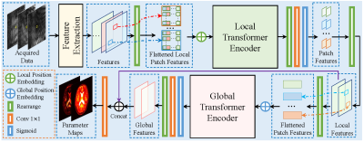 |
45 |
Deep Magnetic Resonance Fingerprinting Based on local and global
vision transformer
Peng Li1,
Xiaodi Li1,
Xin Lu2,
and Yue Hu1
1Harbin Institute of Technology, Harbin, China, 2De Montfort University, Leicester, United Kingdom Keywords: Image Reconstruction, MR Fingerprinting Magnetic resonance fingerprinting (MRF) can achieve simultaneous imaging of multiple tissue parameters. However, the size of the tissue fingerprint dictionary used in MRF grows exponentially as the number of tissue parameters increases, which may result in prohibitively large dictionaries that require extensive computational resources. Existing CNN-based methods obtain parameter reconstruction patch-wisely, using only local information and resulting in limited reconstruction speed. In this paper, we propose a novel end-to-end local and global vision transformer (LG-VIT) for MRF parameter reconstruction. The proposed method enables significantly fast and accurate end-to-end parameter reconstruction while avoiding the high computational cost of high-dimensional data. |
|
3101.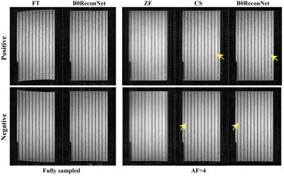 |
46 |
B0 Inhomogeneity Distortion Corrected Image Reconstruction with
Deep Learning on An Open Bore MRI-Linac
Shanshan Shan1,2,
Yang Gao3,4,
Meng Ma5,
Hongping Gan5,
David Waddington2,
Brendan Whelan2,
Paul Liu2,
Chunyi Liu1,
Mingyuan Gao1,
and Feng Liu4
1Center for Molecular Imaging and Nuclear Medicine, State Key Laboratory of Radiation Medicine and Protection,School for Radiological and Interdisciplinary Sciences (RAD-X), Soochow University, Suzhou, China, 2ACRF Image X Institute, Faculty of Medicine and Health, The University of Sydney, Sydney, Australia, 3School of Computer Science and Engineering, Central South University, Changsha, China, 4School of Information Technology and Electrical Engineering, University of Queensland, Brisbane, Australia, 5School of Software, Northwestern Polytechnical University, Suzhou, China Keywords: Image Reconstruction, Brain MRI-Linac systems require real-time anatomical images with high geometric fidelity to localize and track tumours during radiotherapy treatments. Image distortions caused by B0 field inhomogeneity and slow MR acquisition hinder the application of real-time MRI-guided radiotherapy. Here, we develop and investigate a deep learning-based reconstruction pipeline to reconstruct B0 inhomogeneity distortion-corrected images (B0ReconNet) directly from k-space. MR acceleration techniques such as compressed sensing (CS) were integrated into B0ReconNet to further reduce acquisition time. Simulated and experimental data with fully sampled and retrospectively subsampled acquisitions on a 1T open bore MRI-Linac were used to validate the proposed method. |
|
3102.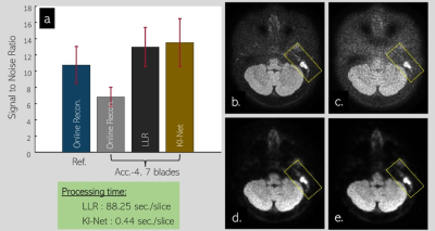 |
47 |
Accelerated Propeller FSE-DWI with Unrolled Deep Learning
Reconstruction at 1.5T Clinical MRI
Uten Yarach1,
Atita Suwannasak1,
and Prapatsorn Sangpin2
1Radiologic Technology, Chiang Mai University, Chiang Mai, Thailand, 2Philips Healthcare (Thailand), Bangkok, Thailand Keywords: Image Reconstruction, Brain Fast spin echo diffusion magnetic resonance imaging (FSE-DWI) is often referred to as a standard for MRI diagnosis of Cholesteatoma. However, the acquired data require multiple steps during image reconstruction which turn out high residual artifacts. In this work, we develop rapid reconstruction for propeller FSE-DWI to improve its signal-to-noise ratio (SNR) through unrolled deep learning (DL) framework. Results show that the proposed unrolled DL reconstruction enables increasing bout 2x SNR compared to SNR obtained by online reconstructed images. Moreover, its speed is about 200x faster than conventional locally low rank constraint reconstruction. |
|
3103.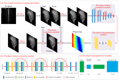 |
48 |
Unsupervised model for removing Nyquist/motion artifacts in
SPatiotemporal Encoding MRI
Qingjia Bao1,
Liyang Xia2,
Kewen Liu2,
Xinjie Liu1,
Peng Sun3,
Lucio Frydman4,
and Chaoyang Liu1
1Innovation Academy for Precision Measurement Science and Technology, Wuhan, China, 2Wuhan University of Technology, Wuhan, China, 3Philips Healthcare, Beijing, China, 4Weizmann Institute of Science, Rehovot, Israel Keywords: Image Reconstruction, Susceptibility Although a major advantage of SPEN vs EPI is a higher immunity to artifacts, it suffers from Nyquist or motion artifacts. We proposed a new unsupervised CNN model that takes advantage of both physical model and Deep learning. The model consists of three parts: phase feature extraction module, which can extract the phase features of even/odd phase differences or motion-caused phase differences in multi-shot echo data. Then, the phase maps are generated with these phase difference features. Lastly, the phase correction modules to remove artifacts. The results show that the proposed model can effectively correct Nyquist/motion artifacts in single-shot/multi-shot SPEN. |
|
3104.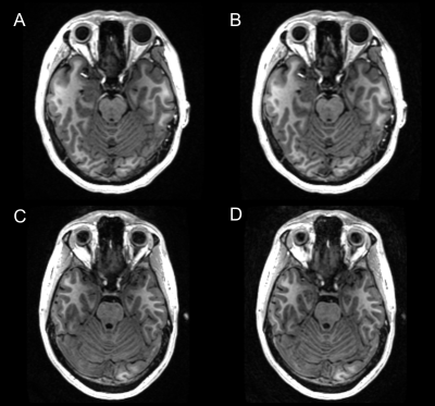 |
49 |
Task-based evaluation of deep learning-based reconstruction for
highly-accelerated 3D T1-weighted brain MRI scans
Sangtae Ahn1,
Chitresh Bhushan1,
John Huston2,
J. Kevin DeMarco3,
Robert Y. Shih3,4,
Joshua D. Trzasko2,
Rafi Brada5,
Graeme Mckinnon6,
Isabelle Heukensfeldt Jansen1,
Dan Rettmann7,
Brian Burns8,
Ty A. Cashen6,
Nir Mazor5,
Xucheng Zhu8,
and Thomas K. Foo1
1GE Research, Niskayuna, NY, United States, 2Mayo Clinic College of Medicine, Rochester, MN, United States, 3Walter Reed National Military Medical Center, Bethesda, MD, United States, 4Uniformed Services University of the Health Sciences, Bethesda, MD, United States, 5GE Research, Herzliya, Israel, 6GE HealthCare, Waukesha, WI, United States, 7GE HealthCare, Rochester, MN, United States, 8GE HealthCare, Menlo Park, CA, United States Keywords: Image Reconstruction, Machine Learning/Artificial Intelligence, Brain, Neuro 3D MRI enables thin slices at the cost of long scan times, causing practical challenges. Recently, deep-learning (DL) techniques have successfully accelerated MR scans. However, it is challenging to characterize the image quality (IQ) performance of DL methods by conventional metrics because IQ depends on applications, i.e., how images are used. We evaluate the IQ performance of DL-Speed, our DL-based acceleration method, for 3D T1-weighted MPRAGE brain scans, in 1) post-reconstruction subcortical structure segmentation, and 2) a reader study. The results imply DL-Speed can accelerate scans with reduction factor R=10 while maintaining IQ comparable to standard parallel imaging with R=2.1. |
|
3105.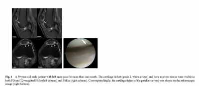 |
50 |
Improved diagnostic efficacy on structural abnormalities of knee
using high-resolution deep learning-based 2D FSE images
Xiaxia Wu 1,
Weiyin Vivian Liu2,
and Yunfei Zha1
1Renmin Hospital of Wuhan University, Wuhan, China, 2GE Healthcare,MR Reaearch China,Beijing, Wuhan, China Keywords: Image Reconstruction, Cartilage Diagnostic performance was limited to image resolution and contrast between target tissues and surrounding tissues. A rapid knee imaging has been perused but no loss of image quality is critical. This study proposed a rapid knee imaging based on two-dimensional fast spin echo sequence and examined the reliability and diagnostic performance of deep learning-based reconstruction T1-, T2- and PD- weighted images on knee joint pathology via comparison of images with and without deep-learning reconstruction algorithm (DLR). Diagnostic efficacy on knee structural abnormalities of 2D DLR FSE sequence elevated using knee arthroscopy results as the gold standard. |
|
3106.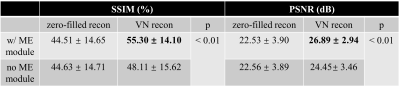 |
51 |
Motion-mitigated reconstruction of accelerated MRI by using an
unfolded variational network
Zijian Zhou1,2,
Haikun Qi1,2,
and Peng Hu1,2
1School of Biomedical Engineering, ShanghaiTech University, Shanghai, China, 2Shanghai Clinical Research and Trial Center, Shanghai, China Keywords: Image Reconstruction, Motion Correction Motion-mitigated reconstruction of highly undersampled MRI was achieved by adding a motion estimation module to the data consistency part of the model-based unfolded variation network. The motion estimation module consisted of a pair of convolutional blocks with residual inputs and added only limited number of trainable parameters to the network. The network was trained and tested on synthesized motion-corrupted images from a publicly available knee dataset. The reconstructed images with the proposed motion estimation module were sharper, and details were better recovered, with the structural similarity and peak signal-to-noise ratio significantly improved. |
|
3107.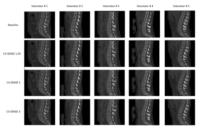 |
52 |
Does CS-SENSE acceleration influence the performance of an AI
based synthetic CT algorithm? A volunteer study in the lumbar
spine.
Yulia Shcherbakova1,
Tijl A van der Velden1,2,
and Peter R Seevinck1,2
1Imaging Division, UMC Utrecht, Utrecht, Netherlands, 2MRIguidance B.V., Utrecht, Netherlands Keywords: Image Reconstruction, Skeletal, AI, acceleration, CS-SENSE, spine, sCT, bone In this work, we investigated the performance of an AI based algorithm - synthetic CT generation - when subjected to compressed sensing-sensitivity encoding (CS-SENSE) accelerated gradient echo images. We performed MR experiments in five volunteers, using different CS-SENSE acceleration factors for the MR acquisitions. Our results showed that using CS-SENSE factors of 1.45 and 2 increased noise in the MR source images but did not compromise the sCT reconstruction on visual inspection, which was confirmed by quantitative metrics. However, CS-SENSE with a factor of 3 caused artifacts in the sCT images which may affect the safety and diagnostic performance of the product. |
|
3108.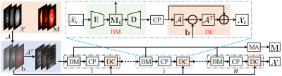 |
53 |
Learned Tensor Low-CP-Rank and Bloch response manifold priors
for Non-Cartesian MRF Reconstruction
Peng Li1,
Xiaodi Li1,
Xin Lu2,
and Yue Hu1
1Harbin Institute of Technology, Harbin, China, 2De Montfort University, Leicester, United Kingdom Keywords: Image Reconstruction, MR Fingerprinting, Tensor Low-rank, CP Decomposition, Bloch Response Manifold, non-Cartesian We propose a deep unrolled network for non-Cartesian MRF reconstruction by unrolling the MRF reconstruction model regularized by the tensor low-rank and the Bloch resonance manifold priors. To avoid computationally burdensome singular value decomposition, we propose a learned CP decomposition module to exploit the tensor low-rank priors of MRF data. Inspired by the MRF imaging mechanism, we also propose a Bloch response manifold module to learn the mapping between reconstructed MRF data and the multiple parameter maps. Numerical experiments show that the proposed network can improve the reconstruction quality of MRF data and multi-parameter maps within significantly reduced computational time. |
|
3109.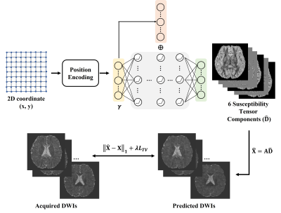 |
54 |
DTI-Net: Unsupervised Diffusion Tensor Reconstruction Using
Implicit Neural Representation
Yuting Shi1,
Yuyao Zhang2,
and Hongjiang Wei1
1Shanghai Jiao Tong University, Shanghai, China, 2ShanghaiTech University, Shanghai, China Keywords: Image Reconstruction, Machine Learning/Artificial Intelligence Diffusion tensor imaging (DTI) needs a large number of diffusion-weighted images (DWIs) to reliably reconstruct the diffusion measurements of the brain white matter, making the data acquisition time-consuming. Deep learning has emerged as a powerful technique to reduce the number of acquired DWIs. While most existing deep learning methods are supervised and need high-quality ground truth data as the training labels. Here, we proposed an unsupervised and subject-specific DTI reconstruction method called DTI-Net to significantly reduce the required number of DWIs, while also can simultaneously conduct the super-resolution reconstruction of the tensors. |
|
3110.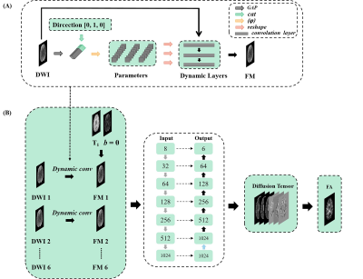 |
55 |
High efficient DTI reconstruction network with flexible
diffusion directions
Zejun Wu1,
Jiechao Wang1,
Zunquan Chen1,
Zhigang Wu2,
Jianfeng Bao3,
Shuhui Cai1,
and Congbo Cai1
1Department of Electronic Science, Xiamen University, Xiamen, China, 2Philips Healthcare, Shenzhen, China, 3the First Affiliated Hospital of Zhengzhou University, Zhengzhou, China Keywords: Image Reconstruction, Diffusion Tensor Imaging Deep learning has been used in diffusion tensor imaging (DTI) to fast reconstruct diffusion parameters. However, diffusion-weighted images (DWIs) as network input must maintain diffusion gradient direction consistency during training and testing for deep-learning-based DTI parameter mapping. A dynamic-convolution-based network was developed to achieve generalized DTI parameter mapping for flexible diffusion gradient directions. This proposed method uses dynamic convolution kernels to embed diffusion gradient direction information into feature maps of the corresponding diffusion signal. The results indicate that the proposed method can reconstruct high-quality DTI-derived maps from six diffusion gradient directions. |
|
3111.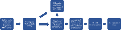 |
56 |
GROG Gridding using modified VGG-16 CNN model for Non-Cartesian
MR Image Reconstruction
Muhammad Atif1,
Madiha Arshad1,
Yumna Bilal1,
Omair Inam1,
Hassan Shahzad2,
and Hammad Omer1
1Medical Image Processing Research Group (MIPRG), Department of Electrical and Computer Engineering, COMSATS University, Islamabad, Pakistan, 2National Centre of Physics (NCP), Islamabad, Pakistan Keywords: Image Reconstruction, Image Reconstruction Self-Calibrating GROG (SC-GROG) is a gridding algorithm that maps the k-space MRI data from non-Cartesian to Cartesian domain. The main limitation of SC-GROG is its computational cost to calculate the GROG weights. This paper proposes a customized deep learning framework (based on VGG-16 CNN model) to calculate the 2D-Gridding weight sets for SC-GROG. Initially, the proposed model is trained on human head images, and later fine-tuning is performed using Golden-angle radial Liver Perfusion datasets. The results show that the proposed method significantly reduces the computation time for the estimation of GROG weights while maintaining the image quality. |
|
3112.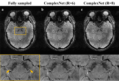 |
57 |
Accelerating 7T susceptibility-weighted imaging with
complex-valued convolutional neural network
Caohui Duan1,
Xiangbing Bian1,
Kun Cheng1,
Xiaoyu Wang1,
Jinhao Lyu1,
Xueyang Wang1,
Jianxun Qu2,
Xin Zhou3,
and Xin Lou1
1Department of Radiology, Chinese PLA General Hospital, Beijing, China, 2MR Collaboration, Siemens Healthineers Ltd., Beijing, China, 3Key Laboratory of Magnetic Resonance in Biological Systems, State Key Laboratory of Magnetic Resonance and Atomic and Molecular Physics, National Center for Magnetic Resonance in Wuhan, Wuhan Institute of Physics and Mathematics, Innovation Academy for Precision Measurement Science and Technology, Chinese Academy of Sciences‒Wuhan National Laboratory for Optoelectronics, Wuhan, China Keywords: Image Reconstruction, Machine Learning/Artificial Intelligence Ultra-high field 7T susceptibility-weighted imaging (SWI) has shown great potential in visualizing and evaluating a broad range of pathology, but suffers from long acquisition times. In this study, a complex-valued convolutional neural network (ComplexNet) model was proposed to reconstruct highly accelerated 7T SWI data. The average reconstruction time of ComplexNet was 0.56 seconds per slice (45.16 seconds per participant). Meanwhile, ComplexNet can provide high-quality 7T SWI for visualizing subtle pathology, including cerebral microbleeds, asymmetric deep medullary veins, and swallow tail sign. |
|
3113. |
58 |
Parallel non-Cartesian Spatial-Temporal Dictionary Learning
Neural Networks (stDLNN) for Accelerating Dynamic MRI
Zhijun Wang1,
Huajun She1,
and Yiping P. Du1
1School of Biomedical Engineering, Shanghai Jiao Tong University, Shanghai, China Keywords: Image Reconstruction, Machine Learning/Artificial Intelligence Dynamic MRI shows promising clinical values and several applications have been investigated such as cardiac, pulmonary, and hepatic imaging. However, a successful application of dynamic MRI is hampered by its time-consuming acquisition. To improve the performance and interpretability for the accelerating reconstruction methods, we proposed the parallel non-Cartesian Spatial-Temporal Dictionary Learning Neural Networks (stDLNN), which combines the traditional spatial-temporal dictionary learning methods with the deep neural networks for accelerating dynamic MRI. It has favorable interpretability and provides better image quality than the state-of-the-art CS methods (L+S, BCS) and deep learning methods (DCCNN, PNCRNN), especially at high acceleration rate at R=25. |
|
3114. |
59 |
Dynamic MRI using Learned Transform-based Tensor Low-Rank
Network (LT$$$^2$$$LR-Net)
Yinghao Zhang1,
Xiaodi Li1,
Peng Li1,
and Yue Hu1
1School of Electronics and Information Engineering, Harbin Institute of Technology, Harbin, China, China Keywords: Image Reconstruction, Machine Learning/Artificial Intelligence Tensor low-rank models have recently emerged as powerful alternative representations for three-dimensional dynamic MR datasets. In this paper, we introduce a novel learned transform-based tensor low-rank network for dynamic MRI based on the tensor singular value decomposition (t-SVD). Instead of manually designing the t-SVD-based transform, we propose to utilize CNN to adaptively learn the relatively optimal transformation from the dynamic MR dataset for more robust and accurate tensor low-rank representations. Experimental results on cardiac cine MRI reconstruction demonstrate the superior performance of the proposed framework compared with the state-of-the-art methods. |
|
3115. |
60 |
Self-Supervised Image Reconstruction of 7T MP2RAGE for Multiple
Sclerosis: 0.5mm Isotropic Resolution in 10 Minutes
Thomas Yu1,2,3,
Francesco La Rosa4,
Gian Franco Piredda1,5,6,
Jonadab Dos Santos Silva4,
Faye Bourie4,
Henry Dieckhaus7,
Govind Nair7,
Patrick Liebig8,
Jean-Philippe Thiran2,3,5,
Tobias Kober1,2,3,
Erin Beck4,7,
and Tom Hilbert1,2,3
1Advanced Clinical Imaging Technology, Siemens Healthineers International AG, Lausanne, Switzerland, 2Signal Processing Laboratory 5 (LTS5), Ecole Polytechnique Fédérale de Lausanne (EPFL), Lausanne, Switzerland, 3Department of Radiology, Lausanne University Hospital and University of Lausanne, Lausanne, Switzerland, 4Department of Neurology, Icahn School of Medicine at Mount Sinai, New York, NY, United States, 5Centre d’Imagerie Biomédicale (CIBM), EPFL, Lausanne, Switzerland, 6Human Neuroscience Platform, Fondation Campus Biotech Geneva, Geneva, Switzerland, 7National Institute of Neurological Disorders and Stroke, National Institutes of Health, Bethesda, MD, United States, 8Siemens Healthineers International AG, Erlangen, Germany Keywords: Image Reconstruction, Machine Learning/Artificial Intelligence, Ultra-High Field MRI High-resolution 3D MR imaging is necessary for the detailed assessment of focal pathologies, such as cortical lesions. However, high-resolution demands tradeoffs with acceleration and SNR, which is difficult to address with standard machine learning reconstructions due to the infeasibility of collecting large datasets of fully sampled data. Using a dataset of high-resolution (0.5mm isotropic), 3D, 7T MP2RAGE scans of multiple sclerosis patients, we show that a self-supervised reconstruction from one scan, requiring no fully sampled data, has higher apparent SNR than a median of three scans, currently used for assessment, with comparable tissue contrast and lesion conspicuity. |
|
 Back to Meeting Home
 Back to the Program-at-a-Glance
The International Society for Magnetic Resonance in Medicine is accredited by the Accreditation Council for Continuing Medical Education to provide continuing medical education for physicians.
 Back to Meeting Home
 Back to the Program-at-a-Glance
 View Presentation Video