Digital Poster
Hyperpolarization & Non-Proton
ISMRM & ISMRT Annual Meeting & Exhibition • 03-08 June 2023 • Toronto, ON, Canada

| Computer # | |||
|---|---|---|---|
4091.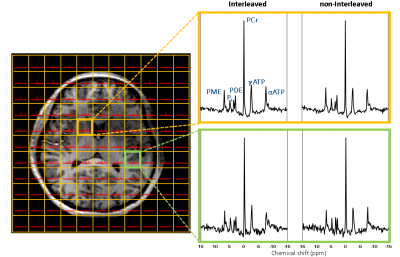 |
1 |
Interleaved Whole Brain 23Na MRI and 31P MRSI Acquisitions at 7T
Zahra Shams1,
Jiying Dai1,
Mark Gosselink1,
Wybe W.J.M. van der Kemp1,
Fredy Visser1,
Dennis W.J. Klomp1,
Hans J.M. Hoogduin1,
Tijl A. van der Velden1,
Giel Mens1,
Jannie P. Wijnen1,
and Evita C. Wiegers1
1Department of Radiology and Nuclear Medicine, University Medical Center Utrecht, Utrecht, Netherlands Keywords: Data Acquisition, Data Acquisition The purpose of this study is to assess the feasibility of an interleaved acquisition of 31P MRSI and 23Na MRI. Triple-nuclei [metabolic] imaging of the brain was performed in one scan session with an interleaved acquisition of 3D 31P MRSI and 3D radial UTE 23Na MRI at 7 Tesla. The results of the interleaved 23Na-31P were compared with those acquired from non-interleaved runs, concluding no influence of each nucleus on the SNR or spectra quality. In conclusion, we showed that these two nuclei pools do not interfere with each other during interleaved acquisition. |
|
4092.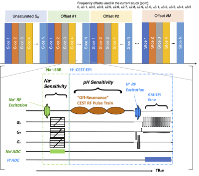 |
2 |
Interleaved multinuclear Na+-H+-CEST-EPI metabolic MRI for
simultaneously quantifying salinity and acidity
Chencai Wang1,2,
Xiaodong Zhong3,
Alfredo L. Lopez Kolkovsky4,
Sonoko Oshima1,2,
Saneel Khairnar2,5,
and Benjamin M Ellingson1,2,6,7
1Department of Radiological Sciences, UNIVERSITY OF CALIFORNIA, LOS ANGELES, Los Angeles, CA, United States, 2Brain Tumor Imaging Laboratory, University of California, Los Angeles, Los Angeles, CA, United States, 3MR R&D Collaborations, Siemens Medical Solutions USA, Los Angeles, CA, United States, 4Institute of Myology, Paris, France, 5Department of Molecular, Cell, and Developmental Biology, UNIVERSITY OF CALIFORNIA, LOS ANGELES, Los Angeles, CA, United States, 6Department of Neurosurgery, University of California, Los Angeles, Los Angeles, CA, United States, 7Department of Psychiatry and Biobehavioral Sciences, University of California, Los Angeles, Los Angeles, CA, United States Keywords: Pulse Sequence Design, Non-Proton, Sodium, 2-Nuclei Extensive evidence suggests abnormal metabolism, sodium homeostasis, and tumor acidity are interconnected and play critical roles in brain tumor formation, progression, seizure activity, treatment resistance, and immune suppression. While Na+ and advanced H+ -based MRI techniques can provide this information, Na+ and H+ images are traditionally acquired sequentially, resulting in a total scan time exceeding what is clinically reasonable. In the current study, we demonstrate utility of a new interleaved sodium gradient echo and proton-based pH-sensitive amine chemical exchange saturation transfer echoplanar imaging (Na+-GRE/H+-CEST-EPI) sequence in 15 minutes, making it feasible to study patients with brain tumors. |
|
4093.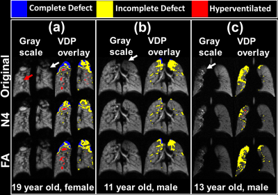 |
3 |
Improved efficiency and quantitative accuracy in hyperpolarized
129Xe ventilation imaging using 2D spiral acquisition
Riaz Hussain1,
Joseph W Plummer1,2,
Abdullah S Bdaiwi1,2,
Matthew M. Willmering1,
Laura L Walkup1,2,3,4,
and Zackary I Cleveland1,2,3,4
1Center for Pulmonary Imaging Research, Division of Pulmonary Medicine, Cincinnati Children's Hospital Medical Center, Cincinnati, OH, United States, 2Department of Biomedical Engineering, University of Cincinnati, Cincinnati, OH, United States, 3Imaging Research Center, Department of Radiology, Cincinnati Children’s Hospital Medical Center, Cincinnati, OH, United States, 4Department of Pediatrics, University of Cincinnati, Cincinnati, OH, United States Keywords: Data Acquisition, Hyperpolarized MR (Gas) Despite yielding high signal-to-noise ratio, conventional gradient recalled echo (GRE) sequence requires a relatively long breath-hold for 129Xe ventilation imaging. 2D-spiral sequence enables faster imaging and offers possibility to correct regional B1/flip-angle inhomogeneities and signal decay using keyhole reconstruction without making assumptions about bias texture that can obscure the physiology of interest. Here, GRE and 2D-spiral showed comparable image quality and revealed regional ventilation impairment in cystic fibrosis. Furthermore, flip-angle correction preserved signal variation due to underlying lung pathophysiology, including gravitation ventilation gradients and subtle defects. Thus, flip-angle corrections in 2D-spiral sequence may detect early and reversible disease-induced ventilation impairment. |
|
4094.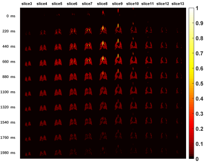 |
4 |
3D Pulmonary Dynamic Ventilation Imaging with High Spatial and
Temporal Resolution Using Hyperpolarized 129Xe MRI
Hongchuang Li1,2,
Haidong Li1,2,
Ming Zhang1,2,
Xiaoling Liu1,2,
Xiuchao Zhao1,2,
Yeqing Han1,2,
Chaohui Ye1,2,
and Xin Zhou1,2
1Key Laboratory of Magnetic Resonance in Biological Systems, State Key Laboratory of Magnetic Resonance and Atomic and Molecular Physics, National Center for Magnetic Resonance in Wuhan, Wuhan Institute of Physics and Mathematics, Innovation Academy for Precision Measurement Science and Technology, Chinese Academy of Sciences- Wuhan National Laboratory for Optoelectronics, Wuhan, China, 2University of Chinese Academy of Sciences, Beijing, China Keywords: Pulse Sequence Design, Hyperpolarized MR (Gas) The mutual restriction of temporal and spatial resolution is a challenge for hyperpolarized gas dynamic MRI, especially for 3D acquisition. Herein, we proposed a methodology to image the pulmonary dynamic ventilation with high temporal and spatial resolution using hyperpolarized gas MRI based on a 3D gradient echo sequence. Furthermore, we observed the difference of pulmonary dynamic ventilation in the posterior-anterior direction. |
|
4095.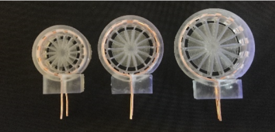 |
5 |
Rapid Radiofrequency Coil Prototyping for Hyperpolarized
Carbon-13 Imaging
Alexander Isaac Zavriyev1,
John Choi1,
Bukola Yetunde Adebesin1,
Molly M Sheehan1,
David J Tischfield1,
and Terence P. F. Gade1
1University of Pennsylvania, Philadelphia, PA, United States Keywords: Data Acquisition, Non-Array RF Coils, Antennas & Waveguides Dynamic nuclear polarization magnetic resonance spectroscopic imaging (DNP-MRSI) of hyperpolarized carbon-13 (HP 13C) is an exciting new imaging technique that provides valuable information about the metabolism of disease states. Though there are many hyperpolarized substrates that can be used to investigate metabolism in a variety of models, the application of this technology often requires custom radiofrequency (RF) coil design. Furthermore, different coils within the same project may provide different results. In this study, we utilized 3D printing molds to generate inductors along with printed circuit board (PCB) designs to create a high reproducibility (<1% variation) pipeline for MR coils. |
|
4096.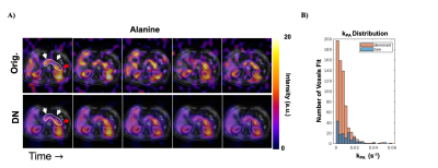 |
6 |
Hyperpolarized 13C Metabolic Imaging of the Human Abdomen with
Spatiotemporal Denoising
Tanner M. Nickles1,2,
Yaewon Kim 1,
Philip M. Lee1,
Hsin-Yu Chen1,
Michael Ohliger1,
Peder E. Z. Larson1,2,
Zhen J Wang1,
Daniel B. Vigneron 1,2,
and Jermey W. Gordon1
1Department of Radiology and Biomedical Imaging, University of California San Francisco, San Francisco, CA, United States, 2UC Berkeley-UCSF Bioengineering Program, University of California San Francisco, San Francisco, CA, United States Keywords: Data Processing, Hyperpolarized MR (Non-Gas), Pancreas, Abdomen, Cancer A substantial challenge in hyperpolarized (HP) 13C MRI is the limited signal-to-noise ratio (SNR) of downstream metabolites, which restricts the achievable spatial resolution. To overcome this for large coverage abdominal studies, a patch-based spatiotemporal denoising approach was applied to denoise dynamic imaging data in [1-13C]pyruvate echo-planar imaging (EPI) human datasets. With denoising, a 11.4 ± 1.8 and 8.7 ± 2.4 fold sensitivity gain was achieved for [1-13C]alanine and [1-13C]lactate, along with improved spatial coverage. These results support the potential of spatiotemporal denoising to improve quantification in HP 13C MRI for normal and cancer studies. |
|
4097.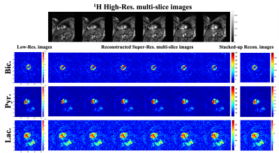 |
7 |
Effects of patch size on 3D patch-based super-resolution
reconstruction of hyperpolarized 13C cardiac MRI
Sung-Han Lin1,
Junjie Ma2,
and JaeMo Park1,3
1Advanced Imaging Research Center, UT Southwestern Medical Center, Dallas, TX, United States, 2GE Healthcare, New York, NY, United States, 3Radiology, UT Southwestern Medical Center, Dallas, TX, United States Keywords: Data Processing, Cardiomyopathy The volumetric patch-based super-resolution method can reconstruct a single-slice low-resolution hyperpolarized 13C MRI to multiple 13C slices with enhanced spatial resolution by exploiting the corresponding high-resolution structural 1H MRI. In this study, the overall performance of this method and the effects of patch size were evaluated using a simulated digital phantom and an anthropomorphic cardiac 13C MR phantom. The optimal patch size for this reconstruction method was also applied to in-vivo human 13C cardiac images acquired with an injection of hyperpolarized [1-13C]pyruvate. |
|
4098.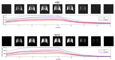 |
8 |
Lung Multi-Breath Wash in/out MRI with 19F with Sub 0.5 Second
Scan Time
Sang Hun Chung1,
Khoi Minh Huynh1,
Yong Chen2,
Pew-Thian Yap1,
Jennifer Goralski1,
Scott Donaldson1,
and Yueh Z Lee1
1University of North Carolina Chapel Hill, chapel hill, NC, United States, 2Case Western Reserve University, Cleveland, OH, United States Keywords: Data Acquisition, Lung, 19F fluorine We studied the feasibility of multi breath wash-in/out lung acquisition with 19F gas and a sub 0.5 second acquisition spiral sequence. We compared the sub 0.5 seconds acquired data ability to measure wash-in/out time constants to a regular breath-hold 19F lung imaging and report correlation as well as differences in mean. Our results yielded high correlations (r > 0.88) for both wash-in and wash-out time constant calculations. The results indicate our proposed method yield similar calculated time wash-in/out time constants without the need for breath-holds and show promise for the use of free breathing 19F gas lung MRI. |
|
4099.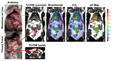 |
9 |
Improved Dynamic In Vivo pH Imaging Using Hyperpolarized
13C-bicarbonate
Xiaoxi Liu1,
Changhua Mu1,
Ying-Chieh Lai1,2,
Robert A. Bok1,
Romelyn Delos Santos1,
Yaewon Kim1,
Avantika Sinha1,
Jeremy Gordon1,
Robert Flavell1,
and Peder Larson1
1Department of Radiology & Biomedical Imaging, University of California, San Francisco, San Francisco, CA, United States, 2Department of Medical Imaging and Intervention, Chang Gung Memorial Hospital at Linkou, Taiwan, Taiwan Keywords: Data Acquisition, Hyperpolarized MR (Non-Gas) We present a dynamic pH measurement using metabolite-specific gradient-echo spiral sequence with flow suppression to improve the SNR of bicarbonate and CO2 with short T2* and used bipolar gradients to suppress the high bicarbonate signal in the vessel. With the improved image quality and high temporal resolution, it is possible to measure the dynamic pH in vivo. Future studies will work to increase the sample size to confirm these results. |
|
4100.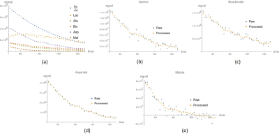 |
10 |
Estimation of Metabolite Signals for In Vitro MRI of Glycolytic
Flow in Hyperpolarized Carbon-13
Ching-Yi Hsieh1,2,
Ying-Chieh Lai3,
Kuan-Ying Lu3,
and Gigin Lin1,2,3
1Institute for Radiological Research, Chang Gung University, Taoyuan, Taiwan, 2Clinical Metabolomics Core Laboratory, Chang Gung Memorial Hospital, Linkou, Taoyuan, Taiwan, 3Department of Medical Imaging and Intervention, Chang Gung Hospital, Linkou, Taoyuan, Taiwan Keywords: Data Processing, Hyperpolarized MR (Non-Gas), metabolites; apparent exchange rate; kinetic model Downstream metabolites may have a poor SNR in hyperpolarized carbon-13 MRI, generating apparent exchange rate constant discrepancies when studying glycolytic flow in vivo or in vitro dynamically in real-time. We developed a method for estimating metabolite signal. This approach estimates metabolite signals using kinetic modeling and noise. The process was evaluated using simulations and in vitro studies. In vitro findings were also compared to 13C NMR cell media AUC. Comparing in vitro data from our method and NMR, both demonstrated consistency when uncertainty was included, suggesting that our method can accurately quantify metabolite signals and indicate how glycolytic flow changes. |
|
4101.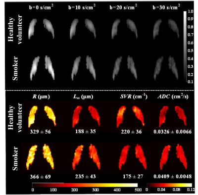 |
11 |
Assessment of Pulmonary Morphometry using Hyperpolarized 129Xe
DWI with Variable-Sampling-Ratio Compressed Sensing Patterns
Qian Zhou1,2,
Haidong Li1,2,
Qiuchen Rao1,
Ming Zhang1,2,
Xiuchao Zhao1,2,
Luyang Shen1,
Yuan Fang1,
Hongchuang Li1,2,
Xiaoling Liu1,2,
Sa Xiao1,2,
Lei Shi1,2,
Yeqing Han1,2,
Chaohui Ye1,2,
and Xin Zhou1,2
1Key Laboratory of Magnetic Resonance in Biological Systems, State Key Laboratory of Magnetic Resonance and Atomic and Molecular Physics, National Center for Magnetic Resonance in Wuhan, Wuhan Institute of Physics and Mathematics, Innovation Academy for Precision Measurement Science and Technology, Chinese Academy of Sciences- Wuhan National Laboratory for Optoelectronics, Wuhan, China, 2University of Chinese Academy of Sciences, Beijing, China Keywords: Data Acquisition, Hyperpolarized MR (Gas) The long acquisition time of hyperpolarized 129Xe multiple b-values DWI made it hard to apply in patients with severe pulmonary diseases. Herein, we proposed a method of variable-sampling-ratio compressed sensing patterns for accelerating hyperpolarized 129Xe DWI. A four-fold reduction in acquisition time was achieved using the proposed method while preserving good image quality. Meanwhile, the method can be used for evaluating pulmonary injuries caused by cigarette smoking. |
|
4102.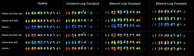 |
12 |
Measuring Air Flow Dynamics in the Lungs Using Hyperpolarized
129Xe MRI
Mostafa K Ismail1,
Steve Kadlecek1,
Hooman Hamedani1,
Faraz Amzajerdian1,
Ryan Baron1,
Luis Loza1,
Madeline Boyes1,
Klaus Hopster1,
Rachel Hilliard1,
Thomas Schaer1,
Benjamin Sinder2,
Patrick Cahill2,
Brian Snyder3,
Kai Ruppert1,
and Rahim Rizi1
1University of Pennsylvania, Philadelphia, PA, United States, 2Children’s Hospital of Philadelphia, Philadelphia, PA, United States, 3Boston Children’s Hospital, Boston, MA, United States Keywords: Data Analysis, Hyperpolarized MR (Gas), Xenon The ability to non-invasively measure air flow in the lungs could enable the early detection and assessment of abnormal air flow resulting from various obstructive and restrictive diseases. Here, we present a novel method for both obtaining and analyzing ventilation maps using hyperpolarized 129Xe MRI (HXe). In addition to assessing tidal volume and fractional ventilation, we introduce a novel sigmoid function describing inhalation and exhalation that enables the assessment of how fast air flows in the lungs and apply this method in a preclinical model of thoracic insufficiency syndrome (TIS) as well as three human lung transplant (LTx) recipients. |
|
4103.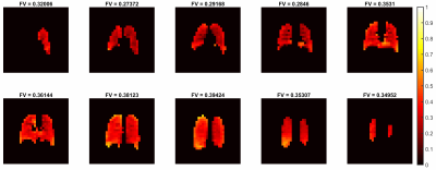 |
13 |
Multiple-Slice, Multiple-Breath Washout Hyperpolarized 129Xe
Ventilation Mapping of the Lung using Accelerated Data
Acquisition
Faiyza Shoaib Alam1,2,
Marcus John Couch3,
Brandon Zanette2,
Daniel Li2,
Felix Ratjen2,4,
and Giles Santyr1,2
1Medical Biophysics, University of Toronto, Toronto, ON, Canada, 2Translational Medicine, The Hospital for Sick Children, Toronto, ON, Canada, 3Siemens Healthcare Limited, Montreal, QC, Canada, 4Division of Respiratory Medicine, The Hospital for Sick Children, Toronto, ON, Canada Keywords: Parallel Imaging, Hyperpolarized MR (Gas), multiple-breath washout, multiple-slice, fractional ventilation Multiple-breath washout hyperpolarized 129Xe MRI (MBW Xe-MRI) results in a regional map of fractional ventilation (FV), measuring percent gas clearance/breath. One limitation of previous work from our group is the single thick coronal slice (200mm) centered on the chest cavity results in significant partial volume effects. Acquiring multiple slices using parallel imaging adds spatial resolution in the slice direction. Feasibility of multiple-slice FV mapping using parallel acceleration is investigated in one adult. Median [IQR] FV was 0.35 [0.30 0.36]. Gravitational dependence was observed in posterior-anterior direction (-0.0122 cm-1). Multiple-slice MBW Xe-MRI is feasible in healthy adults using parallel image acquisition. |
|
4104.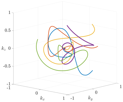 |
14 |
Highly accelerated 3D hyperpolarized MRI at ultra-high fields
using Seiffert Spirals
Tobias Speidel1,
Stephan Knecht2,
Klaus Kolb1,
Martin Gierse2,
and Volker Rasche1
1Internal Medicine II, Ulm University Medical Center, Ulm, Germany, 2NVision Imaging Technologies GmbH, Ulm, Germany Keywords: New Trajectories & Spatial Encoding Methods, Data Acquisition Isotropic 3D imaging of hyperpolarized media is generally challenging due to the extremely limited lifetime of the hyperpolarized phase. Fortunately, images of hyperpolarized media are often intrinsically sparse, resulting in an excellent precondition for non-linear reconstruction techniques such as Compressed Sensing. To preserve this condition, a 3D Seiffert Spiral trajectory was implemented at 11.7 T, yielding low-coherent sampling properties for an arbitrary number of excitations, until depletion of the hyperpolarized signal. We show that qualitative images with a resolution of <500 $$$\mu$$$m can be reconstructed from continuous data that was acquired in less than one second. |
|
4105.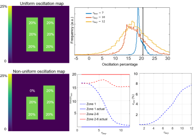 |
15 |
Optimizing Hyperpolarized 129Xe MRI of cardiopulmonary
oscillations using a Digital Phantom
Junlan Lu1,
Elianna Bier2,
Suphachart Leewiwatwong2,
David Mummy3,
Sakib Kabir3,
Fawaz Alanezi4,
Sudarshan Rajagopal4,
Scott Haile Robertson5,
Peter J Niedbalski6,
and Bastiaan Driehuys3
1Medical Physics, Duke University, Durham, NC, United States, 2Biomedical Engineering, Duke University, Durham, NC, United States, 3Radiology, Duke University, Durham, NC, United States, 4Cardiology, Duke University, Durham, NC, United States, 5Clinical Imaging Physics Group, Duke University, Durham, NC, United States, 6Pulmonary, Critical Care, and Sleep Medicine, University of Kansas Medical Center, Kansas City, KS, United States Keywords: Data Analysis, Hyperpolarized MR (Gas), Keyhole reconstruction, Simulation Cardiogenic red blood cell (RBC) signal oscillations in 129Xe whole-lung dynamic spectroscopy provide a promising biomarker for identifying patients with pulmonary hypertension (PH). However, to detect more complex and heterogeneous diseases, it is necessary to move from simple global metrics to spatially resolved mapping. This has been demonstrated using keyhole reconstruction methods but little work has yet been done to optimize the reconstruction and visualization of these maps. Here, we introduce a digital phantom to investigate the effects of radial views, key radius, and SNR. From these simulations, we deduce a key radius of 9 points is optimal for minimizing radial undersampling-based heterogeneity and maximizing sensitivity. |
|
4106.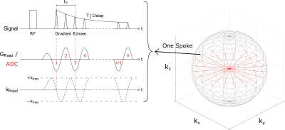 |
16 |
Three-dimensional radial echo-planar spectroscopic imaging for
in vivo hyperpolarized 13C MRSI at 3 T
Marcel Awenius1,2,3,
Philipp Biegger1,
Christin Glowa4,
Melanie Müller1,
Renate Bangert1,
Helen Abeln1,3,5,
Dominik Ludwig1,
Tristan A. Kuder1,
Mark E. Ladd1,2,6,
Peter Bachert1,2,
and Andreas Korzowski1
1Division of Medical Physics in Radiology, German Cancer Research Center (DKFZ), Heidelberg, Germany, 2Faculty of Physics and Astronomy, University of Heidelberg, Heidelberg, Germany, 3Max-Planck-Institute for Nuclear Physics, Heidelberg, Germany, 4Division of Medical Physics in Radiation Oncology, German Cancer Research Center (DKFZ), Heidelberg, Germany, 5Faculty of Chemistry and Earth Sciences, University of Heidelberg, Heidelberg, Germany, 6Faculty of Medicine, University of Heidelberg, Heidelberg, Germany Keywords: Pulse Sequence Design, Hyperpolarized MR (Non-Gas), Carbon-13 In this study, a novel radially-sampled echo-planar spectroscopic imaging (rEPSI) sequence was implemented at a clinical 3T MR scanner to enable the non-invasive investigation of metabolic processes via hyperpolarized 13C substrates in real-time. Customized data analysis pipelines yielded high quality spectra and volumetric intensity maps for in vivo experiments using hyperpolarized [1-13C]pyruvate. Data extracted from k-space center points enabled a non-localized quantification of T1 values. |
|
4107.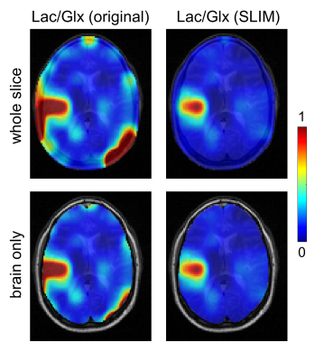 |
17 |
Robust and Automated Processing of Deuterium Metabolic Imaging
(DMI) using Spatial Prior Knowledge
Robin A de Graaf1,
Yanning Liu1,
Zachary A Corbin1,
and Henk M De Feyter1
1Yale University, New Haven, CT, United States Keywords: Image Reconstruction, Deuterium Deuterium Metabolic Imaging (DMI) is a novel method to generate spatial maps of dynamic metabolism. While DMI acquisition methods are simple and robust, DMI processing still requires expert user interaction, for example in the removal of extracranial natural abundance 2H lipid signals that interfere with metabolism-linked 2H-lactate formation. Here we pursue the use of MRI-based spatial prior knowledge to provide automated and objective lipid removal. Adequate lipid suppression without perturbation of brain voxels is achieved, thereby allowing the generation of distinct and reliable metabolic maps on patients with brain tumors. |
|
4108.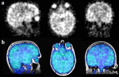 |
18 |
Regional 17O T2* in Human Brain at 3T
Hao Song1,
Hamidreza Saeidi1,
Johannes Fisher1,
Ali Caglar Özen1,
Stefan Schumann2,
and Michael Bock1
1Dept. of Radiology, Medical Physics, Medical Center University of Freiburg, Faculty of Medicine, University of Freiburg, Freiburg, Germany, 2Dept. of Anesthesiology and Critical Care, Medical Center University of Freiburg, Faculty of Medicine, University of Freiburg, Freiburg, Germany Keywords: Data Analysis, Non-Proton Information about T2* relaxation time plays an important role in data acquisition for 17O-MRI. In this work, we investigated the in vivo T2* relaxation time constants of 17O in regional brain tissue at 3T. Overall, regional T2* relaxation times are in a range of 1.5 - 2ms (except for CSF). Additionally, a T2* variation of 0.12ms was found from superficial to deep layers in the insular cortex. Our results indicate that short-TE acquisition strategies are favorable for 17O-MRI of the brain. |
|
4109.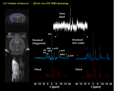 |
19 |
Signal-to-noise Ratio Enhancement of 31P Magnetic Resonance
Spectroscopy using a Pre-trained Deep Learning Model
Yeong-Jae Jeon1,2,
Kyung Min Nam3,4,5,
Alex Bhogal3,
and Hyeon-Man Baek1,2
1Department of Health Sciences and Technology, GAIHST, Gachon University, Incheon, Korea, Republic of, 2Lee Gil Ya Cancer & Diabetes Institute, Gachon University, Incheon, Korea, Republic of, 3Department of Radiology, University Medical Centre Utrecht, Utrecht, Netherlands, 4Institute for Diagnostic and Interventional Neuroradiology, Support Center for Advanced Neuroimaging (SCAN), University of Bern, Bern, Swaziland, 5Translational Imaging Ceter, sitem-insel AG, Bern, Switzerland Keywords: Data Processing, Spectroscopy, Non-Proton, Animals, Brain, Precision & Accuracy We demonstrate the feasibility of a novel denoising approach utilizing a pretrained deep learning model with multiscale local polynomial smoothing for single voxel 31P MRS data in the mice brain at 9.4T. We evaluated the low-rank denoising, one of the popular methods and the proposed method using LCModel to compare their performance. Both methods resulted in improved signal-to-noise ratio and decreased uncertainty (Cramer-Rao Lower Bounds). In this work, the suggested method outperformed in signal-to-noise ratio enhancement. |
|
 Back to Meeting Home
 Back to the Program-at-a-Glance
The International Society for Magnetic Resonance in Medicine is accredited by the Accreditation Council for Continuing Medical Education to provide continuing medical education for physicians.
 Back to Meeting Home
 Back to the Program-at-a-Glance
 View Presentation Video