Digital Poster
Data Analysis & Processing II
ISMRM & ISMRT Annual Meeting & Exhibition • 03-08 June 2023 • Toronto, ON, Canada

| Computer # | |||
|---|---|---|---|
3116.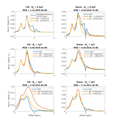 |
61 |
Improving the accuracy of Multipool-Lorentzian fitting in CEST
MRI by use of a Pseudo-Voigt lineshape for direct water
saturation
Markus Huemer1,
Clemens Stilianu1,
and Rudolf Stollberger1,2
1Institute of Biomedical Imaging, Graz University of Technology, Graz, Austria, 2BioTechMed Graz, Graz, Austria Keywords: Data Analysis, CEST & MT In Chemical Exchange Saturation Transfer (CEST) MRI one common analysis technique is Multipool-Lorentzian fitting of the resulting Z-Spectrum. Multiple Lorentzian lineshapes are fitted to the measured data. The lineshape of the direct water saturation (DS) can deviate substantially from a Lorentzian depending on the saturation scheme and $$$B_1^+$$$. To prevent a cross-influence of the DS on the CEST and increase the fit accuracy we propose the use of a Pseudo-Voigt lineshape for the water line. |
|
3117.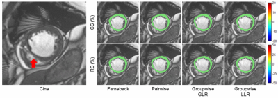 |
62 |
Deformable Groupwise Registration Using a Locally Low-Rank
Dissimilarity for Myocardial Strain Estimation from Cine MRI
Images
Haiyang Chen1 and
Chenxi Hu1
1The Institute of Medical Imaging Technology, School of Biomedical Engineering, Shanghai Jiao Tong University (SJTU), Shanghai, China Keywords: Data Analysis, Data Processing, registration We propose a deformable groupwise registration method using a locally low-rank (LLR) dissimilarity to estimate myocardial strain from cine MRI images. The proposed method eliminates the drift effect commonly observed in the optical flow and sequentially pairwise registration, facilitating more accurate strain estimation in the diastolic phase. Compared to the globally low-rank dissimilarity, LLR dissimilarity shows slightly better tracking accuracy by imposing the low-rank property in local image regions rather than the whole image. Experiments on a large public cine MRI dataset demonstrates the accuracy of the proposed method on tracking and strain estimation. |
|
3118. |
63 |
Digital Reference Objects with BART
Nick Scholand1,2,
Martin Schilling3,
Martin Heide3,
and Martin Uecker1,2,3
1Institute of Biomedical Imaging, Graz University of Technology, Graz, Austria, 2German Centre for Cardiovascular Research (DZHK), Partner site Göttingen, Göttingen, Germany, 3Department of Interventional and Diagnostic Radiology, University Medical Center Göttingen, Göttingen, Germany Keywords: Software Tools, Software Tools Digital Reference Objects (DRO) are important for developing imaging and reconstruction techniques especially in medical imaging applications. They allow a stepwise increase in complexity during testing of methods by simulating reproducible datasets.
In this work we present a variety of tools the BART toolbox
provides for the creation and simulation of DROs. We present
a workflow for designing DROs with Bézier curves using
vector graphics applications which are then imported into
BART. |
|
3119.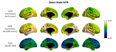 |
64 |
Tract-specific myelin mapping using magnetization
transfer-prepared diffusion imaging: comparison with
conventional MTR tractometry
Wen Da Lu1,2,
Mark Cameron Nelson2,3,
Ilana Ruth Leppert2,
Pietro Bontempi4,
Simona Schiavi4,5,
Christopher Dennis Rowley1,2,
Alessandro Daducci4,
and Christine Lucas Tardif1,2,3
1Department of Biological and Biomedical Engineering, McGill University, Montreal, QC, Canada, 2McConnell Brain Imaging Centre, Montreal Neurological Institute and Hospital, Montreal, QC, Canada, 3Department of Neurology and Neurosurgery, McGill University, Montreal, QC, Canada, 4Department of Computer Science, University of Verona, Verona, Italy, 5Department of Neuroscience, Rehabilitation, Ophthalmology, Genetics, Maternal and Child Health (DINOGMI), University of Genoa, Genova, Italy Keywords: Data Analysis, Tractography & Fibre Modelling, Magnetization Transfer, Diffusion-Weighted Imaging, Tractometry Tractometry is a technique used to investigate the microstructure variations along white matter tracts or to reconstruct microstructure-weighted connectivity matrices. However, partial volume effects from crossing fibers bias the individual fiber measurements and conceal subtle differences. Here, we compare MTR tractometry to a novel approach using dual-encoded MT-weighted DWI analyzed using COMMIT to disentangle the MTR signal of individual white matter fibers. The results show a broader distribution in edge MTR values for the dual-encoding approach in comparison to tractometry. Both techniques show similar spatial MTR patterns, however the dual-encoded MT-weighted DWI approach shows a higher pattern of inter-individual variability. |
|
3120.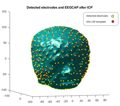 |
65 |
Automatic Localization of EEG Electrodes from MR Structural
Images
Mohammadreza Rezaei-Dastjerdehei1 and
Pierre LeVan2
1Department of Biomedical Engineering, University of Calgary, Calgary, AB, Canada, 2Department of Radiology, University of Calgary, Calgary, AB, Canada Keywords: Data Analysis, Segmentation, EEG Electrodes Localization, T1-weighted MRI The localization of EEG sources is one of the fundamental approaches to facilitate the interpretation of EEG data. However, the accuracy of source localization depends on the exact knowledge of the position of the electrodes on the scalp, which currently requires time-consuming and/or expensive approaches. Here, an automatic method is proposed that retrieves the electrode positions by localizing the curvature changes in T1-weighted MRI images caused by electrodes. The results show an average detection sensitivity of ~96.4%, with an average position error of 4.23 mm for all subjects. |
|
3121. |
66 |
Habitats Analysis in Hepatocellular Carcinoma to Predict
Microvascular Invasion by Intravoxel Incoherent Motion: A Pilot
Study
Chenhui Li1,
Jinhuan Xie1,
Liling Long1,
Huiting Zhang2,
and Yang Song2
1The First Affiliated Hospital of Guangxi Medical University, Nanning, China, 2MR Scientific Marketing, Siemens Healthineers, Shanghai, China Keywords: Data Analysis, Segmentation, Habitats The method of delineating the ROI of whole lesions on quantitative parameter images and then averaging them for comparison did not accurately quantify heterogeneity. In this study, we adopted a habitats analysis method combined with tissue cellularity and blood flow information from IVIM model to segment whole tumor to four subregions to predict microvascular invasion (MVI) in hepatocellular carcinoma. The results show that habitats analysis predicts MVI positivity with an accuracy of 70.19%, and the averaged value of each parameter in whole tumor was not predictive for MVI. This provides a good starting point for further application of this method. |
|
3122.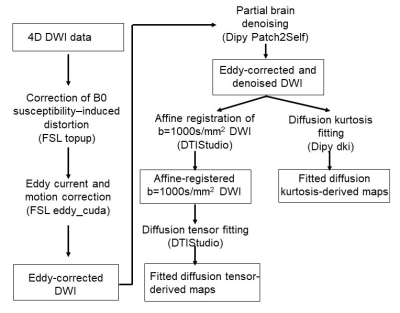 |
67 |
A robust image analysis pipeline for high fidelity kurtosis and
tensor fitting of 1.2mm isotropic infant brain diffusion MRI
Tianjia Zhu1,2,
Minhui Ouyang1,3,
Kay Sindabizera1,
Juri Kim2,
and Hao Huang1,3
1Department of Radiology, Children's Hospital of Philadelphia, Philadelphia, PA, United States, 2Department of Bioengineering, University of Pennsylvania, Philadelphia, PA, United States, 3Department of Radiology, University of Pennsylvania, Philadelphia, PA, United States Keywords: Data Processing, Pediatric, Neuro High-resolution infant diffusion MRI (dMRI) data poses specific challenges such as severe motion artifacts, long acquisition time for multi-shell data, and low signal-to-noise (SNR). We present a robust image analysis pipeline for high fidelity kurtosis and tensor fitting of 1.2mm isotropic infant brain dMRI that allows us to efficiently analyze in-vivo dMRI data despite the considerable technical challenges specific to infant imaging. State-of-the-art preprocessing including slice-to-volume motion correction and susceptibility-by-movement correction is combined with advanced self-supervised learning-based denoising to produce high fidelity diffusion tensor and diffusion kurtosis fitting. |
|
3123.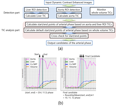 |
68 |
Arterial phase detection when utilizing stack-of-stars dynamic
Data Set with various pseudo contrast enhancement
Yutaka Hoshiyama1,
Chunqi Wang2,
Hong Yang2,
Hideki Ota3,4,
Atsuro Masuda4,
Masahiro Kawabata4,
Hideaki Kutsuna1,
Kensuke Shinoda1,
and Yoshimori Kassai1,3
1MRI Systems Division, Canon Medical Systems Corporation, Ohtawara, Japan, 2Research & Development Center, Canon Medical Systems (China) Co., Ltd, Beijing, China, 3Department of Advanced MRI Collaboration Research, Tohoku University Graduate School of Medicine, Sendai, Japan, 4Department of Diagnostic Radiology, Tohoku University Hospital, Sendai, Japan Keywords: Data Analysis, Contrast Agent, Dynamic We propose an automated method to present candidates of the arterial phase as the optimal time points from the liver contrast-enhanced MRI images based on the time intensity curve (TIC) analysis of the several ROIs. In clinical situation, there may be some differences of TIC due to a speed of uptake and washout and a quality of the image. In this study, the robustness of our proposed automated method was investigated by utilizing stack-of-stars dynamic images with various pseudo contrast enhancement effects. The results showed that our proposed automated method was applicable for such effects. |
|
3124.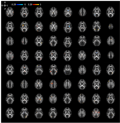 |
69 |
Subject-specific analysis reveals spatially heterogeneous white
matter abnormalities in sports-related concussion
Ho-Ching Yang1,
Mario Dzemidzic1,2,
Qiuting Wen1,
Larry D Riggen Jr3,
Steven P Broglio4,
Michael A McCrea5,
Thomas W McAllister6,
Jaroslaw Harezlak7,
and Yu-Chien Wu1,8
1Department of Radiology and Imaging Sciences, Indiana University School of Medicine, Indianapolis, IN, United States, 2Department of Neurology, Indiana University School of Medicine, Indiana University School of Medicine, Indianapolis, IN, United States, 3Department of Biostatistics and Health Data Science, Indiana University School of Medicine, Indianapolis, India, 4Michigan Concussion Center, University of Michigan, Ann Arbor, MI, United States, 5Department of Neurosurgery, Medical College of Wisconsin, Milwaukee, WI, United States, 6Department of Psychiatry, Indiana University School of Medicine, Indianapolis, IN, United States, 7Department of Epidemiology and Biostatistics, Indiana University School of Public Health, Bloomington, IN, United States, 8Stark Neurosciences Research Initiative, Indiana University School of Medicine, Indianapolis, IN, United States Keywords: Data Analysis, Traumatic brain injury, Subject-specific analysis, Heterogeneous, Concussion Sport-related concussion (SRC) injury inclines to cause subject-specific brain region abnormalities. This study applied subject-specific analysis that accounts for inter-subject variation to investigate heterogeneity of white matter alterations in a sample from a large national multicenter study, the Concussion Assessment, Research and Education Consortium. Our results demonstrated that subject-specific white matter abnormalities in SRC can be uncovered by calculating the extreme Z-score maps based on a template generated from normally distributed diffusion tensor imaging metric values in non-contact sports controls. Our finding indicates that the SRC-induced white matter abnormalities show heterogeneous spatial distribution across participants. |
|
3125.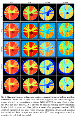 |
70 |
Multidimensional optimal denoising with block matching is robust
to misalignment and contrast changes
Khoi Minh Huynh1,
Sang Hun Chung1,
Yueh Lee1,
and Pew-Thian Yap1
1Department of Radiology and Biomedical Research Imaging Center, UNC Chapel Hill, Chapel Hill, NC, United States Keywords: Data Processing, Lung Denoising methods can leverage common information from multiple image volumes for better noise reduction. However, performance can degrade with inter-volume misalignment or contrast changes. We proposed a block-matching denoising method for effective noise removal and robustness to inter-volume differences. |
|
3126.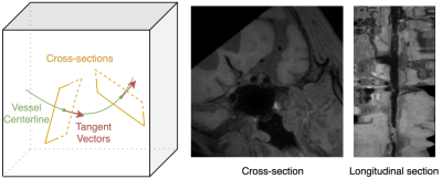 |
71 |
Improving interpretation accuracy for curved planar reformation
of intracranial vessel wall MRI by reducing intra-plane rotation
Xin Wang1,
Yin Guo2,
Kaiyu Zhang2,
Gador Canton3,
Niranjan Balu3,
Thomas S. Hatsukami4,
Mahmud Mossa-Basha5,
and Chun Yuan3
1Department of Electrical and Computer Engineering, University of Washington, Seattle, WA, United States, 2Department of Bioengineering, University of Washington, Seattle, WA, United States, 3Department of Radiology, University of Washington, Seattle, WA, United States, 4Department of Surgery, University of Washington, Seattle, WA, United States, 5Department of Radiology, University of North Carolina-Chapel Hill, Chapel Hill, NC, United States Keywords: Data Processing, Visualization This work aims to develop an algorithm for straightened curved planar reformation (CPR), i.e., extracting the 2D cross-sectional images along a vessel centerline in a vessel wall MRI scan. Previous methods determine the orientation of CPR images in a fixed way, leading to undesired rotation along the centerline. Our method instead calculates the local coordinate system with respect to previous slices, thus keeping the continuity of cross-sections. Our CPR results show a better readability of the straightened vessel image, especially when one wants to utilize the spatial information among adjacent CPR slices for a more comprehensive and accurate medical analysis. |
|
3127.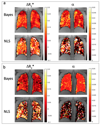 |
72 |
Improved pulmonary oxygen-enhanced MRI parameter precision
enabled by hierarchical Bayesian inference
Josephine H Naish1,2,
Marta Tibiletti1,
Christopher Short3,4,
Tom Semple3,4,
Simon Padley3,4,
Jane C Davies3,4,
and Geoff JM Parker1,5
1Bioxydyn Ltd, Manchester, United Kingdom, 2MCMR, Manchester University NHS Foundation Trust, Manchester, United Kingdom, 3National Heart & Lung Institute, Imperial College London, London, United Kingdom, 4Royal Brompton Hospital, Guy's & St Thomas’ Trust, London, United Kingdom, 5Centre for Medical Image Computing, Department of Medical Physics and Biomedical Engineering, University College London, London, United Kingdom Keywords: Data Analysis, Lung, Bayes Lung oxygen-enhanced MRI can provide regional information relating to lung function, but voxel-wise parameter estimation is hampered by low SNR. Here we present a hierarchical Bayesian approach to voxel-wise parameter estimation implemented in R and the probabilistic programming language Stan. In both simulations and in OE-MRI data acquired in patients with cystic fibrosis, the Bayesian approach results in substantially less noisy parameter maps compared to conventional non-linear least squares estimation. |
|
3128.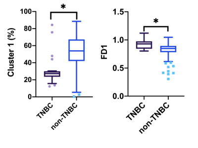 |
73 |
Assessment of tumor subregion complexity using fractal analysis
in breast cancer
Run Xu1,
Guanwu Li1,
Dan YU2,
Yongming Dai2,
and Xinyue Liang2
1Department of Radiology, Yueyang Hospital of Integrated Traditional Chinese and Western Medicine, Shanghai University of Traditional Chinese Medicine, Shanghai, China, 2Central Research Institute, United Imaging Healthcare, Shanghai, China Keywords: Data Analysis, Cancer This study aimed to develop multiparametric physiologic MRI-based spatial habitat analysis and to validate the association between the habitats and the molecular subtypes. The distinct tumor habitats were identified using multiple MRI-parameter maps, then applied to calculate the volume fraction and fractal dimension (FD) and to investigate their diagnosis value. The combination of FD and volume fraction improves the discriminatory ability for TNBC from non-TNBC with an AUC to 0.951 from 0.853. |
|
3129.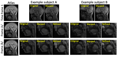 |
74 |
Identification of Subject-Specific Motion Variations via
Atlas-Based Deformation Refinement from Real-Time MRI
Fangxu Xing1,
Riwei Jin2,
Imani Gilbert3,
Georges El Fakhri1,
Jamie L. Perry3,
Bradley P. Sutton2,
and Jonghye Woo1
1Radiology, Harvard Medical School, Boston, MA, United States, 2University of Illinois at Urbana-Champaign, Champaign, IL, United States, 3East Carolina University, Greenville, NC, United States Keywords: Data Analysis, Data Analysis, Speech, motion analysis, atlas Dynamic magnetic resonance imaging has become increasingly efficient at capturing speech deformations of the velopharyngeal region in real time. With previously developed dynamic vocal tract atlases, quantification of group deformation statistics of a population in speech has become possible in a common atlas space. However, subject-specific deformation characteristics as an underlying property tend to be hidden in the spatial and temporal alignment process of atlas construction. We present a registration-based deformation characterization method that extracts subject-specific motion variations in two layers of registration steps. A dataset of fifteen human subjects is processed to reveal unique deformation patterns of each subject. |
|
3130.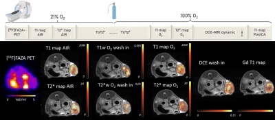 |
75 |
MRI for tissue classification of subcutaneous colon cancer using
oxygen enhanced (OE), dynamic contrast enhanced (DCE) MRI and
[18F]FAZA PET
Marta Vuozzo1,
Max Zimmermann2,
Mystele Tendonge3,
Petros Martirosian2,
Manfred Kneilling4,
Fritz Schick2,
Bernd Pichler3,
and Andreas Schmid2
1Werner Siemens Imaging Center, University Hospital Tübingen, Tübingen, Germany, 2University Hospital Tübingen, Tübingen, Germany, 3Univeristy Hospital Tübingen, Tübingen, Germany, 4Univerity Hospital Tübingen, Tübingen, Germany Keywords: Data Analysis, Cancer, Multimodal Solid tumors exhibit intratumoral heterogeneity, which is related to therapy efficacy. Since biopsies represent a small part of the tumor, multimodal imaging techniques provide panoptic cancer characterization. We developed an acquisition and analysis protocol based on multiparametric MRI to classify and provide a holistic characterization of intratumoral heterogeneity and correlate it with positron emission tomography and histology. We applied MRI to characterize phenotypic changes induced by antiPD-L1 therapy response. Results reveal that models trained exclusively with MRI data provide biologically relevant maps of phenotypes showing intratumoral heterogeneity, but also allow non-invasive identification of tumors that respond or resist to therapy. |
|
3131.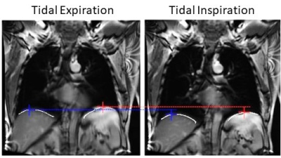 |
76 |
Diaphragm Excursion Measured by Free-breathing 1H MRI in
Patients with Asthma: Repeatability and Bronchodilator Response
Yonni Friedlander1,2,
Dante P.I. Capaldi3,
Norm Konyer1,
Melanie Kjarsgaard2,
Carmen Venegas2,4,
Parameswaran Nair2,4,
and Sarah Svenningsen1,2,4
1Imaging Research Centre, St. Joseph's Healthcare Hamilton, Hamilton, ON, Canada, 2Firestone Institute of Respiratory Health, St. Joseph's Healthcare Hamilton, Hamilton, ON, Canada, 3Deparment of Radiation Oncology, University of California San Francisco, San Francisco, CA, United States, 4Division of Respirology, McMaster University, Hamilton, ON, Canada Keywords: Data Analysis, Lung Using free-breathing 1H MRI, diaphragm excursion was measured in 9 patients with severe asthma at two visits 4-months apart. At each visit, patients with asthma were imaged before and after administration of a bronchodilator. Diaphragm excursion measured at the two visits were not significantly different from one another (mean bias=0.34cm, p=0.07) and were well correlated (r=0.53, p=0.004), demonstrating repeatability of the measurement. In addition, diaphragm excursion was significantly increased following bronchodilator administration (p=0.01). |
|
3132.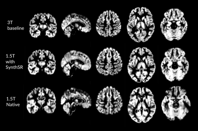 |
77 |
Improving Low Resolution MRI Contrast for Brain-Environment
Neuroepidemiology
Jasmine D Cakmak1,
Reza Azarpazhooh2,
Alexander Khaw2,
and Udunna Anazodo1,2
1McGill University, Montreal, QC, Canada, 2Western University, London, ON, Canada Keywords: Data Processing, Segmentation, Synthetic MRI Recycling large-scale clinical data to retrospectively study associations of brain imaging variables with emerging environmental risk factors is a sustainable approach in the growing field of neuroepidemiology. Here, we evaluated the utility of a deep learning tool to increase the resolution of clinical (1.5T) T1-weighted MRI by comparing global assessment of gray matter volume (GMV) and cortical thickness (CT) to 3T research scans. Overall, the resolved 1.5T images had higher (18%) GMV and CT compared to 3T and were more biased than unresolved images. Further analysis in larger cohorts using improved segmentation approaches could validate recycling of enhanced clinical scans. |
|
3133.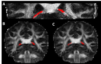 |
78 |
Head-to-head Comparison of Free Water Elimination Methods in
Fornix
Ken Sakaie1,
Katherine Koenig1,
and Mark J. Lowe2
1The Cleveland Clinic, Cleveland, OH, United States, 2Imaging Institute, The Cleveland Clinic, Cleveland, OH, United States Keywords: Data Analysis, Alzheimer's Disease Diffusion Tensor Imaging measures in fornix may serve as a biomarker in Alzheimer’s disease. However, partial volume averaging between the fornix and surrounding cerebrospinal fluid imposes systematic bias that can conflate tissue microstructure with atrophy. Free Water Elimination methods have been proposed as a solution, but comparison between these methods is limited. We perform direct comparison between the approaches in terms of repeatability and bias. |
|
3134.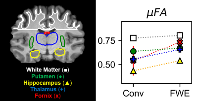 |
79 |
Microscopic fractional anisotropy with free water elimination
Nico J J Arezza1,2,
Mohammad Omer1,
and Corey A Baron1,2
1Medical Biophysics, Western University, London, ON, Canada, 2Centre for Functional and Metabolic Mapping, Robarts Research Institute, London, ON, Canada Keywords: Data Processing, Neuro Water diffusion anisotropy is a diffusion MRI metric that is sensitive to brain injury and neurodegeneration. Microscopic fractional anisotropy (μFA) is a new metric of diffusion anisotropy that is immune to crossing fiber effects, unlike traditional FA, which enables probing of axon integrity in gray matter and crossing white matter tracts. However, μFA is underestimated in voxels adjacent to cerebrospinal fluid (CSF) due to partial volume contamination, which is particularly problematic in cortical gray matter and tissue adjacent to ventricles. Here, we demonstrate a free water elimination method to remove the CSF signal from μFA measurements in four healthy volunteers. |
|
3135.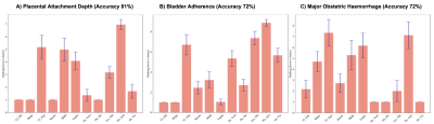 |
80 |
Feature selection to facilitate surgical planning from MRI of
Placenta Accreta Spectrum Disorder
Joanna Chappell1,
Nada Mufti1,2,
Hassna Irzan1,
Pat O’Brien3,
George Attilakos3,
Magda Sokolska4,
Priya Narayanan3,
Trevor Gaunt5,
Paul Humpheries5,
Premal Patel5,
Elspeth Whitby6,
Eric Jauniaux2,3,
J. Ciaran Hutchinson5,
Neil Sebire5,
David Atkinson7,
Giles Kendall2,4,
Sebastien Ourselin 1,
Tom Vercauteren1,
Anna L David2,3,
and Andrew Melbourne1
1School of Biomedical Engineering and Imaging Sciences (BMEIS), Kings College London, London, United Kingdom, 2Elizabeth Garret Anderson Institute for Women’s Health, University College London, London, United Kingdom, 3University College London Hospital, London, United Kingdom, 4Department of Medical Physics and Biomedical Engineering, University College London Hospitals, London, United Kingdom, 5Great Ormond Street Hospital for Children, London, United Kingdom, 6Sheffield Teaching Hospitals NHS Foundation Trust, Sheffield, United Kingdom, 7Centre for Medical Imaging, University College London, London, United Kingdom Keywords: Data Processing, Placenta Feature Selection Models provide a ranking of pathological MRI markers able to predict the outcome of Placenta Accreta Spectrum Disorder, which could be used to aid in clinical decision-making and improve maternal outcome. The potential being to reduce the workload of radiologists by establishing the most clinically relevant pathological MRI markers that predict outcome. Our results found three pathological markers to have the highest ranking to the outcomes with an average accuracy of 75% using a Random Forest Selection Model and Boruta algorithm. |
|
 Back to Meeting Home
 Back to the Program-at-a-Glance
The International Society for Magnetic Resonance in Medicine is accredited by the Accreditation Council for Continuing Medical Education to provide continuing medical education for physicians.
 Back to Meeting Home
 Back to the Program-at-a-Glance
 View Presentation Video