
Digital Poster
Advanced Image Reconstruction Techniques
 Back to Meeting Home
 Back to the Program-at-a-Glance
| Computer # | |||
|---|---|---|---|
4947.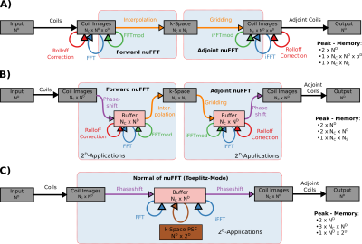 |
1 |
Scaling nuFFT Memory-Overhead Down to Zero: Computational
Trade-Offs and Memory-Efficient PICS-Reconstructions with BART
Moritz Blumenthal1,2,
H. Christian M. Holme2,
and Martin Uecker1,2
1Institute for Diagnostic and Interventional Radiology, University Medical Center Göttingen, Göttingen, Germany, 2Institute of Biomedical Imaging, Graz University of Technology, Graz, Austria Keywords: Image Reconstruction, Software Tools We propose a new decomposition of the nuFFT algorithm, allowing for a zero-memory-overhead implementation of the (adjoint-)nuFFT. With one grid-sized buffer, the decomposition allows for a memory efficient nuFFT with negligible computational overhead compared to a two-fold oversampled conventional nuFFT - to the prize of less efficient parallelization on many-threads CPU systems. We reduce memory requirements of 3D non-Cartesian PICS-reconstructions in BART by a factor up to ten, allowing for GPU acceleration of reconstructions with eight coils and matrix size 256x256x256 on a 4GB consumer-level GPU. |
|
4948.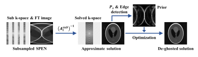 |
2 |
A k-space insight of aliasing effects and their removal of SPEN
MRI
Sijie Zhong1,2,
Minjia Chen3,
Ke Dai1,4,
Hao Chen1,2,
Lucio Frydman4,
and Zhiyong Zhang1,2
1School of Biomedical Engineering, Shanghai Jiao Tong University, Shanghai, China, 2Institute of Medical Robotics, Shanghai Jiao Tong University, Shanghai, China, 3Department of Engineering, University of Cambridge, Cambridgeshire, United Kingdom, 4Department of Chemical and Biological Physics, Weizmann Institute of Science, Rehovot, Israel Keywords: Image Reconstruction, Artifacts, SPEN The reconstruction theory of Spatio-temporal encoding MRI is not complete at present. Aliasing artifacts in SPEN MRI reconstructions can be traced to image contributions corresponding to high-frequency k-space signals. The k-space picture provides the spatial displacements, phase offsets and linear amplitude modulations associated to these artifacts, as well as routes to removing these from the reconstruction results. These new ways to estimate the artifact priors were applied to reduce SPEN reconstruction artifacts on simulated, phantom and human brain MRI data. |
|
4949.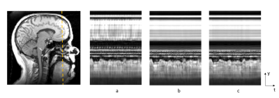 |
3 |
Locally high rank reconstruction through Partial Separability
model (PS-LHR) with regional optimized temporal basis (ROT) of
dynamic speech MRI
Riwei Jin1,
Yudu Li2,
Fangxu Xing3,
Imani Gilbert4,
Jamie Perry4,
Jonghye Woo3,
Zhi-pei Liang5,
and Brad Sutton1
1Department of Bioengineering, University of Illinois Urbana-Champaign, Champaign, IL, United States, 2National Center for Supercomputing Applications, Champaign, IL, United States, 3Gordon Center for Medical Imaging, Department of Radiology, Massachusetts General Hospital/Harvard Medical School, Boston, MA, United States, 4Department of Communication Sciences and Disorders, East Carolina University, Greenville, NC, United States, 56 Department of Electrical and Computer Engineering, University of Illinois Urbana-Champaign, Champaign, IL, United States Keywords: Image Reconstruction, Sparse & Low-Rank Models To optimize the reconstruction quality of isotropic 3D dynamic speech magnetic resonance imaging with large scan volume, we applied two novel methods based on the Partial Separability model theory: 1. Locally High-Rank reconstruction through Partial Separability model (PS-LHR) which enables higher rank to be devoted to the dynamic speech region. 2. Implementation of Regional-Optimized Temporal basis (ROT) to focus the temporal navigator information on the speech region. The improvement in reconstruction quality was seen to decrease the noise of regions of the image outside the area of interest and increase dynamic smoothness in the speech region. |
|
4950.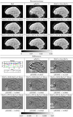 |
4 |
Efficient correction for MRI artifacts due to nonlinear
variations in magnetic field using principal component analysis
Daniel I Gendin1 and
Jiaen Liu2
1Advanced Imaging Research Center, UT Southwestern Medical Center, Dallas, TX, United States, 2Advanced Imaging Research Center and Radiology, UT Southwestern Medical Center, Dallas, TX, United States Keywords: Image Reconstruction, Brain Head motion can lead to artifacts in MR images. One source of motion artifacts is dynamic nonlinear variation in the background magnetic field (B0). Fully accounting for this variation is computationally intractable. We proposed an efficient method of performing this correction by making use of principal component analysis and considering only a few dominant modes. We applied our method to measured data and compared the results to the case with no correction as well correction using a previous K-means based method. |
|
4951.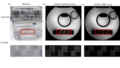 |
5 |
MUSIC-MRI: unleash the resolving power of k-space beyond the
Fourier transform
Dongbiao Sun1,2,
Yan Zhuo1,2,
Lin Chen2,3,
and Zihao Zhang1,3
1State Key Laboratory of Brain and Cognitive Science, Beijing MR Center for Brain Research, Institute of Biophysics, Chinese Academy of Sciences, Beijing, China, 2University of Chinese Academy of Sciences, Beijing, China, 3Institute of Artificial Intelligence, Hefei Comprehensive National Science Center, Hefei, China Keywords: Image Reconstruction, Image Reconstruction, Non-Fourier transform reconstruction Modern MRI reconstructs images by performing Fourier transform (FT) on k-space to decompose the signal on an orthogonal basis composed of trigonometric functions. We are inspired by the problem of estimating the Direction of Arrival in radar theory and propose a novel route for MRI reconstruction based on multiple signal classification (MUSIC). MUSIC-MRI overcomes the sidelobe problem of FT and significantly promotes the actual resolution in the meaning of full width at half maximum (FWHM). Our phantom experiments show that the FWHMs are 0.45mm by MUSIC-MRI and 1.50mm by FT, while the nominal resolution of the k-space data is 0.94mm. |
|
4952.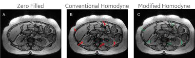 |
6 |
Modified Homodyne reconstruction using a high-resolution phase
in magnetic resonance images
Xinzeng Wang1,
Daniel Litwiller2,
Arnaud Guidon3,
Tim Sprenger4,
and Robert Marc Lebel5
1GE Healthcare, Houston, TX, United States, 2GE Healthcare, Denver, CO, United States, 3GE Healthcare, Boston, MA, United States, 4GE Healthcare, Stockholm, Sweden, 5GE Healthcare, Calgary, AB, Canada Keywords: Image Reconstruction, Artifacts, Partial Fourier A partial Fourier acquisition has been widely used for fast MR imaging. To reduce the truncation artifacts in partial Fourier image, Homodyne reconstruction is often used, and it exploits the conjugate symmetry in real-valued signal to recover the full k-space. However, the MR signal is complex-valued. Artifacts are commonly observed in Homodyne images in the regions of rapid phase change due to the interference of imaginary components of adjacent pixels. In this work, we proposed a modified Homodyne reconstruction to reduce the conventional Homodyne artifacts and truncation artifacts by using a high-resolution phase from a pre-trained deep-learning network. |
|
4953.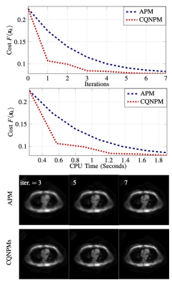 |
7 |
Complex Quasi-Newton Proximal Methods for the Image
Reconstruction in Compressed Sensing MRI
Tao Hong1,
Jeffrey A. Fessler2,
and Luis Hernandez-Garcia 1
1Department of Radiology, University of Michigan, Ann Arbor, MI, United States, 2Department of Electrical and Computer Engineering, University of Michigan, Ann Arbor, MI, United States Keywords: Image Reconstruction, Data Processing, Fast reconstruction algorithm This work studies a complex quasi-Newton proximal method (CQNPM) for MRI reconstruction using wavelets or total variation (TV) based regularization. Our experiments show that our method is faster than the accelerated proximal method [1,2] in terms of iteration and CPU time. |
|
4954.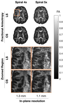 |
8 |
High-resolution single-shot spiral diffusion-weighted imaging at
7T using the expanded encoding model and compressed sensing
Paul I. Dubovan*1,2,
Gabriel Varela-Mattatall*1,2,
Tales Santini1,2,
Kyle M. Gilbert1,2,
Ravi S. Menon1,2,
and Corey A. Baron1,2
1Centre for Functional and Metabolic Mapping (CFMM), Robarts Research Institute, Western University, London, ON, Canada, 2Department of Medical Biophysics, Schulich School of Medicine and Dentistry, Western University, London, ON, Canada Keywords: Image Reconstruction, Image Reconstruction Reconstruction problems involving the expanded encoding model and field monitoring are typically solved using least squares optimization with the conjugate gradient method and early stopping as an implicit form of regularization. However, this is likely a suboptimal strategy for low SNR acquisitions, such as accelerated or high-resolution diffusion MRI. Hence, in this work we present an extension to the matMRI reconstruction toolbox that incorporates compressed sensing regularization. Results demonstrate that the expanded encoding model and compressed sensing regularization are complementary tools that mitigate artifacts from phase perturbations while permitting lower SNR conditions for high-resolution single-shot spiral diffusion-weighted imaging. |
|
4955.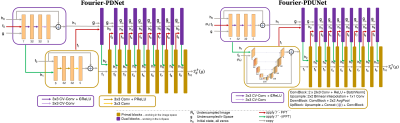 |
9 |
Complex-Valued Fourier Primal-Dual: Undersampled MRI
Reconstruction in Hybrid-space
Soumick Chatterjee1,2,3,
Philipp Ernst1,2,
Oliver Speck3,4,5,
and Andreas Nürnberger1,2,5
1Faculty of Computer Science, Otto von Guericke University Magdeburg, Magdeburg, Germany, 2Data and Knowledge Engineering Group, Otto von Guericke University Magdeburg, Magdeburg, Germany, 3Department of Biomedical Magnetic Resonance, Otto von Guericke University Magdeburg, Magdeburg, Germany, 4German Center for Neurodegenerative Disease, Magdeburg, Germany, 5Center for Behavioral Brain Sciences, Magdeburg, Germany Keywords: Image Reconstruction, Artifacts Iterative undersampled MRI reconstructions, such as compressed sensing, can reconstruct undersampled MRIs - but due to their slow execution speed, they are not suitable for real-time applications. Several deep learning approaches have been proposed, mostly working in image space. Some of the approaches, which work on the k-space or in a mix of spaces, employ real-valued convolutions splitting the complex k-space into real and imaginary parts for processing - destroying the geometric relationship within the data. This research proposes Fourier-PD and Fourier-PDUNet models using complex-valued convolutions, which attempt to predict missing k-space frequencies and also to reduce artefacts in the image space. |
|
4956.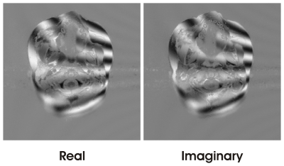 |
10 |
Statistical Approach to Single-Point Water-Fat Imaging with
Independent Component Analysis (ICA)
Qing-San Xiang1
1Radiology, University of British Columbia, Vancouver, BC, Canada Keywords: Image Reconstruction, Image Reconstruction, water-fat imaging Water-Fat Imaging with only a single acquisition is desirable for its pulse sequence simplicity and scan time efficiency. However, robust reconstruction is more challenging due to limited available information. In this work, it is discovered that some previously unused information can be found by statistical analysis, leading to effective reconstruction of water and fat images. In particular, Independent Component Analysis (ICA) was used to determine the phase error. Well separated water and fat images were subsequently obtained after phase correction. |
|
4957.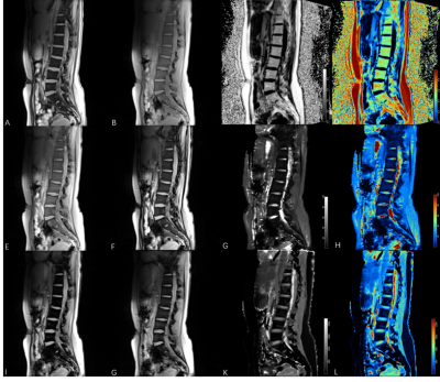 |
11 |
Feasibility study of a simulTaneous multi-relaXation-time
Imaging (TXI) method in Intervertebral Disc
Jie Yang1,
Yujian liu1,2,
Wenjia Hang1,
Meining Chen3,
Jianqi Li4,
Yinqiao Yi4,
Haodong Zhong4,
and Xu Yan3
1Department of Radiology, Zigong First People's Hospital, Zigong, China, 2Sichuan Vocational College of Health and Rehabilitation, Zigong, China, 3MR Scientific Marketing, Siemens Healthineers, Shanghai, China, 4Shanghai Key Laboratory of Magnetic Resonance, East China Normal University, Shanghai, China Keywords: Image Reconstruction, MSK Quantitative imaging of intervertebral disc degeneration is important for the diagnosis of lower back pain. In our study, a simulTaneous multi-relaXation-time Imaging (TXI) method was evaluated, which generated bone marrow fat fraction (PDFF), R2* and R1 mapping within a single scan. The results showed that the BMFF, T2*, T1 mapping calculated from TXI method were close to the values reported in other studies for lumbar discs. |
|
4958.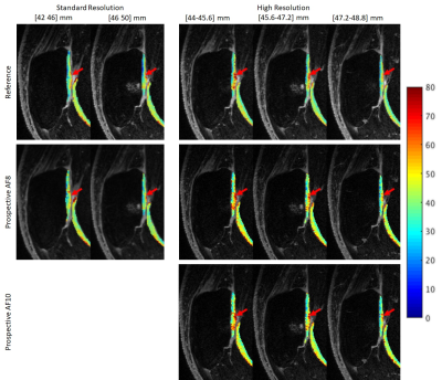 |
12 |
Retrospective and prospective accelerated T1ρ/T2 mapping with
Compressed Sensing: high resolution T1ρ mapping and simultaneous
T1ρ/T2 mapping
Jeehun Kim1,2,
Zhiyuan Zhang1,3,
Ruiying Liu4,
Brendan Eck1,
Mingrui Yang1,
Hongyu Li4,
Mei Li1,
Richard Lartey1,
Carl S. Winalski1,5,
Leslie Ying4,6,
and Xiaojuan Li1,5
1Department of Biomedical Engineering, Program of Advanced Musculoskeletal Imaging (PAMI), Cleveland Clinic, Cleveland, OH, United States, 2Department of Electrical, Computer, and Systems Engineering, Case Western Reserve University, Cleveland, OH, United States, 3Department of Biomedical Engineering, Case Western Reserve University, Cleveland, OH, United States, 4Electrical Engineering, University at Buffalo, State University of New York, Buffalo, NY, United States, 5Department of Diagnostic Radiology, Imaging Institute, Cleveland Clinic, Cleveland, OH, United States, 6Biomedical Engineering, University at Buffalo, State University of New York, Buffalo, NY, United States Keywords: Image Reconstruction, Cartilage Quantitative MR T1rho T2 imaging shows promising results on detecting early-stage osteoarthritis, but long scan time limits the spatial resolution, making it vulnerable to partial volume averaging. Such effect reduces the sensitivity to small focal degeneration. In this study, compressed sensing reconstruction with spatio-temporal finite difference regularization was used to accelerate high-resolution (slice thickness < 2mm) T1rho imaging and standard-resolution simultaneous T1rho and T2 imaging, and evaluated the result comparing with reference imaging, retrospective and prospective reconstruction, and scan-rescan repeatability. |
|
4959.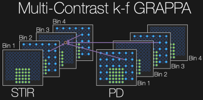 |
13 |
Accelerating Multi-Contrast Imaging Near Metallic Implants with
Variable Resolution Sampling and Joint Reconstruction
Nikolai J Mickevicius1,
Andrew S Nencka1,
and Kevin M Koch1
1Medical College of Wisconsin, Milwaukee, WI, United States Keywords: Image Reconstruction, Image Reconstruction In multi-spectral imaging near metallic implants, redundant information is present in neighboring spectral bins and contrasts due to the overlapping and smooth spectral profiles. This redundancy is exploited here to reduce scan time when multiple image contrasts are desired. |
|
4960.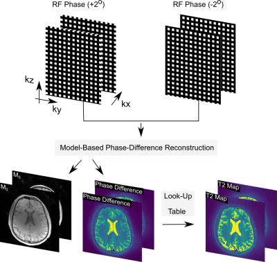 |
14 |
Model-based phase-difference reconstruction for accelerated
phase-based T2 mapping
Xiaoqing Wang1,2,
Jaejin Cho1,2,
Yohan Jun1,2,
Borjan Gagoski3,
and Berkin Bilgic1,2
1Athinoula A. Martinos Center for Biomedical Imaging, Massachusetts General Hospital, Charlestown, MA, United States, 2Department of Radiology, Harvard Medical School, Boston, MA, United States, 3Fetal-Neonatal Neuroimaging and Developmental Science Center, Boston Children's Hospital, Harvard Medical School, Boston, MA, United States Keywords: Image Reconstruction, Brain, model-based recontruction Phase-based T2 mapping acquires two GRE volumes with opposite RF phase increments, then computes the phase difference and relates this to T2 values through Bloch modeling. We exploit the redundancy in these two acquisitions by posing this as a nonlinear inverse problem, and directly estimate the phase difference from k-space. This reduces the problem to the estimation of a complex M0 volume, and a real-valued phase difference, thus reducing the number of real-valued unknowns from 4 to 3 per voxel. The proposed reconstruction flexibly admits additional regularization, and yields higher quality images from undersampled acquisitions for rapid T2 mapping. |
|
4961.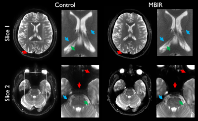 |
15 |
Evaluation of a model-based image reconstruction technique for
accelerated point spread function encoded echo planar imaging
Nolan Meyer1,2,
Myung-Ho In1,
David F Black1,
Norbert G Campeau1,
Kirk M Welker1,
John Huston III1,
Matt A Bernstein1,
and Joshua D Trzasko1
1Radiology, Mayo Clinic, Rochester, MN, United States, 2Mayo Clinic Graduate School of Biomedical Sciences, Rochester, MN, United States Keywords: Image Reconstruction, Neuro, Clinical, radiologist, evaluation A model-based image reconstruction (MBIR) platform was developed for accelerated point spread function encoded echo planar imaging. Beginning with retrospectively truncated data, images were reconstructed with the enhanced MBIR framework. These were compared by a team of neuroradiologists with images obtained using standard reconstructions from non-truncated datasets. Scores were assigned for evaluation criteria including high-contrast resolution (HCR), low-contrast detectability (LCD), signal to noise ratio (SNR), artifacts, and overall preference. Nonparametric statistical test results indicate preference for MBIR in HCR, LCD, and overall preference, with comparable results for SNR and artifacts. MBIR thus facilitates PSF-EPI acceleration, enhancing clinical practicality. |
|
4962.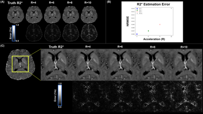 |
16 |
Fast $$$T_{2}^{*}$$$ and QSM using locally low rank methods for
complex-valued, multi-echo GRE reconstruction
Charles Iglehart1,
Ali Bilgin1,2,3,
and Manojkumar Saranathan4,5
1Electrical and Computer Engineering, The University of Arizona, Tucson, AZ, United States, 2Biomedical Engineering, The University of Arizona, Tucson, AZ, United States, 3Medical Imaging, The University of Arizona, Tucson, AZ, United States, 4Radiology, UMass Chan Medical School, Worcester, MA, United States, 5Neuroscience, Morningside Graduate School of Biomedical Sciences, Worcester, MA, United States Keywords: Image Reconstruction, Brain $$$T_{2}^{*}$$$ and quantitative susceptibility mapping are of increasing clinical interest, but typically require lengthy ME-GRE scans. To accelerate such acquisitions, we propose and implement a volumetric reconstruction method employing locally low rank (LLR) techniques to recover complex-valued ME images from highly undersampled k-space data that is suitable for parameter mapping. We present results for reconstructed magnitude and phase across multiple datasets and acceleration ratios, as well as computed $$$R_{2}^{*}$$$ and QSM from reconstructed images. |
|
4963.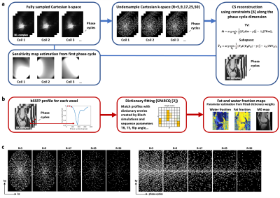 |
17 |
Low Rank Subspace-Constrained Compressed Sensing Reconstruction
of Highly Accelerated Phase-Cycled bSSFP MRI for Fat Fraction
Quantification
Eva S Peper1,2,
Adèle LC Mackowiak1,2,3,
Berk C Açıkgöz1,2,
Nils Plähn1,2,
Yasaman Safarkhanlo4,
Li Feng5,
and Jessica AM Bastiaansen1,2
1Department of Diagnostic, Interventional and Pediatric Radiology (DIPR), Inselspital, Bern University Hospital, University of Bern, Bern, Switzerland, 2Translation Imaging Center (TIC), Swiss Institute for Translational and Entrepreneurial Medicine, Bern, Switzerland, 3Department of Diagnostic and Interventional Radiology, Lausanne University Hospital (CHUV) and University of Lausanne (UNIL), Lausanne, Switzerland, 4Department of Cardiology, University Hospital Bern, Bern, Switzerland, 5BioMedical Engineering and Imaging Institute (BMEII), Department of Radiology, Icahn School of Medicine at Mount Sinai, New York, NY, United States Keywords: Image Reconstruction, Sparse & Low-Rank Models, bSSFP, phase-cycled bSSFP, subspace reconstruction Fat fraction (FF) maps can be estimated from phase-cycled (PC) balanced steady-state free precession (bSSFP) imaging using a dictionary fitting approach (SPARCQ). Three-dimensional PC bSSFP imaging, however, is time consuming, since around 16 PCs must be sampled to fully describe a voxels’ PC profile. Compressed sensing (CS) has been proven to accelerate MRI scans. This study has two aims 1) to evaluate the performance of two CS algorithms (total variation (TV)- vs. subspace-constrained) taking regularisation along the PC dimension into account and 2) to investigate the upper limits of acceleration using these techniques on FF quantification using SPARCQ. |
|
4964.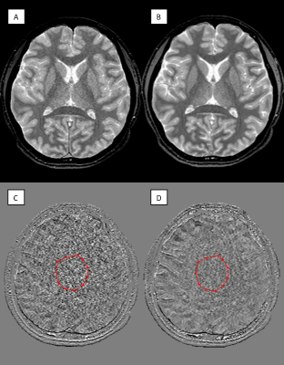 |
18 |
Constrained Reconstruction of White Noise (CROWN) for
Strategically Acquired Gradient Echo (STAGE) Imaging
Paul Kokeny1,
Qiuyun Xu1,
Sara Gharabaghi1,
Sean Sethi1,2,
Yu Liu3,
Youmin Zhang3,
Peng Liu3,
Naying He3,
Fuhua Yan3,
and E. Mark Haacke1,2,4
1SpinTech MRI, Bingham Farms, MI, United States, 2Radiology, Wayne State University, Detroit, MI, United States, 3Shanghai Jiao Tong University School of Medicine, Ruijin Hospital, Shanghai, China, 4Neurology, Wayne State University, Detroit, MI, United States Keywords: Image Reconstruction, Quantitative Imaging By using the inherent relationship between proton spin density (PSD) and T1, we propose a new image-processing approach to reduce noise called CROWN (Constrained Reconstruction of White Noise). Firstly, we established a linear relationship between these two parameters, then applied a cost function to constrain simulated Strategically Acquired Gradient Echo (STAGE) PSD map and T1 map data in the presence of noise. Secondly, we applied this approach to in vivo STAGE images to reduce noise and improve SNR without the loss of image detail. CROWN has the potential to make higher resolution or faster imaging viable with improved SNR. |
|
4965.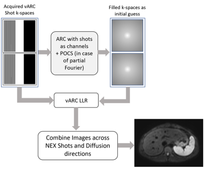 |
19 |
Diffusion Weighted MR Imaging Using Low Rank Reconstruction for
Multi-shot Variable Auto-Calibrating (vARC) Sampling with Volume
Coil
Nitin Jain1,
Ashok Kumar P Reddy1,
Rajdeep Das1,
Sajith Rajamani1,
Rajagopalan Sundaresan1,
Harsh Kumar Agarwal1,
M Ramasubba Reddy2,
and Ramesh Venkatesan1
1GE Healthcare, Bangalore, India, 2Indian Institute of Technology Madras, Chennai, India Keywords: Image Reconstruction, Body Diffusion weighted MR imaging (DWI) is key to pathology detection in anatomies such as brain, abdomen and prostate. Echo planar imaging (EPI) provides a rapid means to acquire DWI. EPI with variable k-space sampling scheme and an auto-calibrating image reconstruction technique, vARC, has recently been shown to reduce distortion in DWI and improve the image quality in single channel volume coil/body coil acquisitions. Here, we propose a new low rank reconstruction technique for robust reconstruction and improved image quality for DWI acquired using vARC’s EPI multi-shot acquisition scheme with single channel body coil. |
|
4966.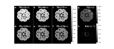 |
20 |
Joint K-TE reconstruction for T2 mapping with the weighted
linear regression fitting
Yan Dai1,
Jie Deng1,
and Xun Jia2
1University of Texas Southwestern Medical Center, Dallas, TX, United States, 2Johns Hopkins University, Baltimore, MD, United States Keywords: Image Reconstruction, Quantitative Imaging We developed a joint quantitative imaging reconstruction algorithm to reconstruct T2-weighted (T2W) images and T2 mapping simultaneously through an iterative optimization process, which integrated the weighted linear regression fitting. The proposed method achieved accurate T2 measurements and improved signal-to-noise (SNR) of both T2W images and the T2 map. |
|
 View Presentation Video
 Back to Meeting Home
 Back to the Program-at-a-Glance