Digital Poster
Sequence Design for Quantitative Imaging I
ISMRM & ISMRT Annual Meeting & Exhibition • 03-08 June 2023 • Toronto, ON, Canada

Digital Poster
Sequence Design for Quantitative Imaging I
| Computer # | |||
|---|---|---|---|
2195.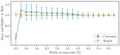 |
21 | Comparison of k-space Sampling Patterns for MR Fingerprinting
Felix Horger1,2,3, Raphael Tomi-Tricot1,2,3,4, Pierluigi Di Cio1,3, Joseph Hajnal1,2,3, and Shaihan Malik1,2,3
1Biomedical Engineering Department, School of Biomedical Engineering & Imaging Sciences, King's College London, London, United Kingdom, 2Centre for the Developing Brain, School of Biomedical Engineering & Imaging Sciences, King's College London, London, United Kingdom, 3London Collaborative Ultra high field System (LoCUS), London, United Kingdom, 4MR Research Collaborations, Siemens Healthcare Limited, Frimley, United Kingdom Keywords: MR Fingerprinting/Synthetic MR, In Silico The temporal low-rank property of signals produced by MR Fingerprinting can be used to circumvent reconstruction of individual time frames. Instead, temporally compressed components are estimated. Since every acquired sample then contributes to every compressed component, it is unclear which k-space sampling pattern is optimal. This work collects evidence for higher robustness of radial sampling towards measurement errors and its superior ability to resolve rapidly varying temporal signals, compared to a Cartesian pattern, simulated in a realistic scenario and supported by an in vivo observation. The aim is making informed decisions about optimal sampling patterns for MRF. |
|
2196.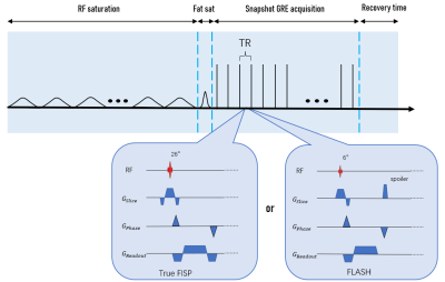 |
22 | Snapshot CEST at 3T using 3D True FISP
Yupeng Wu1, Zhichao Wang2, Qifan Pang1, Gaiying Li1, Mengying Chen1, Xu Yan3, Caixia Fu4, Yang Song3, and Jianqi Li1
1Shanghai key lab of magnetic resonance, East China Normal University, Shanghai, China, 2Zhejiang Lab, Hangzhou, China, 3MR Scientific Marketing, Siemens Healthineers, Shanghai, China, 4MR Application Development, Siemens Shenzhen Magnetic Resonance Ltd, Shenzhen, China Keywords: Pulse Sequence Design, CEST & MT, GRE FLASH TrueFISP For clinical implementation, chemical exchange saturation transfer (CEST) imaging must be fast with high signal‐to‐noise ratio (SNR), 3D coverage, and produce robust contrast. In general, CEST imaging with fast low angle shot GRE sequence (FLASH) can achieve fast acquisition, but may yield low SNR efficiency. This study evaluated APT and NOE maps for single saturation 3D CEST imaging with True FISP and FLASH sequences, respectively. We found that True FISP could achieve better SNR of Z-spectrum and obtain more reliable CEST effects contrast. 3D True FISP sequence is expected to become a more promising 3D CEST-GRE sequence for clinical application. |
|
2197.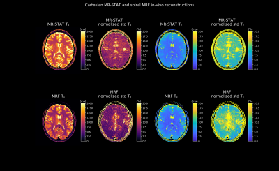 |
23 | Cartesian MR-STAT vs spiral MR Fingerprinting: a comparison
Oscar van der Heide1,2, Mariya Doneva3, Peter Koken3, Jakob Meineke3, Miha Fuderer1,2, Cornelis A.T. van den Berg1,2, and Alessandro Sbrizzi1,2
1Computational Imaging Group for MR Diagnostics and Therapy, Center for Image Sciences, University Medical Center Utrecht, Utrecht, Netherlands, 2Department of Radiology, Division of Imaging and Oncology, University Medical Center Utrecht, Utrecht, Netherlands, 3Philips Research, Hamburg, Germany Keywords: MR Fingerprinting/Synthetic MR, Quantitative Imaging MR Fingerprinting (“MRF”) and MR Spin Tomography in Time-domain (“MR-STAT”) are quantitative MRI techniques that allow multiple quantitative tissue parameter maps (e.g. T1, T2 and proton density) to be estimated from a single short scan. In this work, we compare the accuracy and precision of Cartesian MR-STAT and spiral MRF on gel phantoms and in-vivo. On gel phantoms we get excellent agreement with reference measurements. In-vivo we observe differences in reconstructed T1 values. As for the precision, a more in-depth study is required to take into account differences in noise-suppression mechanisms (i.e. k-space apodization and spiral sampling window).
|
|
2198.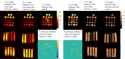 |
24 | 3D Extension of the multi parametric acquisition Multi-Phase balanced non-Steady State Free Precession
Riwaj Byanju1, Gyula Kotek1, Mika W. Vogel2, Juan A Hernandez-Tamames1, and Dirk H. J. Poot1
1Erasmus MC, Rotterdam, Netherlands, 2GE Healthcare, Hoevelaken, Netherlands Keywords: Pulse Sequence Design, Quantitative Imaging We propose a 3D extension of the novel MP-b-nSSFP sequence, which interleaves RF pulse types for Multiparametric mapping from the transient response. Comparing selective versus non-selective refocusing pulses we observe lower bias with non-selective pulses, despite modelling the spatially varying effect of the pulses in the fitting process. Phantom and in-vivo comparison to QRAPTEST (MAGIC) are performed. |
|
2199.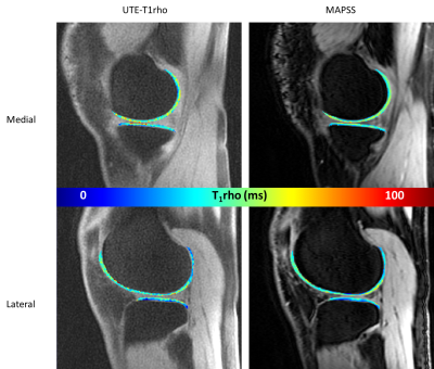 |
25 | Comparison of UTE-T1ρ vs. MAPSS-T1ρ Sequences in In-Vivo Knees
Sara E Sacher1, Michael Carl2, Hollis G Potter1, and Matthew F Koff1
1Hospital for Special Surgery, New York, NY, United States, 2GE Healthcare, San Diego, CA, United States Keywords: Pulse Sequence Design, Quantitative Imaging, Ultrashort echo time The relative accuracy of an ultrashort echo time (UTE) based T1ρ sequence was compared to a MAPSS based T1ρ sequence in the evaluation of articular cartilage. Scanning with similar parameters between the acquisitions was performed, producing similar T1ρ values in all cartilage sub-compartments except for the femoral trochlea (TrF). TrF MAPSS-T1ρ values were shorter than TrF UTE-T1ρ values (10.91% difference, p = 0.0028). Overall, there was reasonable agreement between the two sequences indicating that UTE-T1ρ may be a promising method to use in place of conventional MAPSS sequences to quantify T1ρ values of articular cartilage. |
|
2200.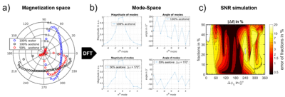 |
26 | Decoding of 3T and 7T bSSFP profile asymmetries for T1, T2, and fraction quantification in two-compartment systems
Nils Marc Joel Plähn1, Adèle Mackowiak2, Berk Açikgöz3, and Jessica Bastiaansen3
1Department of Diagnostic, Interventional and Pediatric Radiology (DIPR), Inselspital, Bern Universit, Bern, Switzerland, 2Department of Radiology, Lausanne University Hospital (CHUV) and University of Lausanne (UNIL), Lausanne, Switzerland, 3Department of Diagnostic, Interventional and Pediatric Radiology (DIPR), Inselspital, Bern University Hospital, University of Bern, Bern, Switzerland Keywords: Data Analysis, High-Field MRI, phase-cycled bSSFP, Aceton fraction quantification A novel Off-Resonant encoded Analytical parameter quantification using Complex Linearized Equations (ORACLE) method using phase-cycled bSSFP profiles was developed. The approach decodes complex asymmetry profiles in multi-compartment systems for simultaneous proton fraction, T1/T2 ratio, T1 and T2 quantification. The approach was validated in simulations and in an acetone-water phantom at 3T and 7T. Simulations and experiments validated the proposed method for multi-parameter quantification using phase-cycled bSSFP for two compartment singlet systems with high accuracy and precision. This provides the first step towards proton fraction quantification of more complex multiplet-systems, such as fat or myelin, exploiting complex asymmetry profiles. |
|
2201.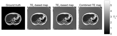 |
27 | Split spin-echo acquisition for ultrashort T2* mapping: a simulation study
Mikhail Zubkov1 and Irina Melchakova1
1School of Physics and Engineering, ITMO University, Saint Petersburg, Russian Federation Keywords: Data Acquisition, Pulse Sequence Design MRI serves as a non-invasive way of assessing the degree of iron overload. This is done via T2*-mapping commonly performed with muti-echo gradient echo sequences. This approach fails in cases with extreme iron overload where the relaxation times drop down to submillisecond range. A method allowing ultra-short T2* mapping is simulated, employing a temporal acquisition offset in a spin-echo pulse sequence. Two acquisitions are suggested: one – before the spin-echo centre, and another – after, resulting in two different T2*-weighted images, acquired with half-line radial encoding. This method allows obtaining T2*-weighted images with weightings starting effectively at zero echo time. |
|
2202.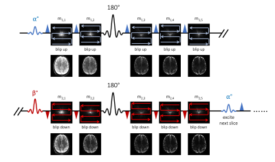 |
28 | VUDU-SAGE: Efficient T2 and T2* Mapping using Joint Reconstruction for Motion-Robust, Distortion-Free, Multi-Shot, Multi-Echo EPI
Jaejin Cho1,2, Tae Hyung Kim3, Avery JL Berman4, Yohan Jun1,2, Xiaoqing Wang1,2, Borjan Gagoski2,5, and Berkin Bilgic1,2,6
1Athinoula A. Martinos Center for Biomedical Imaging, Charlestown, MA, United States, 2Harvard Medical School, Boston, MA, United States, 3Computer Engineering, Hongik University, Seoul, Korea, Republic of, 4Carleton University, Ottawa, ON, Canada, 5Fetal-Neonatal Neuroimaging & Developmental Science Center, Boston Children’s Hospital, Boston, MA, United States, 6Harvard/MIT Health Sciences and Technology, Cambridge, MA, United States Keywords: Data Acquisition, Quantitative Imaging, T2 and T2* mapping We demonstrate T2 and T2* mapping and joint reconstruction for multi-echo, multi-shot EPI data from a variable flip angle, blip-up and -down undersampling (VUDU) for spin and gradient echo (SAGE) acquisition. VUDU-SAGE employs a blip-up and -down acquisition strategy for correcting B0 distortion, and FLEET-ordering for motion-robust multi-shot EPI while maximizing signal using variable flip angles. VUDU-SAGE acquires five echoes consisting of two gradient-echo, two mixed, and one spin-echo contrasts. We jointly reconstruct all echoes and estimate T2 and T2* maps using Bloch dictionary matching. In-vivo experiment presents T2 and T2* map at 1x1x4mm3 resolution with a 9-second acquisition. |
|
2203.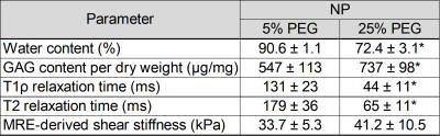 |
29 | Effects of Nucleus Pulposus Tissue Hydration on T1ρ and T2 Relaxation Times and Mechanical Properties
Megan Co1, Brian Raterman2, Arunark Kolipaka1,2, and Benjamin A Walter1,3
1Department of Biomedical Engineering, The Ohio State University, Columbus, OH, United States, 2Department of Radiology, The Ohio State University, Columbus, OH, United States, 3Spine Research Institute, The Ohio State University, Columbus, OH, United States Keywords: Data Acquisition, Quantitative Imaging, Spine, Intervertebral Disc T1ρ and T2 mapping have been developed to quantitively assess intervertebral disc (IVD) degeneration by quantifying proteoglycan and water content, respectively. In addition, magnetic resonance elastography (MRE) has been validated to quantify shear stiffness. This study determines how water content affects MRI relaxation times and MRE-derived mechanical properties. Our results showed that hydration has an influence on T1ρ and T2 relaxation times and MRE-derived shear stiffnesses and that there is a correlation between relaxation times and shear stiffnesses. This highlights MRI as a non-invasive technique to quantify tissue composition and mechanical properties to assess degenerative changes within the IVD. |
|
2204.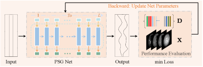 |
30 | Unsupervised Learning-based Pulse Sequence Optimization framework for Magnetic Resonance Fingerprinting
Peng Li1, Yinghao Zhang1, Xin Lu2, and Yue Hu1
1Harbin Institute of Technology, Harbin, China, 2De Montfort University, Leicester, United Kingdom Keywords: Pulse Sequence Design, MR Fingerprinting The optimal design of the Magnetic resonance fingerprinting (MRF) sequence is still challenging due to the optimization of high-degrees-of-freedom acquisition parameters. In this paper, we propose a novel unsupervised learning-based pulse sequence design framework for efficient MRF sequence optimization. Specifically, we propose a novel pulse sequence generation network (PSG-Net) that fully exploits the sequence correlation to generate the optimal pulse sequence from a zero-initialized input. To achieve improved precision of parameter estimation, we use a predefined pulse sequence performance evaluation function that can directly represent tissue quantification separability as the loss function to update the parameters of the PSG-Net. |
|
2205.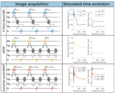 |
31 | Phase-based T2 mapping using dual phase-cycled balanced SSFP imaging
Merijn Berendsen1, Maša Bozic-Iven1,2, Joao Tourais1, Chiara Coletti1, Ingo Hermann1,2, and Sebastian Weingärtner1
1Delft University of Technology (TU Delft), Delft, Netherlands, 2Computer Assisted Clinical Medicine, Medical Faculty Mannheim, Heidelberg University, Mannheim, Germany Keywords: Pulse Sequence Design, Pulse Sequence Design Phased-based techniques have shown promise for scan time efficient quantification of T2 relaxation times. However, previously proposed methods use an unbalanced Gradient Echo approach, which shows residual sensitivity to B1+ and T1 changes. In this work, a dual phase-cycled balanced SSFP approach is proposed. Simulations and phantom measurements show its resilience against field inhomogeneities. Excellent agreement between phase values was obtained in phantom and simulations. Initial phantom acquisitions at 3T yield T2 maps with visually high map quality and close agreement to a spin-echo reference. Future evaluation of in vivo robustness and inter- and intra-subject repeatability is warranted. |
|
2206.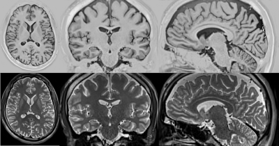 |
32 | Fast, High-Resolution, and Optimal Contrast MRI
Yao Sui1,2, Onur Afacan1,2, Camilo Jaimes1,2, Ali Gholipour1,2, and Simon K Warfield1,2
1Harvard Medical School, Boston, MA, United States, 2Boston Children's Hospital, Boston, MA, United States Keywords: Image Reconstruction, Quantitative Imaging Any MRI practices prefer high resolution, high signal-to-noise ratio, short scan time, and high contrast. Unfortunately, fast scan leads to low resolution while high-resolution scan results in a reduced signal-to-noise ratio. In particular, it is challenging for radiologists and technologists, who perform MRI scans, to find optimal sequence parameters for each patient, leading to sub-optimal contrast. We developed a new methodology that enables fast and high-resolution brain MRI with improved signal-to-noise ratio and optimal contrast between white-matter and gray-matter for each individual patient, based on quantitative imaging and reconstruction techniques. Experiments on clinical data demonstrated the advantages of our approach. |
|
2207.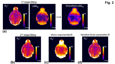 |
33 | An improved postprocessing method for more accurate R2* measurements in the presence of macroscopic B0 field variations
Chu-Yu Lee1, Olivia Lullmann2, Emily J Steinbach2, Daniel R Thedens1, Lyndsay A Harshman2, and Vincent A Magnotta1
1Department of Radiology, The University of Iowa, Iowa City, IA, United States, 2Department of Pediatrics, The University of Iowa, Iowa City, IA, United States Keywords: Data Processing, Relaxometry, R2*; Macroscopic field; Correction Macroscopic field variations result in an overestimate of R2*. One common approach to correct the bias is to utilize a three-parameter fit; however, the fitting is subject to overfitting. This study presents a new two-stage fitting of the three-parameter model by assuming that the macroscopic field variation is slowly varying in space. Using a numerical simulation and in vivo mice data, we demonstrate that the proposed method allows for a more accurate R2* measurement, and is less sensitive to noise. |
|
2208.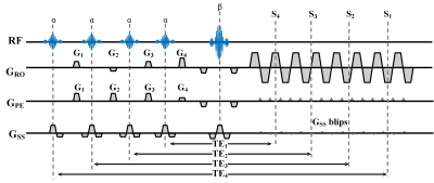 |
34 | Sub-second T2 mapping of the whole brain via multiband SENSE multiple overlapping-echo detachment imaging and deep learning
Simin Li1, Taishan Kang2, Jian Wu1, Weikun Chen1, Zhigang Wu3, Jiazheng Wang3, Congbo Cai1, and Shuhui Cai1
1Department of Electronic Science, Xiamen University, Xiamen, China, 2Department of Radiology, Zhongshan Hospital of Xiamen University, School of Medicine, Xiamen University, Xiamen, China, 3MSC Clinical & Technical Solutions, Philips Healthcare, Beijing, China Keywords: Image Reconstruction, Quantitative Imaging Most quantitative magnetic resonance imaging (qMRI) methods are time-consuming. Multiple overlapping-echo detachment (MOLED) imaging can achieve quantitative parametric mapping for a single slice within hundred milliseconds. To further accelerate MOLED, we combine MOLED with multiband SENSE (MB-SENSE) technology to achieve simultaneous multi-slice T2 mapping. To solve the problem of reconstructed image quality degraded caused by a high multiband factor MB, a plug-and-play (PnP) approach with prior denoisers was applied for image restoration to realize denoising at a high MB. The proposed multiband multiple overlapping-echo detachment (MB-MOLED) imaging can achieve sub-second T2 mapping of the whole brain with a high MB. |
|
2209.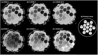 |
35 | Reduction of ADC bias with deep learning-based acceleration in diffusion-weighted MRI: A phantom validation study
Teresa Nolte1, Masami Yoneyama2, Chiara Morsch1, Alexandra Barabasch1, Maximilian Schulze-Hagen1, Johannes M. Peeters3, Christiane Kuhl4, and Shuo Zhang5
1Diagnostic and Interventional Radiology, Uniklinik RWTH Aachen University, Aachen, Germany, 2Philips Japan, Tokyo, Japan, 3Philips Healthcare, Best, Netherlands, 4Diagnostic and Interventional Radiology, University Hospital RWTH Aachen, Aachen, Germany, 5Philips GmbH Market DACH, Hamburg, Germany Keywords: Image Reconstruction, Diffusion/other diffusion imaging techniques, Noise Further acceleration of diffusion MRI in clinical examinations is desired but challenging mainly due to low signal and associated potential bias in the quantitative apparent diffusion coefficient (ADC) values. Artificial intelligence-based denoising and image reconstruction may provide a solution to address this challenge. We investigate and compare different image reconstruction methods, including conventional parallel imaging, compressed sensing, and a deep learning-based technique, in ADC accuracy and precision using a diffusion phantom with illustration of the principle in numeric simulation. |
|
2210.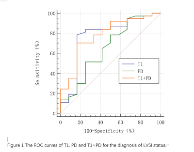 |
36 | Synthetic MRI derived quantitative mapping in predicting Lymphovascular interstitial infiltration status of cervical cancer
Zebo Huang1, Weiqiang Dou2, and Wenwei Tang1
1Nanjing Maternity and Child Health Care Hospital, Nanjing, China, 2GE Healthcare, MR Research China, Beijing, P.R. China, Beijing, China Keywords: MR Fingerprinting/Synthetic MR, Uterus, LVSI This study aimed to investigate whether synthetic-MRI derived quantitative maps can predict lymphovascular interstitial infiltration (LVSI) status in cervical cancer. 49 patients with cervical cancer were recruited with the status of LVSI confirmed by pathology. Synthetic MRI derived T1, T2 and PD mapping were obtained for each patient. Statistical differences were shown in T1 and PD values between LVSI-positive and LVSI-negative patients. An optimal diagnostic efficacy was further shown for T1+PD with high AUC of 0.777. With these findings, it can be concluded that relaxation maps derived from synthetic MRI may be helpful for predicting LVSI status in cervical cancer. |
|
2211.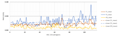 |
37 | Reliability and reproducibility of whole-brain quantitative MR relaxometry using different-channel-number coils
Ling Sang1, Weiyin Vivian Liu2, Hu Chen3, and Wen Chen1
1Department of Radiology, Taihe Hospital, Wuhan, China, 2GE Healthcare, Beijing, China, 3Department of Radiology,Taihe Hospital, Wuhan, China Keywords: Data Acquisition, Relaxometry Brain MAGiC imaging became widely applied in multi-center cooperation and in an individual longitudinal follow-up; however, acquisition with a backup coil at an emergency situation (i.e., a sudden coil breakdown) or a coil with different numbers of channels has not been explored on a 1.5T scanner yet. In contrast to small variation of PD values in the whole brain, T2 values varies relatively dramatically especially in cerebellum and showed statistically different between scans using different coils. It is worth noting that existence of inter-coil relaxometry difference should be careful for diagnosis. |
|
2212.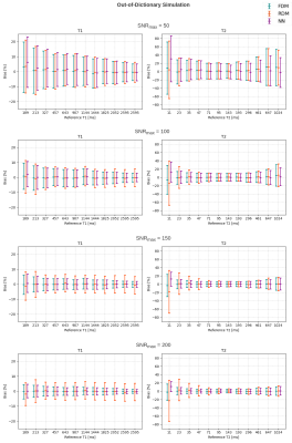 |
38 | Analysis of the Discretization Error vs. Estimation Time Tradeoff of MRF Dictionary Matching and the Advantage of the Neural Net-based Approach
Chinmay Rao1, Jakob Meineke2, Nicola Pezzotti3, Marius Staring1, Matthias van Osch1, and Mariya Doneva2
1Leiden University Medical Center, Leiden, Netherlands, 2Philips Research, Hamburg, Germany, 3Philips Research, Eindhoven, Netherlands Keywords: MR Fingerprinting/Synthetic MR, MR Fingerprinting Traditional MR fingerprinting involves matching the acquired signal evolutions against a dictionary of expected tissue fingerprints to obtain the corresponding tissue parameters. Since this dictionary is essentially a discrete representation of a physical model and the matching process amounts to brute-force search in a discretized parameter space, there arises a tradeoff between discretization error and parameter estimation time. In this work, we investigate this tradeoff and show via numerical simulation how a neural net-based approach solves it. We additionally conduct a phantom study using 1.5T and 3T data to demonstrate the consistency of neural net-based estimation with dictionary matching. |
|
2213.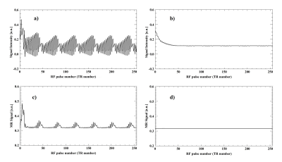 |
39 | Reproducing the effect of Steady-State stabilization on MR signals and images and its relation to pulse sequences by MRI simulation
Noriyuki Tawara1 and Daiki Tamada2
1Department of Radiological Sciences, Faculty of Health Sciences, Japan Healthcare University, Sapporo, Japan, 2Department of Radiology, Yamanashi University, Yamanashi, Japan Keywords: Visualization, Visualization The objective of this study was to reproduce the generation process of Steady-State and FLASH Band in MRI phenomena that cannot be reproduced by the actual equipment because of the restrictions imposed by venders, in order to directly confirm their relationship with pulse sequences. MRI simulation can reproduce Bloch equation faithfully, and thus it is possible to reproduce the relation between pulse sequences and the phenomena related to MRI as numerical data. |
|
 Back to Meeting Home
 Back to the Program-at-a-Glance
The International Society for Magnetic Resonance in Medicine is accredited by the Accreditation Council for Continuing Medical Education to provide continuing medical education for physicians.
 Back to Meeting Home
 Back to the Program-at-a-Glance
 View Presentation Video