Digital Poster
Accelerating Acquisitions
ISMRM & ISMRT Annual Meeting & Exhibition • 03-08 June 2023 • Toronto, ON, Canada

| Computer # | |||
|---|---|---|---|
4635.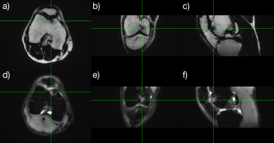 |
41 |
Fat/water separated & SMS accelerated pseudo 3D PROPELLER
Ola Norbeck1,2,
Henric Rydén1,2,
Adam van Niekerk1,
Enrico Avventi1,2,
Tim Sprenger3,
Stefan Skare1,2,
Johan Berglund4,
and Mikael Skorpil 1,2
1Karolinska Instituet, Stockholm, Sweden, 2Karolinska University Hospital, Stockholm, Sweden, 3MR Applied Science Laboratory Europe, GE Healthcare, Munich, Germany, 4Uppsala University Hospital, Uppsala, Sweden Keywords: Data Acquisition, Data Acquisition, MSK, Dixon, SMS By combining thin slices, PROPELLER acquisition, SMS acceleration and chemical shift encoding with asymmetrical gradients reformattable pseudo 3D volumes can be acquired. These volumes are, in contrast to 3D RARE volumes, efficiently chemical shift encoded, less susceptible to T2-blurring and less sensitive to motion. |
|
4636.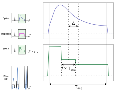 |
42 |
FSE Dixon with bandwidth-matched asymmetric readouts tailored
for real-valued fat/water estimates
Henric Rydén1,
Mikael Skorpil2,
Matea Borbas3,
and Adam van Niekerk1
1Clinical Neuroscience, Karolinska Institutet, Stockholm, Sweden, 2Karolinska Institutet, Stockholm, Sweden, 3Karolinska University Hospital, Stockholm, Sweden Keywords: Pulse Sequence Design, Fat, Dixon A novel asymmetric readout waveform for Dixon FSE imaging is presented for efficient sampling, tailored for real-valued estimation of fat/water estimates. |
|
4637.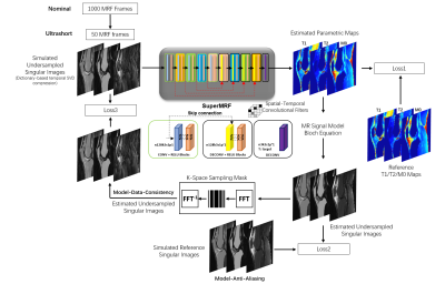 |
43 |
SuperMRF: Deep Robust Acceleration for MR Fingerprinting
Hongyu Li1,
Brendan L. Eck2,
Mingrui Yang2,
Jeehun Kim2,
Ruiying Liu1,
Peizhou Huang3,
Dong Liang4,
Xiaojuan Li2,
and Leslie Ying1,3
1Department of Electrical Engineering, University at Buffalo, State University of New York, Buffalo, NY, United States, 2Department of Biomedical Engineering, Program of Advanced Musculoskeletal Imaging (PAMI), Cleveland Clinic, Cleveland, OH, United States, 3Department of Biomedical Engineering, University at Buffalo, State University of New York, Buffalo, NY, United States, 4Paul C. Lauterbur Research Center for Biomedical Imaging, Medical AI research center, SIAT, CAS, Shenzhen, China Keywords: MR Fingerprinting/Synthetic MR, Image Reconstruction We propose a novel, deep learning-based method, “SuperMRF”, for the reconstruction of MR Fingerprinting (MRF) parametric maps that enables rapid image reconstruction. Built upon a convolutional neural network, SuperMRF uses three loss functions to incorporate additional information from the Bloch equations, estimated maps, de-aliasing, and data consistency losses. We investigate the use of SuperMRF for further acceleration of data acquisition by reducing the number of MRF time frames. Our results demonstrate that proposed SuperMRF is robust to noise and can achieve a 20x reduction in acquired MRF time frames. Tissue property maps can be reconstructed in less than one second. |
|
4638.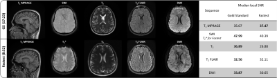 |
44 |
Accelerated MRI using intelligent protocolling and
subject-specific denoising
Keerthi Sravan Ravi1,2,
Gautham Nandakumar3,
Nikita Thomas3,
Mason Lim3,
Enlin Qian2,4,
Marina Manso Jimeno2,4,
Pavan Poojar5,
Zhezhen Jin6,
Patrick Quarterman7,
Maggie Fung7,
Girish Srinivasan3,
John Thomas Vaughan Jr.2,
and Sairam Geethanath5
1Biomedical Engineering, Columbia University, New York, NY, United States, 2Columbia Magnetic Resonance Research Center, Columbia University, New York, NY, United States, 3PhenoMX, Chicago, IL, United States, 4Department of Biomedical Engineering, Columbia University, New York, NY, United States, 5Accessible MR Laboratory, Biomedical Engineering and Imaging Institute, Dept. of Diagnostic, Molecular and Interventional Radiology,, Mt. Sinai, New York, NY, United States, 6Department of Biostatistics, Columbia University Irving Medical Center, New York, NY, United States, 7MR Clinical Solutions, GE, New York, NY, United States Keywords: Data Acquisition, Data Acquisition The routine brain screen protocol employed at our institution was accelerated using Look Up Tables to achieve a 1.94x gain in imaging throughput. Image-denoising was performed on accelerated data by leveraging deep learning models trained on contrast-specific publicly available datasets. These were corrupted by native noise during forward modeling. In addition, subject-specific denoising was demonstrated. The superior performance of denoised data on automated volumetry of Alzheimer’s Disease (AD) relevant brain anatomies on T1w data demonstrated potential for accelerated AD imaging. |
|
4639.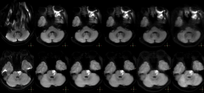 |
45 |
The Impact of Acceleration Factors of Compressed Sensing on the
Image Quality of the Fast Spin Echo Diffusion Weighted Imaging
for the Skull Base
Haonan Zhang1,
Qingwei Song1,
Jiazheng Wang2,
and Ailian Liu1
1Department of Radiology, the First Affiliated Hospital of Dalian Medical University, Dalian, Dalian, China, 2Philips Healthcare, Beijing, China, Beijing, China Keywords: Brain Connectivity, Brain Compared with echo planar imaging diffusion weighted iamging (EPI-DWI), turbo spin echo diffusion weighted imaging (TSE-DWI) can significantly reduce magnetic sensitivity artifacts in skull base imaging. However, the longer scan time limits its clinical promotion. The purpose of this study is to investigate the effect of the compression sensing acceleration factor on the image quality of TSE-DWI in the skull base area. |
|
4640.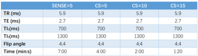 |
46 |
Highly accelerated acquisition of MP2RAGE using Compressed SENSE
Yang Zhao1,
Yishi Wang2,
Guangbin Wang1,
and Weibo Chen2
1Department of Radiology, Shandong Provincial Hospital Affiliated to Shandong First Medical University, Jinan, China, 2Philips Healthcare, Beijing, China Keywords: Data Acquisition, Data Acquisition, compressed sensing The magnetization-prepared 2 rapid acquisition gradient echo (MP2RAGE) sequence provides morphological T1-weighted images and quantitative T1 maps. Compressed sensing (CS) acceleration technique has been drawing extensive attention with its high acquisition efficiency for MRI sequence. In this study, volume measurement and ROI analysis were used to assess the images obtained from CS-MP2RAGE sequence under different acceleration factors. We find that CS-MP2RAGE show high similarity in brain structure volumes measurement and ROI analysis with traditional SENSE-MP2RAGE. Those findings prove that CS-MP2RAGE can be considered as an alternative to the SENSE-MP2RAGE, though the specific acceleration factor still needs to be further studied. |
|
4641.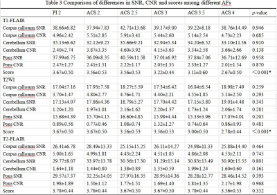 |
47 |
The effect of acceleration factor on brain magnetic resonance
imaging based on artificial intelligence compressed sensing
technology
shuai hu1,
haonan zhang1,
nan wang1,
qingwei song1,
and ailian liu1
1the First Affiliated Hospital of Dalian Medical University, dalian, China Keywords: New Devices, Brain ACS based cranial MRI has good feasibility in clinical use. Scanning time is reduced by 37%, 40%, and 71% for recommended AF in T1-FLAIR, T2WI, and T2-FLAIR, while the image quality meets the requirement of diagnosis. |
|
4642.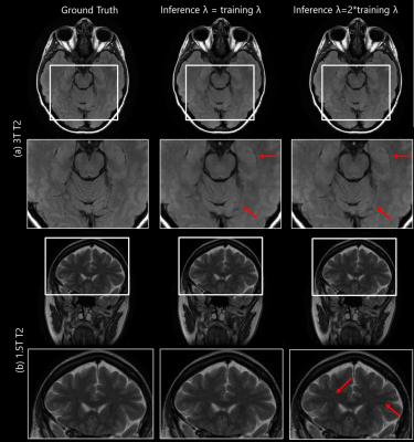 |
48 |
Effect of regularization parameter on model-based deep learning
framework for accelerated MRI
Sampada Bhave1,
Saurav Sajib1,
Aniket Pramanik2,
Mathews Jacob2,
and Samir Sharma1
1Canon Medical Research USA Inc, Mayfield, OH, United States, 2University of Iowa, Iowa City, IA, United States Keywords: Image Reconstruction, Image Reconstruction Model-based deep learning algorithms offer high quality reconstructions for accelerated acquisitions. Training the regularization parameter λ can lead to instabilities during training. In this work, we evaluated effect of fixing λ parameter while training. We observed no difference in image quality when the network was trained with a fixed λ parameter when the fixed value was equal to the value learned from training. We observed that IQ is dependent on the fixed λ values used during training. Furthermore, we observed that tuning the λ parameter during inference adapts the framework to the SNR of the testing dataset, yielding improved performance. |
|
4643.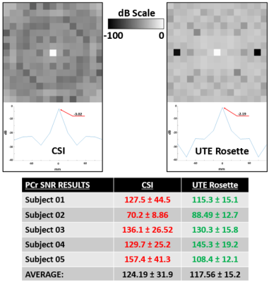 |
49 |
Comparison of Compressed Sensing Accelerated Rosette UTE and
Conventional 31P 3D MRSI at 3T in Leg Muscle
Brian Bozymski1,
Xin Shen2,
Ali Caglar Ozen3,
Serhat Ilbey3,
Albert Thomas4,
Mark Chiew5,
William Clarke5,
Ulrike Dydak1,6,
and Uzay Emir1,7
1School of Health Sciences, Purdue University, West Lafayette, IN, United States, 2Department of Radiology and Biomedical Imaging, University of California San Francisco, San Francisco, CA, United States, 3Department of Radiology and Radiotherapy, Medical Center - University of Freiburg, Freiburg, Germany, 4Department of Radiology, University of California, Los Angeles, CA, United States, 5Wellcome Centre for Integrative Neuroimaging, University of Oxford, Oxford, United Kingdom, 6Department of Radiology and Imaging Sciences, Indiana University School of Medicine, Indianapolis, IN, United States, 7Weldon School of Biomedical Engineering, Purdue University, West Lafayette, IN, United States Keywords: Data Acquisition, Muscle Quantitative comparison of novel, rosette trajectory UTE (70 μs) and conventional weighted Cartesian 3D 31P MRSI sequences is performed at 3T in quadriceps muscle. After previous validation, five healthy volunteers were scanned by both sequences without interruption. Fitted metabolite maps and SNR calculations of PCr and ATP signals across selected slices and voxels displayed competitive performance between each acquisition. Retrospective compressed sensing (CS) acceleration results suggest this UTE MRSI may enable faster, higher resolution 31P metabolite mapping in leg muscle and other organs of interest. |
|
4644.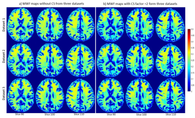 |
50 |
Accelerating myelin water fraction imaging using compressed
sensing and BMC-mcDESPOT
Maryam Alsameen1,
Zhaoyuan Gong1,
John Laporte1,
Mary Faulkner1,
Mohammad Akhonda1,
and Mustapha Bouhrara1
1Laboratory of Clinical Investigations, National Institiute on Aging, National Institutes of Health, Baltimore, MD, United States Keywords: Data Analysis, Brain We demonstrated the feasibility of compressed sensing (CS) to accelerate myelin water fraction (MWF) imaging using the BMC-mcDESPOT method. Our results showed that derived MWF maps using CS were similar to those derived without CS. Findings from this study indicate that whole brain, high resolution, MWF map can be derived within a few minutes. |
|
4645.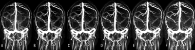 |
51 |
Acceleration of Phase-contrast Magnetic Resonance Venogram by
Compressed SENSE
Yukun Zhang1,
Na Liu1,
Geli Hu2,
Liangjie Lin2,
Yanwei Miao1,
and Qingwei Song1
1the First Affiliated Hospital of Dalian Medical University, Dalian, China, 2Clinical and Technical Support, Philips Healthcare, beijing, China Keywords: Parallel Imaging, Vessels, compressed sensitivity encoding The clinical application of phase-contrast magnetic resonance venogram (PC-MRV) is limited by the long scan time. This study aims to accelerate the acquisition of PC-MRV by the compressed sensitivity encoding (CS-SENSE) technique and find the optimal acceleration factor. Results show that the image quality of fast PC-MRV based on CS-SENSE was significantly higher than that based on conventional sensitivity encoding (SENSE) technology, and the CS-SENSE factor 9 was recommended for mild to moderate patients, and CS AF=12 for critical patients. |
|
4646.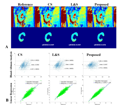 |
52 |
Accelerated parameter mapping in the k-p domain via nonconvex
low rank constraint
Kang Yan1 and
Craig H Meyer1,2
1Biomedical Engineering, University of Virginia, Charlottesville, VA, United States, 2Radiology & Medical Imaging, University of Virginia, Charlottesville, VA, United States Keywords: Image Reconstruction, Relaxometry A nonconvex low rank regularization (NLR) was proposed to accelerate parameter mapping in the k-p domain. The NLR uses weighted nuclear norm minimization (WNNM) to obtain an optimized solution by differently penalizing singular values, in comparison to traditional low rank methods. The performance of the proposed algorithm was demonstrated for T2 mapping of the kidney. Our study demonstrated that the proposed algorithm outperformed k-p domain-based compressed sensing and L&S algorithms. |
|
4647.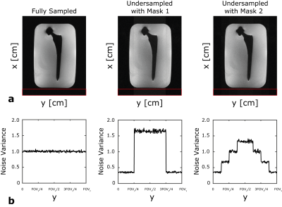 |
53 |
Accelerated MRI near metallic implants at 0.55T using hexagonal
sampling
Bahadir Alp Barlas1,
Kubra Keskin1,
Brian A Hargreaves2,3,4,
and Krishna S Nayak1,5
1Electrical and Computer Engineering, University of Southern California, Los Angeles, CA, United States, 2Radiology, Stanford University, Stanford, CA, United States, 3Electrical Engineering, Stanford University, Stanford, CA, United States, 4Bioengineering, Stanford University, Stanford, CA, United States, 5Biomedical Engineering, University of Southern California, Los Angeles, CA, United States Keywords: Data Acquisition, Low-Field MRI Metallic implants cause severe distortions in the magnetic field that are best mitigated using multi-spectral acquisitions that require longer scan time. At conventional field strengths, this is compensated by aggressive use of parallel imaging. However, at low-field strengths (<1.5T), available parallel imaging factors are reduced because body noise dominance prevents the use of dense arrays with smaller elements. Hexagonal sampling (in ky,kz space) was previously proposed as a way to achieve 2-fold reduction in imaging time without introducing additional artifacts. In this work, we demonstrate applicability of the hexagonal sampling for 0.55T multi-spectral imaging near metal. |
|
4648.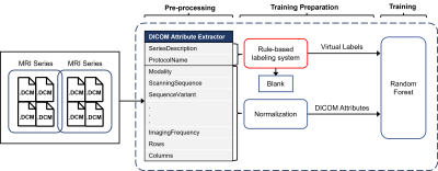 |
54 |
Development of a self-supervised machine learning algorithm for
automatic MRI sequence-type classification
Seongwon Na1,
Yousun Ko2,
Su Jung Ham1,
Mi-Hyun Kim1,
Youngbin Shin1,
Yu Sub Sung1,
Jimi Huh3,
Seung Chai Jung1,
and Kyung Won Kim1
1Asan Medical Center, Seoul, Korea, Republic of, 2University of Ulsan College of Medicine, Seoul, Korea, Republic of, 3Ajou University Hospital, Suwon, Korea, Republic of Keywords: Machine Learning/Artificial Intelligence, Machine Learning/Artificial Intelligence, Sequence; Classification For automatic sequence-type classification of brain MRI, we developed the self-supervised machine learning (ML) algorithm, named ImageSort-net, using a rule-based labeling system based on metadata of Digital Imaging and Communications in Medicine (DICOM) image files. Our rule-based labeling system and ImageSort-net showed high classification performance to predict brain MRI sequence type. ImageSort-net showed reliable performance by appending a new dataset to an existing dataset and without human labeling of the whole dataset. This result indicates that sustainable self-learning ML algorithms using the rule-based virtual label in the new datasets are feasible. |
|
4649. |
55 |
SOC-GRAPPA: An MPSoC based Accelerator for GRAPPA
Abdul Basit1,
Omair Inam1,
and Hammad Omer1
1Electrical Engineering, MIPRG, Comsats University Islamabad, Islamabad, Pakistan Keywords: Parallel Imaging, Cardiovascular, Accelerator, Real-time GRAPPA is a cartesian pMRI method widely adopted for high-speed image reconstruction in many clinical applications e.g. real-time cardiac MRI. However, general-purpose computers have limited processing capabilities to address the computational complexity of GRAPPA in real-time MRI. Recently, multi-processor system-on-chip (MPSoC) has emerged as a potential candidate to meet the rising computational demands of GRAPPA for real-time image reconstruction. In this paper, design and implementation details of a novel MPSoC based GRAPPA accelerator i.e. SOC-GRAPPA are presented. The experimental results of 18-coil cardiac dataset show that the proposed accelerator is capable of reconstructing 30 frames/second in real-time cardiac MRI. |
|
4650.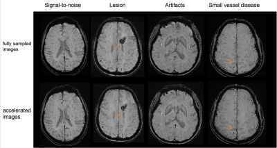 |
56 |
Validation of a deep-learning-based method for accelerating
susceptibility-weighted imaging in clinical subjects
Xiao Wu1,
Shan Xu2,
Yao Zhang2,
Jianzhong Sun2,
and Peiyu Huang2
1Radiology, The Second Affiliated Hospital, Zhejiang University School of Medicine, Hangzhou, China, 2The Second Affiliated Hospital, Zhejiang University School of Medicine, Hangzhou, China Keywords: Data Acquisition, Machine Learning/Artificial Intelligence, deep-learning ,susceptibility-weighted imaging,magnetic resonance imaging In this study, we validated a deep-learning-based method for accelerating susceptibility-weighted imaging (SWI) in 31 clinical subjects. Compared to the fully sampled images, the accelerated SWI images had less noise and imaging artifacts. Although the images had decreased sharpness, the anatomical details of the lesions were mostly kept, and we had not observed false negative\positive lesions. This method could be useful for clinical situations that need timely imaging results. |
|
4651.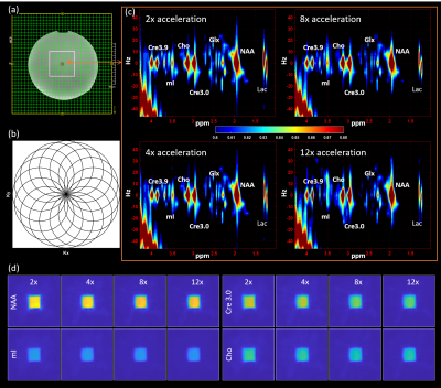 |
57 |
Accelerated Four-Dimensional Rosette J-resolved Spectroscopic
Imaging (4D ROSE-JRESI) with semi-LASER Localization: A pilot
study
Ajin Joy1,
Uzay Emir2,3,
Paul M. Macey4,
and M. Albert Thomas1
1Radiological Sciences, University of California, Los Angeles, Los Angeles, CA, United States, 2School of Health Sciences, Purdue University, West Lafayette, IN, United States, 3Weldon School of Biomedical Engineering, Purdue University, West Lafayette, IN, United States, 4School of Nursing and Brain Research Institute, University of California, Los Angeles, Los Angeles, CA, United States Keywords: Data Acquisition, Brain Undersampling spatial and spectral dimensions is essential to achieve clinically feasible scan times in multi-dimensional spectroscopic imaging. Sampling pattern of rosette trajectory has higher incoherence than that of the other non-Cartesian trajectories like spiral and radial, and can achieve higher compressed-sensing reconstruction performance. While rosette spectroscopic imaging has been attempted for 2D (2 spatial+1 spectral) and 3D (3 spatial+1 spectral) spectroscopic imaging, it has thus far not been shown for J-resolved spectroscopic imaging. In this pilot study, we implemented a rosette echo-planar J-resolved spectroscopic imaging (ROSE-JRESI) sequence and studied the feasibility of high acceleration factors for clinically feasible runtimes. |
|
4652.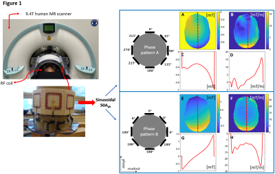 |
58 |
In-vivo image acceleration with an 8-channel local B0 coil array
and parallel imaging in a 9.4T human MR scanner
Rui Tian1,
Theodor Steffen1,
and Klaus Scheffler1,2
1High-Field MR center, Max Planck Institute for Biological Cybernetics, Tuebingen, Germany, 2Department for Biomedical Magnetic Resonance, University of Tübingen, Tuebingen, Germany Keywords: New Trajectories & Spatial Encoding Methods, New Trajectories & Spatial Encoding Methods, nonlinear gradient encoding We further developed a recent idea called spread spectrum MRI to reduce the sampling time by rapidly modulating spins with localized magnetic fields during signal readout. Given phantom experiments tested and safety evaluation for human subjects performed, this time, we started in-vivo measurements of human head with multi-slice FLASH sequence accelerated by local B0 coil modulations, and examined the reconstructed image quality. Sinusoidal modulation schemes in various phase offset patterns and frequencies given different scanner bandwidth were tested and compared, which were shown to boost the image acceleration from 6-fold (i.e., SENSE only) to about 8-fold in one phase-encoded dimension. |
|
4653.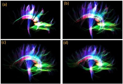 |
59 |
Improved Fast Whole-Brain High Resolution Diffusion Imaging on 7
Tesla MRI
Merry Mani1,
Xinzeng Wang2,
and Baolian Yang3
1University of Iowa, Iowa City, IA, United States, 2GE Healthcare, Houston, TX, United States, 3GE Healthcare, Waukesha, WI, United States Keywords: Brain Connectivity, High-Field MRI Achieving high resolution in diffusion MRI is challenging due to its inherently low SNR, vulnerability to motion and other EPI-related artifacts. On higher field-strengths, the SNR advantage can be exploited to push the resolution if the echo-time can be effectively reduced and the increased number of slices can be efficiently acquired. Combining acceleration techniques such as multi-band and parallel imaging is critical for this approach. Here we combine two deep-learned reconstruction priors, one pertaining to the q-space and another pertaining to image artifact removal, into a model-based iterative reconstruction framework to improve the quality of highly accelerated high-resolution 7T DWIs. |
|
4654.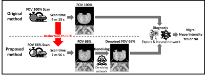 |
60 |
Fast SMWI via denoising for nigral hyperintensity detection in
Parkinson’s disease
Jonghyo Youn1,
Juhyung Park1,
Sooyeon Ji1,
Hongjun An1,
Hwan Heo2,
MyeongOh Lee2,
Soohwa Song2,
Eung Yeop Kim3,
and Jongho Lee1
1Seoul National University, Seoul, Korea, Republic of, 2Heuron Co.Ltd., Incheon, Korea, Republic of, 3Radiology, Samsung Medical Center, Seoul, Korea, Republic of Keywords: Image Reconstruction, Parkinson's Disease Nigral hyperintensity detection in substantia nigra is a potential biomarker for PD. An advanced SWI method, SMWI, has demonstrated reliable detection of the hyperintensity at 3T but it requires a 4 m 15 s scan protocol which is too long for PD patients, suffering from motion artifacts. In this study, we developed a new 2 m 56 s protocol by reducing phase FOV and applying deep learning-powered denoising to compensate for SNR loss from the shorter scan. The new protocol was validated using both simulated and real data via an automated tool and an expert radiologist. |
|
 Back to Meeting Home
 Back to the Program-at-a-Glance
The International Society for Magnetic Resonance in Medicine is accredited by the Accreditation Council for Continuing Medical Education to provide continuing medical education for physicians.
 Back to Meeting Home
 Back to the Program-at-a-Glance
 View Presentation Video