Digital Poster
Image Reconstruction Methods II
ISMRM & ISMRT Annual Meeting & Exhibition • 03-08 June 2023 • Toronto, ON, Canada

| Computer # | |||
|---|---|---|---|
4771.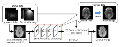 |
1 |
Parallel imaging reconstruction using iterative CNN-based
denoising in image domain
Tomoki Amemiya1,
Atsuro Suzuki1,
Yukio Kaneko1,
Suguru Yokosawa1,
and Toru Shirai1
1Imaging Technology Center, FUJIFILM Corporation, Tokyo, Japan Keywords: Parallel Imaging, Image Reconstruction We propose an iterative reconstruction method of parallel imaging using convolutional neural network (CNN)-based denoising in the image domain and data-consistency processing in k-space. The proposed method reduces the noise and artifacts of a reconstructed image compared with the iterative method using sparsity of wavelet transform, suggesting that using CNN-based denoising in iterative reconstruction is effective in reducing noise in parallel imaging. |
|
4772.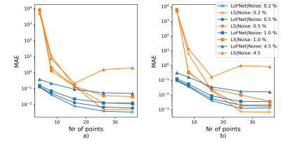 |
2 |
Deep learning-based Lorentzian fitting of WASSR Z-spectra
Sajad Mohammed Ali1,
Nirbhay Yadav2,
Ronnie Wirestam1,
Munendra Singh3,
Hye-Young Heo3,
Peter van Zijl2,
and Linda Knutsson4
1Medical Physics, Lund University, Lund, Sweden, 2Radiology, F.M. Kirby Research Center, Johns Hopkins University, Kennedy Krieger Institute, Baltimore, MD, United States, 3Radiology, Johns Hopkins University, Baltimore, MD, United States, 4F.M. Kirby Research Center, Radiology, Medical Radiation Physics, Kennedy Krieger Institute, Johns Hopkins University, Lund University, Baltimore, MD, United States Keywords: Data Analysis, Machine Learning/Artificial Intelligence, Lorentzian curve fitting Water saturation shift referencing (WASSR) Z-spectra can be used to correct shifts due to B0-field inhomogeneities, for magnetic susceptibility mapping and analysis of relaxation effects. The spectra follow a Lorentzian shape with discrete values. Hence, a Lorentzian fit to retrieve the shape parameters (amplitude A, line width LW and frequency shift ΔfH2O ) simplifies analysis. Conventionally, the least-squares (LS) method is used for such fitting despite being time consuming and sensitive to the unavoidable noise in vivo. We propose a deep learning-based Lorentzian-fitting neural network (LoFNet) that demonstrated improved robustness against noise and sampling density in combination with reduced time consumption. |
|
4773. |
3 |
Accuracy of Deep Learning-based Signal-to-noise Measurements
using Air Recon DL
Evan McNabb1,
Véronique Fortier1,2,3,4,
and Ives R. Levesque3,4
1Medical Imaging, McGill University Health Centre, Montreal, QC, Canada, 2Diagnostic Radiology, McGill University, Montreal, QC, Canada, 3Gerald Bronfman Department of Oncology, McGill University, Montreal, QC, Canada, 4Medical Physics Unit, McGill University, Montreal, QC, Canada Keywords: Data Acquisition, Data Analysis Noise estimates in Deep Learning-based image reconstruction (DLR) from background regions lead to artificially low noise estimates and thus inaccurate SNR. Noise estimate accuracy was improved using the difference between two identical acquisitions. SNR increased in DLR compared to standard reconstruction in clinical sequences over a range of acquired signal averages and voxel sizes. With DLR, varying signal averages had little effect on the measured SNR in fast spin echo acquisitions, while SNR increased by more than three-fold in single-shot fast spin echo. However, with DLR, SNR no longer follows predicable behavior, therefore sequence optimizations need to be performed experimentally. |
|
4774.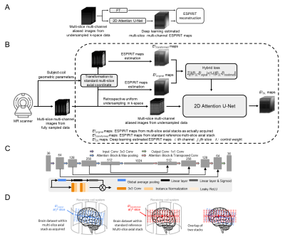 |
4 |
Deep Learning Prediction of Multi-channel ESPIRiT Maps for
Calibrationless MR Image Reconstruction
Junhao Zhang1,2,
Zheyuan Yi1,2,3,
Yujiao Zhao1,2,
Linfang Xiao1,2,
Jiahao Hu1,2,3,
Vick Lau1,2,
Fei Chen3,
Alex T.L.Leong1,2,
and Ed X. Wu1,2
1Laboratory of Biomedical Imaging and Signal Processing, the University of Hong Kong, HongKong, China, 2Department of Electrical and Electronic Engineering, the University of Hong Kong, HongKong, China, 3Department of Electrical and Electronic Engineering, Southern University of Science and Technology, Shenzhen, China Keywords: Parallel Imaging, Data Acquisition, Brain reconstruction, Cardiac reconstruction We present a U-Net based deep learning model to estimate the multi-channel ESPIRiT maps directly from uniformly-undersampled multi-channel multi-slice MR data. The model is trained with a hybrid loss function using fully-sampled multi-slice axial brain datasets from the same MR receiving coil system. The proposed model robustly predicted ESPIRiT maps from uniformly-undersampled k-space brain and cardiac MR data, yielding highly comparable performance to reconstruction using to acquired reference ESPIRiT maps. Our proposed method presents a general strategy for calibrationless parallel imaging reconstruction through learning from coil and protocol specific data. |
|
4775.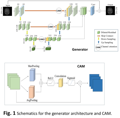 |
5 |
CS-MRI Reconstruction Using an Improved Generative Adversarial
Network with Channel Attention Mechanism
Xia Li1,
Hui Zhang2,
and Tie-Qiang Li3
1Information Engineering, China Jiliang University, Hangzhou, China, 2Information Engineering, China Jiliang University, HANGZHOU, China, 3Karolinska Institute, Stockholm, Sweden Keywords: Image Reconstruction, Brain, Compressed sensing MRI Generative adversarial network (GAN) has emerged as one of the most prominent approaches for fast CS-MRI reconstruction. However, most deep-learning models achieve performance by increasing the depth and width of the networks, leading to prolonged reconstruction time and difficulty to train. We have developed an improved GAN-based model to achieve quality performance without increasing complexity by implementing the following: 1) dilated-residual structure with different dilation rates at different depth of the networks; 2) CAM to adjust the allocation of network resources; 3) multi-scale information fusion module to achieve feature fusion. Experiment data have confirmed the validity for the modules. |
|
4776. |
6 |
A Stochastic Approach for Joint Learning of a Neural Network
Reconstruction and Sampling Pattern in Cartesian 3D Parallel MRI
Marcelo V. W. Zibetti1 and
Ravinder R. Regatte1
1Radiology, NYU Grossman School of Medicine, New York, NY, United States Keywords: Data Acquisition, Image Reconstruction This work proposes a stochastic variation of the bias-accelerated subset selection (BASS) algorithm to learn an efficient sampling pattern (SP) for accelerated MRI. This algorithm is used in the joint learning of an SP and neural network reconstruction. We apply the proposed approach to two different 3D Cartesian parallel MRI problems. The proposed stochastic approach, when used for joint learning, improves the learning speed from 2.5X to 5X, obtaining SPs with similar properties as the non-stochastic approach with nearly the same RMSE and SSIM. |
|
4777.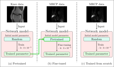 |
7 |
Analysis of Deep Learning-based Reconstruction Models for Highly
Accelerated MR Cholangiopancreatography: to Fine-tune or not to
Fine-tune
Jinho Kim1,2,
Thomas Benkert2,
Bruno Riemenschneider1,
Marcel Dominik Nickel2,
and Florian Knoll1
1Artificial Intelligence in Biomedical Engineering, Friedrich-Alexander-Universität Erlangen-Nürnberg, Erlangen, Germany, 2MR Application Pre-development, Siemens Healthcare GmbH, Erlangen, Germany Keywords: Machine Learning/Artificial Intelligence, Image Reconstruction, Magnetic Resonance Cholangiopancreatography MR cholangiopancreatography (MRCP) is a special MRI technique to visualize the biliary systems. Deep Learning-based (DL) reconstruction models have shown to reduce scan time from many anatomical regions. However, they generally require large training datasets. This is challenging for applications like MRCP, where public datasets are not available. This work analyzes two approaches to training a DL model for highly accelerated MRCP reconstruction: training from scratch using a small MRCP dataset and fine-tuning a model pretrained on a public knee dataset. Results show that despite of the substantial data domain shift between training and testing, fine-tuning outperformed training from scratch. |
|
4778.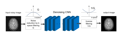 |
8 |
Improvement of CNN Denoising Performance Using Noise Control of
Input Image and Application to Parallelized Image Denoising
Satoshi ITO1 and
Keitaro TAKAHASHI1
1Graduate Program in Information, Electrical and Electronic Systems Engineering, Utsunomiya University, Utsunomiya, Japan Keywords: Data Processing, Data Processing In noise reduction, the lower the amount of noise, the lower the image degradation associated with the noise reduction. Using this property, we propose a new denoising scheme that can improve denoising performance using trained CNN. Noise suppressed image by low-pass filter is inputted to denoising CNN and then output image is enhanced by high-pass filter. Experimental results show that noise suppression of the input image improves both image structure preservation and noise processing performance. The proposed method was applied to a parallel blind image denoising method. As a results, further improvement in performance was shown. |
|
4779.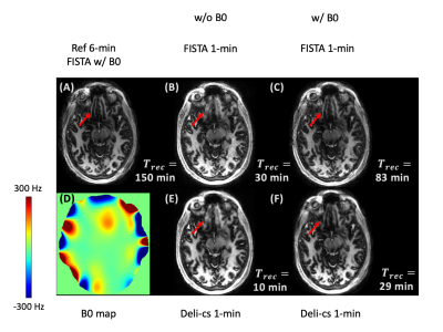 |
9 |
Fast spatio-temporal subspace reconstruction of 3D-MRF with B0
correction and deep-learning-initialized compressed sensing
(Deli-CS)
Natthanan Ruengchaijatuporn1,2,
Siddharth Srinivasan Iyer3,4,5,
Sophie Schauman3,4,
Quan Chen3,4,
Xiaozhi Cao3,4,
Itthi Chatnuntawech6,
and Kawin Setsompop3,4
1Center of Excellence in Computational Molecular Biology, Faculty of Medicine, Chulalongkorn University, Bangkok, Thailand, 2Center for Artificial Intelligence in Medicine, Faculty of Medicine, Chulalongkorn University, Bangkok, Thailand, 3Department of Radiology, Stanford University, Stanford, CA, United States, 4Department of Electrical Engineering, Stanford University, Stanford, CA, United States, 5Department of Electrical Engineering and Computer Science, MIT, Cambridge, MA, United States, 6National Nanotechnology Center, National Science and Technology Development Agency, Pathum Thani, Thailand Keywords: Sparse & Low-Rank Models, Machine Learning/Artificial Intelligence Recent advances in spatio-temporal subspace reconstruction has enabled accurate reconstruction from highly accelerated scans. Nevertheless, such methods suffer from being computationally intensive due to their iterative nature coupled with the large dimensionality of the problem, especially when imperfection correction is incorporated into the formulation. This abstract proposes deep-learning-initialized compressed sensing (Deli-CS) to accelerate such spatio-temporal reconstruction by providing it with a deep-learning-reconstructed initial solution, reducing the number of iterations required. Using MRF as an example, Deli-CS reconstructs data from a rapid 1-mm isotropic whole-brain TGAS-SPI-MRF with time-segmented B0 correction at 3x faster speed compared to FISTA. |
|
4780.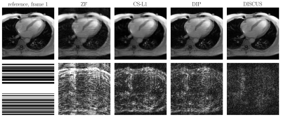 |
10 |
Deep Image Prior with Structured Sparsity (DISCUS) for Dynamic
MRI Reconstruction
Muhammad Ahmad Sultan1,
Chong Chen1,
Yuchi Han1,
and Rizwan Ahmad1
1The Ohio State University, Columbus, OH, United States Keywords: Image Reconstruction, Data Processing, Image Reconstruction, Unsupervised Learning, Deep Image Prior We propose an unsupervised learning method for dynamic MRI reconstruction. Our method, Deep Image prior with StruCtUred Sparsity (DISCUS), is an extension of Deep Image Prior (DIP) and employs joint optimization of network parameters and latent code vectors to recover image series. We enforce group sparsity on code vectors to reveal the underlying low-dimensional manifold of the image series. Using two sets of in vivo measurements and digital phantom simulation, we show that our approach significantly improves the reconstruction quality in terms of Normalized Mean Square Error (NMSE) and Structure Similarity Index Measure (SSIM) compared to compressed sensing and DIP. |
|
4781.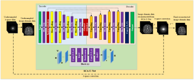 |
11 |
Deep Attention Unfolding CNN Architecture for Parallel MRI
Muhammad Shafique1,2,
Sohaib Ayyaz Qazi3,
Faisal Najeeb1,
and Hammad Omer1
1Electrical and Computer Engineering, COMSATS University Islamabad, Islamabad, Pakistan, 2Electrical Engineering, University of Poonch Rawalakot, Rawalakot AJK, Pakistan, 3Center for Medical Image Science and Visualization, Linköping University, Linköping, Sweden Keywords: Image Reconstruction, Artifacts, Parallel MRI, Deep Learning, Attention Mechanism Deep learning has made significant progress in recent years, however the local receptive field in CNN raises questions about signal synthesis and artefact compensation. This paper proposes a deep learning Spatial-Channel Attention U-Net (SCA-U-Net) to solve the folding problems arising in MR images because of undersampling. The main idea is to enhance local features in the images and restrain the irrelevant features at the spatial and channel levels. The output from SCA-U-Net is further refined by adding a small number of originally acquired low-frequency k-space data. Experimental results show a better performance of the SCA-U-Net model than classical U-Net model. |
|
4782.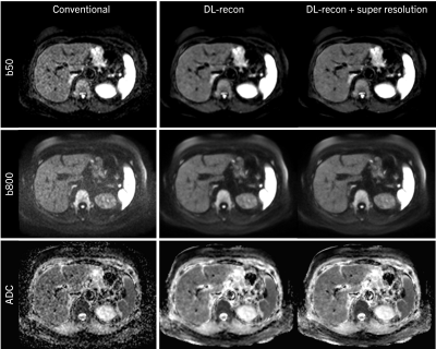 |
12 |
Improved Clinical Diffusion Weighted Imaging by Combining Deep
Learning Reconstruction, Partial Fourier, and Super Resolution
Thomas Benkert1,
Elisabeth Weiland1,
Simon Arberet2,
Majd Helo1,
Fasil Gadjimuradov1,3,
Karl Engelhard4,
Gregor Thoermer1,
and Dominik Nickel1
1MR Application Predevelopment, Siemens Healthcare GmbH, Erlangen, Germany, 2Digital Technology & Innovation, Siemens Medical Solutions USA, Princeton, NJ, United States, 3Pattern Recognition Lab, Friedrich-Alexander-Universität Erlangen-Nürnberg, Erlangen, Germany, 4Institute of Radiology, Martha-Maria Hospital, Nuremberg, Germany Keywords: Diffusion/other diffusion imaging techniques, Translational Studies Diffusion weighted imaging (DWI) has found widespread use in daily clinical routine but can still be limited by long acquisition times and low spatial resolution. In this work, combining deep learning-based k-space to image reconstruction with super resolution processing tailored to support partial Fourier acquisitions is demonstrated to efficiently mitigate these obstacles. The approach is shown for various applications, including liver, breast, prostate, and brain DWI at 0.55T, 1.5T, and 3T. |
|
4783.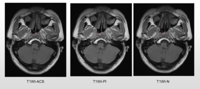 |
13 |
Clinical feasibility of artificial intelligence-assisted
compressed sensing for accelerated MR imaging in nasopharyngeal
carcinoma
Qin Zhao1,
Song Zhang1,
Liyun Zheng2,
and Yongming Dai3
1Department of Radiology, Sun Yat-sen University Cancer Center, State Key Laboratory of Oncology in South China, Collaborative Innovation Center for Cancer Medicine, Guangdong Key Laboratory of Nasopharyngeal Carcinoma Diagnosis and Therapy, Guangzhou, China, 2Shenzhen United Imaging Research Institute of Innovative Medical Equipment, Shenzhen, China, 3MR Collaboration, Central Research Institute, United Imaging Healthcare, Shanghai, China Keywords: Data Acquisition, Machine Learning/Artificial Intelligence To improve the scanning efficiency of magnetic resonance imaging (MRI) for nasopharyngeal carcinoma, this study investigated the value of accelerating technique, artificial intelligence-assisted compressed sensing (ACS), in comparison to conventional sequences without accelerating technique and accelerating MRI using parallel imaging (PI). Eleven patients diagnosed with nasopharyngeal carcinoma were prospectively enrolled. As a result, ACS achieved the shortest acquisition time, with similar or even better image quality and SNR than conventional sequences. ACS has the potential to provide sufficient image quality for T1- and T2-weighted imaging in nasopharyngeal carcinoma and could be an alternative to conventional sequences in clinical practice. |
|
4784.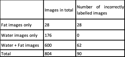 |
14 |
USING DEEP LEARNING WITH AN ACTIVE LEARNING APPROACH TO CORRECT
WATER-FAT MIS-LABELING IN MR THORACIC SPINE IMAGES
Siddhartha Satpathi1,
Jacinta E. Browne1,
AbdulRahman Alfayad1,
Jared T. Verdoorn1,
and James G. Pipe1
1Mayo Clinic, Rochester, MN, United States Keywords: Artifacts, Spinal Cord Upon investigating a dataset of 804 studies for spinal fractures from two major vendors, the authors observed that 11% of the water-fat images in the studies are mis-labelled. This motivated the development of an automated algorithm to correct the mis-labelling. We used a 2D CNN based deep learning model to classify the images correctly with the aim of reducing error and fatigue in clinical diagnosis caused by such mis-labeling, as well as providing correct labels for further AI workflow. We also demonstrated the use of active learning in this problem by achieving the same test-error with fewer labels than using the entire training data. |
|
4785.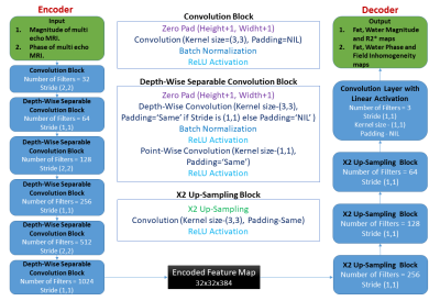 |
15 |
Lightweight encoder-decoder architecture for Fat-Water
separation in MRI using biophysical model-guided deep learning
method
Ganeshkumar M1,
Devasenathipathy Kandasamy2,
Raju Sharma2,
and Amit Mehndiratta1,3
1Centre for Biomedical Engineering, Indian Institute of Technology Delhi, New Delhi, India, 2Department of Radio Diagnosis, All India Institute of Medical Sciences, New Delhi, India, 3Department of Biomedical Engineering, All India Institute of Medical Sciences, New Delhi, India Keywords: Machine Learning/Artificial Intelligence, Image Reconstruction, Deep Learning, Fat-Water seperation In this study, we propose a novel lightweight encoder-decoder architecture for deep learning based Fat-Water separation in multi-echo MRI data. The architecture's performance is evaluated in the biophysical model-guided deep learning-based Fat-Water separation task and compared against the widely used U-Net. This biophysical model-guided deep learning-based Fat-Water separation requires no training data and ground truths, but it involves time-consuming loss minimization for thousands of epochs. Despite having significantly fewer training parameters, the proposed architecture performed equally well in generating the Fat-Water maps compared to the U-Net. So, our proposed architecture aids in the faster generation of Fat-Water maps. |
|
4786.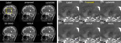 |
16 |
Fast MR imaging with distribution convergence modeling
Jing Cheng1,
Zhuo-Xu Cui1,
Qingyong Zhu1,
and Dong Liang1
1Shenzhen Institutes of Advanced Technology, Chinese Academy of Sciences, Shenzhen, China Keywords: Machine Learning/Artificial Intelligence, Image Reconstruction Existing deep learning-based methods for MR reconstruction mainly use MSE as loss function to train the network under the assumption that MR images follow the sub-Gaussian distribution, without considering the real distribution of the images. In this work, we propose a new DL-based method that models the image distribution with equilibrium Langevin dynamic to converge the distribution, and trains the network with Wasserstein distance to approach the real distribution. Experimental results on highly undersampled MR data demonstrate the superior performance of the proposed method. |
|
4787. |
17 |
Transferable Deep Learning for Fast MR Imaging
Yuxiang Zhou1,
Riti Paul2,
Pak Lun Kevin Ding2,
Leland Hu1,
Ameet C Patel1,
and Baoxin Li2
1Radiology, Mayo Clinic at Arizona, Phoenix, AZ, United States, 2CIDSE, Arizona State University, Tempe, AZ, United States Keywords: Machine Learning/Artificial Intelligence, Data Processing, Reconstruction Artificial intelligence (AI) applications in the field of magnetic resonance imaging have been implemented in routine clinical practice. However, MRI still faces a practical and persistent challenge: its long acquisition time. This has led to two prominent issues in health care: high cost and poor patient experience. Long acquisition time is also a source of degraded imaging quality (e.g., motion artifacts). In the study, we propose to develop novel Deep Learning (DL) architectures combined with transfer learning capabilities to address the above challenge and apply this newly developed AI technique for image reconstruction in MRI with very fast imaging acquisition. |
|
4788.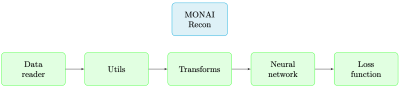 |
18 |
MONAI Recon: An Open Source Tool for Deep Learning Based
Accelerated MRI Reconstruction
Mohammad Zalbagi Darestani1,
Vishwesh Nath2,
Wenqi Li2,
Yufan He2,
Holger Reinhard Roth2,
Ziyue Xu2,
Daguang Xu2,
Reinhard Heckel1,3,
and Can Zhao2
1Rice University, Houston, TX, United States, 2NVIDIA, Santa Clara, CA, United States, 3Technical University of Munich, Munich, Germany Keywords: Software Tools, Machine Learning/Artificial Intelligence, MRI Reconstruction Deep learning models outperform traditional methods in terms of quality and speed for numerous medical imaging applications. A critical application is the acceleration of magnetic resonance imaging (MRI) reconstruction, where a deep learning model reconstructs a high-quality MR image from a set of undersampled measurements. For this application, we present the MONAI Recon Module to facilitate fast prototyping of deep-learning-based models for MRI reconstruction. Our free and open-source software is pre-equipped with a baseline and a state-of-the-art deep-learning-based reconstruction model and contains the necessary tools to develop new models. The developed open-source software covers the entire MRI reconstruction pipeline. |
|
4789. |
19 |
Investigation of DWI with Deep Learning-based Reconstruction in
the Differentiation of Benign and Malignant Breast Lesions
Tiebao Meng1,
Huiming Liu1,
Chuanmiao Xie1,
Jialu Zhang2,
and Long Qian2
1Department of Radiology, Sun Yat-Sen University Cancer Center, Guangzhou, China, 2MR Research, GE Healthcare, Beijing, China Keywords: Machine Learning/Artificial Intelligence, Diffusion/other diffusion imaging techniques As the most common malignant tumor in women, the differentiation of benign lesion from malignant breast tumors is the most essential step in early diagnosis. Recent study demonstrated that DWI could offer great help in differential diagnosis of breast tumors. The novel deep learning-based reconstruction (DLR) technique is able to increase SNR of MRI images. Using DLR, DWI images can be acquired with less NEX (fast DWI) and still maintain the image quality. This study indicated the feasibility of fast DWI protocol with DLR in the differentiation of breast benign lesions and malignant tumors. |
|
4790.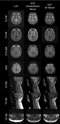 |
20 |
Clinical Viability of AI-enabled 0.5T MRI Scanner to Improve
Access
Arjun Narula1,
Uday Patil2,
Anand SH3,
Harikrishnan Raveendran4,
Shailaja Muniraj5,
Pallavi Rao6,
Anurita Menon6,
Allison Nicole Garza7,
Santosh Kumar7,
Gautam Kumar7,
Srinivas NR7,
Syam Babu7,
Ravi Jaiswal7,
Rajagopalan Sundaresan7,
Ashok Kumar Reddy7,
Sajith Rajamani7,
Rajdeep Das7,
Nitin Jain7,
Sudhir Ramanna7,
Sundar V7,
Florintina Charlaas7,
Sudhanya Chatterjee7,
Rohan Patil7,
Megha Goel7,
Dattesh Shanbhag7,
Vikas Kumar Anand7,
Abhishek Galagali7,
Sathish KV7,
Preetham Shankpal7,
Harsh Kumar Agarwal7,
Suresh Emmanuel Joel7,
and Ramesh Venkatesan7
1Narula Diagnostics, Rohtak, India, 2Manipal Hospitals, Bangalore, India, 3Jivaa Diagnostics, Tumkur, India, 4Core Clinico PET Imaging, Thane, India, 5Prima Diagnostics, Bangalore, India, 6Image Core Lab, Bangalore, India, 7GE Healthcare, Bangalore, India Keywords: Machine Learning/Artificial Intelligence, Low-Field MRI, Accessible MRI We validate the clinical viability of a 0.5T scanner to reduce cost and improve access to quality MRI using AI based IQ enhancement to compensate for IQ reduction due to lower field and other lower hardware specifications. We obtained data from 65 patients from the re-ramped 0.5T and a commercially available 1.5T MRI system for brain and cervical spine. Radiologists compared image quality between the two and rated the image on the ability to perform diagnosis. We observed that more than 90% of the images were rated to be above diagnostic levels and AI reconstruction significantly improved the image quality. |
|
 Back to Meeting Home
 Back to the Program-at-a-Glance
The International Society for Magnetic Resonance in Medicine is accredited by the Accreditation Council for Continuing Medical Education to provide continuing medical education for physicians.
 Back to Meeting Home
 Back to the Program-at-a-Glance
 View Presentation Video