Digital Poster
Acquisition & Analysis Techniques
ISMRM & ISMRT Annual Meeting & Exhibition • 03-08 June 2023 • Toronto, ON, Canada

Digital Poster
Acquisition & Analysis Techniques
| Computer # | |||
|---|---|---|---|
2017.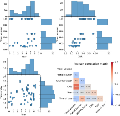 |
21 | The effect of imaging parameters, aging, and circadian rhythm on Freesurfer's estimates: A single subject study at 7T over 7 years
Hendrik Mattern1,2, Falk Lüsebrink1,3,4, and Oliver Speck1,2,5,6
1Biomedical Magnetic Resonance, Otto von Guericke University, Magdeburg, Germany, 2German Center for Neurodegenerative Disease, Magdeburg, Germany, 3Institute of Cognitive Neurology and Dementia Research, Otto von Guericke University, Magdeburg, Germany, 4Max Planck Institute for Human Cognitive and Brain Sciences, Leipzig, Germany, 5Center for Behavioral Brain Sciences, Magdeburg, Germany, 6Leibniz Institute for Neurobiology, Magdeburg, Germany Keywords: Data Analysis, Aging In this explorative study, the effect of imaging parameters, image quality, circadian rhythm, and aging on FreeSurfer’s estimates is investigated. To that end, the human phantom, an openly available data of a single subject scanned at 7T over 7 years with various MR protocols, is used. |
|
2018.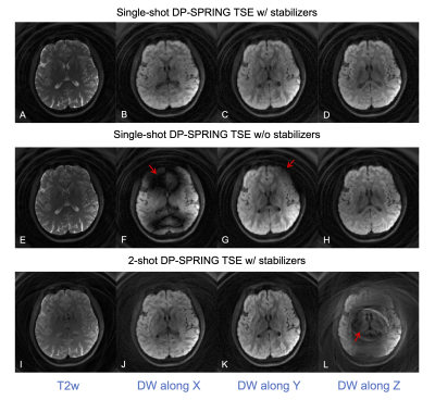 |
22 | Distortion-free diffusion imaging using single-shot diffusion-prepared TSE sequence with spiral-ring readouts and magnitude stabilizers
Zhixing Wang1, Xiaozhi Cao2, Kun Qing3, Xue Feng1, John P. Mugler4, and Craig H. Meyer1,4
1Biomedical Engineering, University of Virginia, Charlottesville, VA, United States, 2Department of Radiology, Stanford University, Stanford, CA, United States, 3Radiation Oncology, City of Hope, Duarte, CA, United States, 4Radiology & Medical Imaging, University of Virginia, Charlottesville, VA, United States Keywords: Data Acquisition, Data Acquisition This study provides an alternative approach to conventional diffusion-weighted (DW) EPI-based sequences. A 2D single-shot (SS) diffusion-prepared (DP) turbo-spin-echo (TSE) sequence, combined with spiral-ring trajectories and magnitude stabilizers, dubbed “SS-DP-SPRING TSE”, was developed for distortion- and motion artifact-free diffusion imaging. Compared to a SS-DW-EPI sequence, this method is less sensitive to B0-inhomogeneity and thus provides DW-images with improved geometric fidelity. |
|
2019.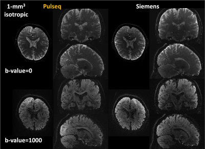 |
23 | Pulseq-gSlider: Scanner-independent high-isotropic-resolution dMRI on an open-source platform
Qiang Liu1,2, Lipeng Ning1, Congyu Liao3,4, Shaik Imam5, Scott Peltier6, Borjan Gagoski7, Berkin Bilgic8,9,10, William A. Grissom5, Maxim Zaitsev11, Jon-Fredrik Nielsen12, and Yogesh Rathi1,13
1Department of Psychiatry, Brigham and Women’s Hospital, Harvard Medical School, Boston, MA, United States, 2School of Biomedical Engineering, Southern Medical University, Guangzhou, China, 3Department of Radiology, Stanford University, Stanford, CA, United States, 4Department of Electrical Engineering, Stanford University, Stanford, CA, United States, 5Institute of Imaging Science, Department of Biomedical Engineering, Department of Electrical Engineering, Vanderbilt University, Nashville, TN, United States, 6Functional MRI Laboratory, University of Michigan, Ann Arbor, MI, United States, 7Fetal-Neonatal Neuroimaging and Developmental Science Center, Boston Children’s Hospital, Harvard Medical School, Boston, MA, United States, 8Athinoula A. Martinos Center for Biomedical Imaging, Massachusetts General Hospital, Charlestown, MA, United States, 9Department of Radiology, Harvard Medical School, Cambridge, MA, United States, 10Harvard-MIT Division of Health Sciences and Technology, Massachusetts Institute of Technology, Cambridge, MA, United States, 11Department of Radiology, Medical Physics, University Medical Center Freiburg, Freiburg, Germany, 12Department of Biomedical Engineering, University of Michigan, Ann Arbor, MI, United States, 13Department of Radiology, Brigham and Women’s Hospital, Harvard Medical School, Boston, MA, United States Keywords: Data Acquisition, Software Tools Using on the open-source pulse sequence programming platform Pulseq, we successfully developed and implemented the gSlider sequence on a 3T scanner without the vendor-specific sequence development environment. One-millimeter whole-brain isotropic dMRI data was acquired using the gSlider implementation from Pulseq and Siemens and test-retest variability was evaluated. The test-retest reliability in fractional anisotropy improved dramatically for the Pulseq implementation while it was comparable for both implementations for mean diffusivity. We demonstrated the feasibility of implementing advanced sequences using the open-source Pulseq platform and expect that it will be easily transferred to and executed on different vendors. |
|
2020.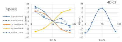 |
24 | Validation of respiratory correlated 4D-MRI for radiotherapy planning, using a motion phantom and comparing to 4D-CT.
Joan Chick1, Evanthia Kousi1, Andreas Wetscherek1, Julie Hughes2, Georgina Hopkinson2, Jessica Gough2,3, Rosalyne Westley2,3, Radhouene Neji4, Alex Dunlop1, Simeon Nill1, Henry Mandeville2,3, Katharine Aitken2,3, Dow-Mu Koh2,3, and Uwe Oelfke1
1Joint Department of Physics, The Institute of Cancer Research and Royal Marsden Hospital, London, United Kingdom, 2Royal Marsden Hospital, London, United Kingdom, 3The Institute of Cancer Research, London, United Kingdom, 4MR Research Collaborations, Siemens Healthcare, Frimley, United Kingdom Keywords: Data Acquisition, Radiotherapy, Respiratory Correlated MRI Respiratory correlated 4D-MRI has the potential to be a valuable tool in radiotherapy. This work aims to optimise and validate a 4D-MRI golden angle stack of stars radial sequence for measuring respiratory motion of abdominal organs. A 4D motion phantom is used to compare 4D-MRI against 4D-CT. For craniocaudal movement, motion is underestimated in 4D-MRI but motion estimation improves for non-axial acquisitions. This could be due to the alignment of the motion direction with the cartesian phase encoding direction for axial stack of stars acquisitions. |
|
2021.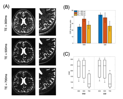 |
25 | Visualization of Perivascular Spaces using an Optimized 3D-TSE Sequence with Reduced Flip Angle at 7T
Gael Saib1, Zeynep Demir1, Paul Taylor2, S. Lalith Talagala3, and Alan P. Koretsky1
1NINDS/LFMI, National Institutes of Health, Bethesda, MD, United States, 2NIMH/SSCC, National Institutes of Health, Bethesda, MD, United States, 3NINDS/NMRF, National Institutes of Health, Bethesda, MD, United States Keywords: Visualization, Neurofluids, CSF Perivascular spaces (PVS) dilatation has been recently linked to aging and neurodegenerative diseases. T2-weighted ultra-high field MRI allows to image the PVS burden at high contrast and spatial resolution non-invasively. However, most of sub-voxel sized PVS cannot be resolved due to partial volume effects and remaining signal from blood and tissue. High-resolution 3D-TSE with reduced flip angle allowed the visualization of PVS with a very high CSF-to-tissue ratio of ~37:1 at 7T. |
|
2022.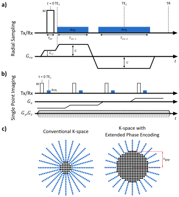 |
26 | XTE: The All-in-One Short-TE Imaging Sequence
Serhat Ilbey1, Michael Bock1, and Ali Caglar Özen1
1Division of Medical Physics, Department of Radiology, University Medical Center Freiburg, Freiburg, Germany Keywords: Pulse Sequence Design, New Trajectories & Spatial Encoding Methods XTE is a general implementation of various sequences, namely UTE, ZTE, SPI, PETRA, and gmPETRA. Each sequence type and their combinations can be realized within XTE by adjusting only a few parameters. We provide phantom and in vivo images acquired with XTE for different sequence settings. |
|
2023.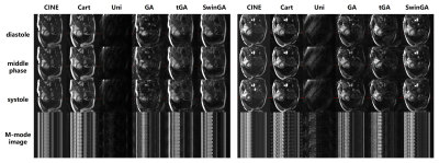 |
27 | Swin golden angle: a radial profile order for golden ratio sampling with navigator compatibility and eddy-current suppression
Zhongsen Li1, Aiqi Sun2, Shuai Wang1, Chuyu Liu1, Sirui Wu1, Haozhong Sun1, Rui Guo3, Haiyan Ding1, and Rui Li1
1Center for Biomedical Imaging Research, Tsinghua University, Beijing, China, 2Institute of Science and Technology for Brain-Inspired Intelligence, Fudan University, Shanghai, China, 3School of Medical Technology, Beijing Institute of Technology, Beijing, China Keywords: Data Acquisition, Cardiovascular, golden angle radial, eddy-current suppresion, profile order Golden angle radial trajectory suffers from eddy-current when combined with trueFISP sequence. If additional navigators are required, how to keep golden ratio sampling while maintain stable steady-state is still a problem to be solved. In this work, we propose a novel radial profile order, named "swin golden angle"(swinGA), which is able to achieve golden ratio sampling, stable trueFISP signal, and navigator acquisition simultaneously. We validated the proposed profile in static phantom study and in-vivo study for free-running cardiac imaging. The results show that the proposed swinGA significantly outperforms other sampling profiles and achieves good image quality. |
|
2024.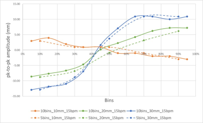 |
28 | Initial observations and potential pitfalls when validating respiratory correlated MRI for radiotherapy planning.
Evanthia Kousi1, Joan Chick1, Andreas Wetscherek1, Julie Hughes2, Georgina Hopkinson2, Jessica Gough2,3, Rosalyne Westley2,3, Radhouene Neji4, Simeon Nill1, Dow-Mu Koh2,3, and Uwe Oelfke1
1Joint Department of Physics, The Institute of Cancer Research and The Royal Marsden NHS Foundation Trust, London, United Kingdom, 2The Royal Marsden NHS Foundation Trust, London, United Kingdom, 3The Institute of Cancer Research, London, United Kingdom, 4MR Research Collaborations, Siemens Healthcare, Frimley, United Kingdom Keywords: Data Analysis, Radiotherapy, 4D-MRI, Respiratory correlated MRI, radiotherapy planning In this abstract we present our initial results from the validation of a respiratory correlated 4D MRI golden angle stack of stars radial sequence prior to its clinical implementation and highlight potential experimental pitfalls to avoid that may impact motion range estimations. Correct phantom set up reduced motion underestimations by 3%-18% for the simulated respiratory waveforms considered. Larger motion discrepancies observed for the low amplitude motion which improved by increasing the number of bins. |
|
2025.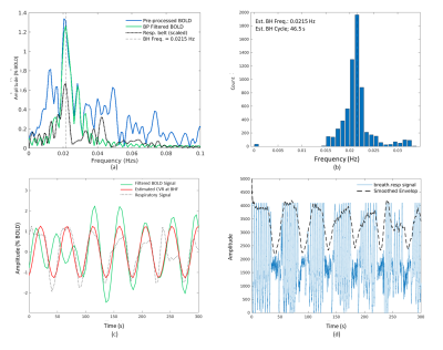 |
29 | A robust method for estimating CVR dynamics from breath-hold BOLD data without end-tidal carbon dioxide recordings
Nuwan D. Nanayakkara1, Liesel-Ann Meusel1, Nicole D. Anderson1,2, and J. Jean Chen1,3,4
1Rotman Research Institute, Baycrest Health Sciences, Toronto, ON, Canada, 2Departments of Psychology and Psychiatry, University of Toronto, Toronto, ON, Canada, 3Department of Medical Biophysics, University of Toronto, Toronto, ON, Canada, 4Department of Biomedical Engineering, University of Toronto, Toronto, ON, Canada Keywords: Data Analysis, fMRI (task based), Cerebrovascular reactivity, CVR Cerebrovascular reactivity (CVR) can be estimated from the BOLD response to a vasoactive stimulus such as a breath-holding (BH) approximated by a sinusoidal regressor. We proposed a robust approach for estimating CVR using the frequency spectrum of BOLD data itself, without regressions or correlations with external recordings. We also estimated regional CVR time delay patterns using the BOLD signal phase relative to a non-brain reference. We demonstrate our pipeline in 18 healthy adults. |
|
2026.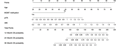 |
30 | A nomogram based on DKI, molecular and clinical risk factors for survival prediction and treatment decision-making in astrocytomas
Yan Tan1, Zeliang Liu2, Xiaochun Wang1, and Hui Zhang1
1First Hospital of Shanxi Medical University, Taiyuan, Shanxi Province, China, China, 2LinFen People's Hospital, Linfen, Shanxi Province, China, China Keywords: Data Analysis, Tumor, diffusion kurtosis imaging, nomogram, astrocytoma, overall survival, treatment To establish a nomogram by integrating DKI, molecular and clinical risk factors for personalized OS estimation in astrocytomas, and explore the nomogram-based treatment benefits. Astrocytomas are the most common brain tumors with poor prognoses. The detection of risk factors may aid in individualizing therapeutic plans and improve survival. It is concluded the nomogram based on DKI, molecular and clinical risk factors could achieve the individualized OS estimation of astrocytomas with excellent performance. For high-risk patients, surgery plus chemo and/or radiotherapy were recommended; while for low-risk patients, additional chemo and/or radiotherapy did not increase survival benefits. |
|
2027.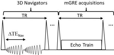 |
31 | Dual-echo 3D Spiral Navigators for the Detection of Temporal Motions and B0 Shifts in Gradient-echo Imaging at 3.0 T
Yuguang Meng1, James J. Lah2, Jason W. Allen1,2, and Deqiang Qiu1
1Department of Radiology and Imaging Sciences, Emory University, Atlanta, GA, United States, 2Department of Neurology, Emory University, Atlanta, GA, United States Keywords: Data Acquisition, Artifacts In gradient-echo imaging, the motion effects on the reconstructed images could be exacerbated by the temporal B0 field changes due to respiration and the subjects’ unintentional position/posture changes during scanning. An efficient 3D spiral navigator independent of the multi-echo gradient-echo (mGRE) acquisition train was designed and implemented in a 3D mGRE sequence. The results showed that although there were unintentional movements during acquisitions, the temporal B0 changes could be significant at 3.0 T. The design provides flexible TE options for mGRE in obtaining simultaneous T1-weighted and T2*-weighted contrasts, quantitative T2* and/or quantitative susceptibility mapping. |
|
2028.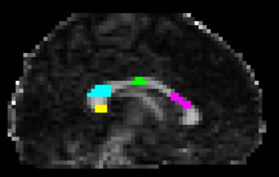 |
32 | Assessing the robustness of the correlation between intra-axonal T2 and axon diameter across participants
Veronica P Dell'Acqua1, Chantal M W Tax1,2, Malwina Molendowska1, Greg D Parker1, Derek K Jones1,3, Muhamed Barakovic1,4,5,6,7, and Erick Jorge Canales-Rodriguez6
1Cardiff University Brain Research Imaging Centre, Cardiff University, Cardiff, United Kingdom, 2University Medical Center Utrecht, Image Sciences Institute, Utrecht, Netherlands, 3Mary MacKillop Institute for Health Research, Australian Catholic University, Melbourne, Australia, 4Translational Imaging in Neurology (ThINk) Basel, University Hospital Basel and University of Basel, Basel, Switzerland, 5MS Center and Research Center for Clinical Neuroimmunology and Neuroscience Basel, University Hospital Basel and University of Basel, Basel, Switzerland, 6Signal Processing Laboratory 5 (LTS5), Ecole Polytechnique Fédérale de Lausanne (EPFL), Lausanne, Switzerland, 7Roche Pharma Research & Early Development, Neuroscience and Rare Diseases, Roche Innovation Center, Basel, Switzerland Keywords: Data Analysis, Relaxometry, Validation, Axon Diameter, Diffusion-Relaxation In-vivo quantification of axon diameter is an attractive and debated topic in the MRI community. The possibility to resolve submicrometric axon diameters non-invasively yields the potential to push further the boundaries in research and clinics but yet, further work is needed to better explore and validate the existing approaches to estimate the inner axon diameter. Recently, the feasibility of estimating the axon diameter from the intra-axonal transverse relaxation time has been investigated combining a diffusion-relaxation protocol and histological data. In the present study, we apply this approach in a larger in vivo population to assess variability across participants. |
|
2029.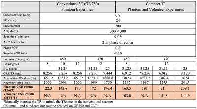 |
33 | Optimized Fast Gray Matter Acquisition T1 Inversion Recovery MRI for Localizing Mammillothalamic Tract in Deep Brain Stimulation Targeting
Kuan Zhang1, Daehun Kang1, Maria A Halverson1, Shengzhen Tao2, Steven A Messina1, MyungHo In1, Joshua D Trzasko1, John III Huston1, Matthew A Bernstein1, and Yunhong Shu1
1Radiology, Mayo Clinic, Rochester, MN, United States, 2Radiology, Mayo Clinic, Jacksonville, FL, United States Keywords: Data Acquisition, Data Acquisition, FGATIR, Deep brain stimulation, Compact 3T, MRI sequence optimization Deep brain stimulation (DBS) is used for neurological disorder treatment but requires precise image-based targeting. The fast gray matter acquisition T1 inversion recovery (FGATIR) sequence was developed to visualize DBS targets, such as the mammillothalamic tract (MTT). We investigated the imaging protocol of FGATIR through simulation, phantom experiment, and volunteer scanning to improve the contrast-to-noise ratio. Adjustments to inversion time, flip angle, and receive bandwidth were made to achieve the highest MTT-thalamus contrast. A compact 3T scanner was also leveraged to further maximize the contrast as the high-performance gradients reduce repetition time, thereby decreased the acquisition window of FGATIR. |
|
2030.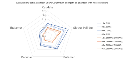 |
34 | Nonsusceptibility frequency contributions and brain size affect quantitative susceptibility mapping in a region-dependent manner
Thomas Jochmann1,2, Fahad Salman2, Ademola Adegbemigun2, Jens Haueisen1, and Ferdinand Schweser2,3
1Department of Computer Science and Automation, Technische Universität Ilmenau, Ilmenau, Germany, 2Buffalo Neuroimaging Analysis Center, Department of Neurology at the Jacobs School of Medicine and Biomedical Sciences, University at Buffalo, The State University of New York, Buffalo, NY, United States, 3Center for Biomedical Imaging, Clinical and Translational Science Institute, University at Buffalo, The State University of New York, Buffalo, NY, United States Keywords: Image Reconstruction, Quantitative Susceptibility mapping We studied the dependency of quantitative susceptibility mapping to scaling the resolution and amplitude in a brain phantom with and without tissue microstructure. We found that nonsusceptibility frequency contributions cause a resolution- and region-dependent bias to quantitative susceptibility mapping. |
|
2031.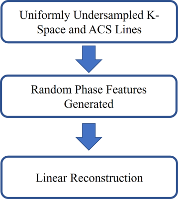 |
35 | Suppressing MRI Background Noise via Modeling Phase Variations
Yuchou Chang1, Gulfam Ahmed Saju1, Jasina Yu1, Reza Abiri2, Zhiqiang Li3, and Tianming Liu4
1Computer and Information Science, University of Massachusetts Dartmouth, North Dartmouth, MA, United States, 2Electrical, Computer and Biomedical Engineering, University of Rhode Island, Kingston, RI, United States, 3Neuroradiology, Barrow Neurological Institute, Phoenix, AZ, United States, 4Computer Science, University of Georgia, Athens, GA, United States Keywords: Image Reconstruction, Parallel Imaging The background phase variations exist in coil sensitivities. Optimized phase distribution can minimize the noise of the parallel MRI reconstruction. However, phase variation may be originated from different factors, and it is difficult to be modeled. A random feature method is proposed to model phase variations in coil sensitivities. Through a linear reconstruction using the random phase feature, background noise can be suppressed. Augmented phase features make the linear reconstruction better remove background noise. |
|
2032.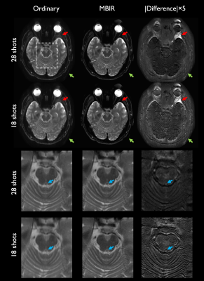 |
36 | Model-based image reconstruction for highly accelerated point spread function encoded echo planar imaging
Nolan Meyer1,2, Myung-Ho In1, Maria Halverson1, John Huston III1, Matt Bernstein1, and Joshua Trzasko1
1Radiology, Mayo Clinic, Rochester, MN, United States, 2Mayo Clinic Graduate School of Biomedical Sciences, Rochester, MN, United States Keywords: Image Reconstruction, Signal Representations A model-based image reconstruction (MBIR) framework is developed for highly accelerated point spread function (PSF) encoded echo planar imaging (EPI). The reconstruction accounts for known physics, and utilizes a subspace representation with local low-rank (LLR) regularization along with a variable splitting solution. Comparisons with images obtained through standard methods demonstrate considerable artifact reduction and sharpness preservation through MBIR. MBIR accommodates arbitrary sampling trajectories along the PSF-encoding dimension, enabling reconstruction of quality images from less than one minute of scan time; and we demonstrate performance boosts with nonstandard trajectories precluded by conventional reconstruction methods. |
|
2033.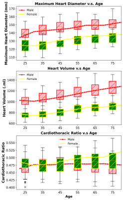 |
37 | Automated Cardiac Quantification and Cardiomegaly Stratification: Feasibility of an AI-based Approach on Large-scale Non-Cardiac-Gated MRI
Thanh-Duc Nguyen1, Saurabh Garg1, Nasrin Akbari1, Saqib Basar1, Sean London2, Yosef Chodakiewitz2, Rajpaul Attariwala1,2, and Sam Hashemi1,2
1Voxelwise Imaging Technology Inc., Vancouver, BC, Canada, 2Prenuvo, Vancouver, BC, Canada Keywords: Machine Learning/Artificial Intelligence, Heart, Deep learning segmentation, cardiomegaly Despite its ubiquity, cardiac assessment at non-cardiac-gated MRI has yet to be standardized, leading to misdiagnosis. This paper examines the feasibility of AI for automatic 3D volumetric cardiac quantification used to detect and stratify cardiomegaly on non-cardiac-gated torso MRI. Using AI, we automatically measured the cardiothoracic ratio and 3D cardiac volumetric features as indicators for detections of cardiomegaly conditions. Large-scale results on 3485 normal-heart individuals revealed the effect of aging on cardiac features in both men and women. AI findings on non-cardiac-gated imaging offer opportunistic useful information, increasing the diagnostic precision, and allowing potentially useful imaging monitoring for cardiomegaly-associated conditions. |
|
2034.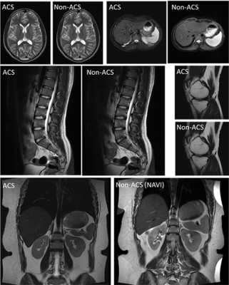 |
38 | An investigation of the performance of T2-weighted MR imaging with AI-assisted compressed sensing in routine clinical settings
Adiraju Karthik1, Apoorwa Devappa2, Aakaar Kapoor3, Dharmesh Singh4, and Dileep Kumar4
1Department of Radiology, Sprint Diagnostics, Jubilee Hills, Hyderabad, India, 2Department of Radiology, Mahadevappa Rampure Medical College, Kalaburagi, India, 3Department of Radiology, City X-Rays Scan & Clinical Private Limited, New Delhi, India, 4Central Research Institute, Global Scientific Collaborations, United Imaging Healthcare, New Delhi, India Keywords: Data Acquisition, Body, AI-assisted compressed sensing, T2-weighted Imaging T2-weighted imaging (T2WI) is an essential diagnostic tool for several diseases. However, one of the challenges faced by patients and radiology departments is the longer scanning time of MR examinations. Recent advancements in artificial intelligence (AI) and deep learning techniques have made it able to acquire images quickly while preserving high-quality image resolution. In this study, the efficacy of a deep learning-based reconstruction technique termed AI-Assisted Compressed Sensing (ACS) was evaluated qualitatively and quantitatively using T2WI in routine clinical settings for brain, spine, knee, kidney and liver. |
|
2035.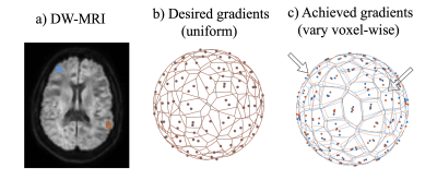 |
39 | Practical Correction of Gradient Nonlinearities in Diffusion-Weighted Imaging
Praitayini Kanakaraj1, Leon Y Cai2, Francois Rheault3, Baxter P Rogers4, Adam Anderson2,4, Kurt G Schilling4, and Bennett A Landman1,2,4,5
1Department of Computer Science, Vanderbilt University, Nashville, TN, United States, 2Department of Biomedical Engineering, Vanderbilt University, Nashville, TN, United States, 3Department of Computer Science, Université de Sherbrooke, Sherbrooke, QC, Canada, 4Department of Radiology and Radiological Sciences, Vanderbilt University Medical Center, Nashville, TN, United States, 5Department of Electrical and Computer Engineering, Vanderbilt University, Nashville, TN, United States Keywords: Data Processing, Diffusion/other diffusion imaging techniques Gradient nonlinearity correction is well-established but not straightforward to implement in existing diffusion software packages due to it producing gradient tables that vary by voxel. We propose a simple, practical approach that approximates full correction by: (1) scaling the diffusion signal and (2) resampling the gradient orientations. Our approach results in uniform gradients across the corrected image and provides the key advantage of seamless integration into current diffusion pipelines. The proposed method resulted in negligible differences in multi- compartment indices from the standard voxel-wise empirical correction. |
|
 Back to Meeting Home
 Back to the Program-at-a-Glance
The International Society for Magnetic Resonance in Medicine is accredited by the Accreditation Council for Continuing Medical Education to provide continuing medical education for physicians.
 Back to Meeting Home
 Back to the Program-at-a-Glance
 View Presentation Video