Digital Poster
Acquisition & Analysis Techniques II
ISMRM & ISMRT Annual Meeting & Exhibition • 03-08 June 2023 • Toronto, ON, Canada

Digital Poster
Acquisition & Analysis Techniques II
| Computer # | |||
|---|---|---|---|
2214.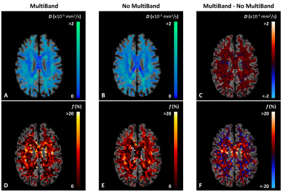 |
41 | The effect of MultiBand acquisition on cerebral inversion recovery intravoxel incoherent motion imaging
Noa van der Knaap1,2,3, Paulien H.M. Voorter1,3, Marcel J.H. Ariës1,2, and Jacobus F.A. Jansen1,3,4
1School for Mental Health & Neuroscience, Maastricht University, Maastricht, Netherlands, 2Department of Intensive Care, Maastricht University Medical Center, Maastricht, Netherlands, 3Department of Radiology and Nuclear Medicine, Maastricht University Medical Center, Maastricht, Netherlands, 4Department of Electrical Engineering, Eindhoven University of Technology, Eindhoven, Netherlands Keywords: Parallel Imaging, Parallel Imaging The MultiBand (MB) imaging technique can reduce scan time considerably, which can be especially relevant for clinical application of techniques with extensive scan protocols, such as intravoxel incoherent motion (IVIM) imaging. However, quantitative IVIM parameter estimates may be affected by the use of MB. This study is a first step to assess the comparability between IVIM acquisitions with and without MB, which has considerable implications for interpretation of IVIM results across different datasets. |
|
2215.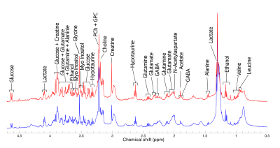 |
42 | Assessment of neurochemistry in human-derived cerebral organoids using high-resolution magic angle spinning NMR
Jamie Near1,2, Vorapin Chinchalongporn3, Rajshree Ghosh Biswas4, Maggie Wu1, Martin Wilson5, Andre Simpson4, and Carol Schuurmans3
1Physical Sciences, Sunnybrook Research Institute, Toronto, ON, Canada, 2Medical Biophysics, University of Toronto, Toronto, ON, Canada, 3Biological Sciences, Sunnybrook Research Institute, Toronto, ON, Canada, 4Chemistry, University of Toronto, Toronto, ON, Canada, 5University of Birmingham, Birmingham, United Kingdom Keywords: Data Acquisition, Tissue Characterization, NMR Cerebral organoids are self-organizing three-dimensional clusters of brain tissue, derived from human pluripotent stem cells. A new and rapidly advancing technology, cerebral organoids serve as an important model system for human brain research. In this study, we used high-resolution magic angle spinning (HR-MAS) NMR to characterize the neurochemical profile of ~100-day old cerebral organoids. High-quality spectra were obtained with excellent spectral resolution. More than 17 metabolites were detected, including many resonances commonly observed in the brain in vivo (e.g. choline, creatine, glutamate, GABA, etc.). Notably, NAA was absent. Future work will assess cerebral organoids at later stages of maturity. |
|
2216.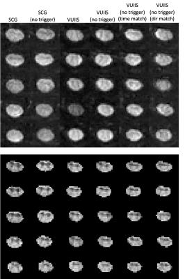 |
43 | To trigger or not to trigger – removing cardiac triggering in diffusion MRI of the cervical spinal cord saves time without sacrificing quality
Kurt G Schilling1, Kristin P O'Grady1, Anna J.E. Combes1, Grace Sweeney1, Logan Prock1, Julien Cohen Adad2, Bennett A Landman3, and Seth A Smith1
1Vanderbilt University Medical Center, Nashville, TN, United States, 2Polytechnique Montreal, Montreal, QC, Canada, 3Vanderbilt University, Nashville, TN, United States Keywords: Data Acquisition, Spinal Cord Cardiac triggering is commonly used in diffusion MRI protocols of the cervical spinal cord to reduce cardiac-related motion. However, this dramatically increases scan time and can limit high angular resolution multi-shell experiments. We test whether advances in preprocessing motion correction and fitting procedures may overcome cardiac-related motion artifacts. We find that removing cardiac triggering regains significant scan time with no increase in prevalence of artifacts, while providing similar quantitative indices with comparable reproducibility. In summary, removing cardiac triggering for cervical spinal cord diffusion saves time without sacrificing image quality. |
|
2217.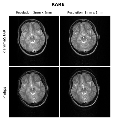 |
44 | Vendor neutral pulse programming: Running gammaSTAR sequences on Philips Hardware
Martijn Nagtegaal1,2, Simon Konstandin3, Daniel Hoinkiss3, Nicolas Gross-Weege4, Volkmar Schulz3,4, Matthias Günther3,5,6, and Matthias J.P. van Osch2
1Department of Imaging Physics, Delft University of Technology, Delft, Netherlands, 2C.J. Gorter MRI Center, Radiology Department, Leiden University Medical Center, Leiden, Netherlands, 3Imaging Physics, Fraunhofer Institute for Digital Medicine MEVIS, Bremen, Germany, 4RWTH Aachen, Aachen, Germany, 5mediri GmbH, Heidelberg, Germany, 6University of Bremen, Bremen, Germany Keywords: Pulse Sequence Design, Pulse Sequence Design, Vendor independent pulse programming Pulse sequence design and development in MR is currently hindered by the possibilities to freely share pulse sequences and test these on scanners of different vendors. Vendor independent pulse programming environments provide a shareable, open, and reproducible way of pulse sequence development. An important step in this is the support on scanners of all main vendors of these protocols. In this work we show for the first time the execution of gammaSTAR imaging protocols on a Philips scanner. The obtained FLASH and RARE based images are very similar to the images obtained using the vendor’s implementation. |
|
2218.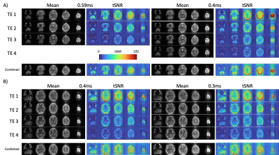 |
45 | Multi-Echo EPI performed on NexGen 7T scanner increases spatial resolution and shortens TE
Alexander JS Beckett1,2, An T Vu3,4, Samantha J Ma5, Essa Yacoub6, and David A Feinberg1,2
1Helen Wills Neuroscience Institute, University of California, Berkeley, CA, United States, 2Advanced MRI Technologies, Sebastopol, CA, United States, 3Radiology, University of California, San Francisco, CA, United States, 4San Francisco Veteran Affairs Health Care System, San Francisco, CA, United States, 5Siemens Medical Solutions USA, Inc., Malvern, PA, United States, 6Center for Magnetic Resonance Research, University of Minnesota, Minneapolis, MN, United States Keywords: Data Acquisition, fMRI Multi-echo (ME) EPI can increase BOLD sensitivity and reduce signal drop-out compared with standard gradient echo (GE) EPI, however spatial resolution and number of echo images are limited by the echo train length of each image. The powerful gradients on the NexGen 7T scanner allow for shorter echo spacing, and hence a greater number of echoes collected at high-resolution (1.6mm isotropic) as compared to standard 7T systems. ME-EPI collected at these resolutions can be separated into TE-dependent and TE-independent components using ME-ICA, showing promise for ME functional connectivity studies at 7T. |
|
2219.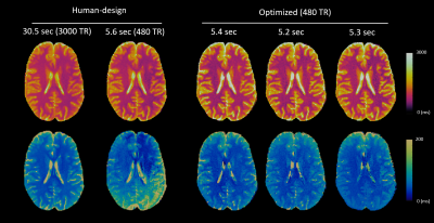 |
46 | Characterizing Artifacts in Multi-dimensional MR Fingerprinting with High Efficiency for Sequence Optimization: Systematic Error Index
Siyuan Hu1, Debra McGivney1, Zhilang Qiu1, and Dan Ma1
1Case Western Reserve University, Cleveland, OH, United States Keywords: Pulse Sequence Design, MR Fingerprinting It is critical to characterize the dominating systematic errors caused by undersampling and field inhomogeneity to design robust MRF scans. However, characterizing such errors by direct simulations of aliasing artifacts is computationally expensive and impractical for sequence optimization for multi-dimensional MRF (mdMRF) scans with higher dimensions. We propose the Systematic Error Index, a model to characterize systematic errors with high computational efficiency. We demonstrate accurate and robust in vivo results from the optimized MRF and mdMRF scans obtained from the proposed SEI-based optimization framework. |
|
2220.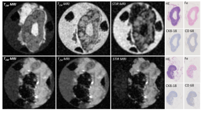 |
47 | Ex vivo lymph node staging by a portable low-field MRI scanner
Anke Christenhusz1, Frank F.J. Simonis1, Bennie ten Haken1, Justin C.A. te Wildt1, Sadaf Sadamzadeh1, Anneriet E. Dassen2, and Lejla Alic1
1Magnetic Detection and Imaging, University of Twente, Enschede, Netherlands, 2Surgery, Medisch Spectrum Twente, Enschede, Netherlands Keywords: Data Acquisition, Cancer, Lymph node, low-field, portable scanner Sentinel lymph node (LN) biopsy facilitated by magnetic nanoparticles (MNP) is introduced for breast cancer patients eligible for breast conserving surgery. As m1etastatic depositions potentially introduce changes in heterogeneity of MNP-enhanced MRI, a portable low-field MRI scanner can be used for ex vivo perioperative LN staging. Therefore, we assessed the changes in T1w, T2w and STIR images due to iron deposition in LN. 2Clinically relevant LN segments, such as fat and iron depositions, identified in the pathology images, were also observable in MR scans. |
|
2221.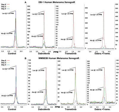 |
48 | Imaging Biomarkers of Therapeutic Response to Melanoma to Kinase Inhibitors
Pradeep Kumar Gupta1, Stepan Orlovskiy1, Jyoti S Tomar1, Fernando Arias-Mendoza1, David S. Nelson1, Stephen Pickup1, Alexander A. Shestov1, Dennis B. Leeper2, Jerry D Glickson1, and Kavindra Nath1
1Departments of Radiology, Perelman School of Medicine, University of Pennsylvania, Philadelphia, PA, United States, 2Department of Radiation Oncology, Thomas Jefferson University, Philadelphia, PA, United States Keywords: Data Acquisition, Cancer In vivo 1H/31P MRS were used to monitor the effects of Kinase Inhibitor (KI) therapy in two metabolically different human melanoma xenografts models. Our goal is to determine the metabolic changes in human melanoma xenograft models when treated with two KIs, a BRAF and a MEK inhibitor. KI combination is more effective than single-drug therapy. Differences in relative levels of metabolites and bioenergetics between two human melanoma xenografts models may produce differential therapeutic responses to BRAF and MEK inhibitors. In melanoma, metabolic changes in response to targeted kinase inhibitor therapy occur rapidly and are connected to subsequent tumor response. |
|
2222.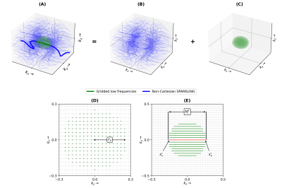 |
49 | Merging Cartesian and non-Cartesian sampling through GoLF-SPARKLING
Chaithya G R1,2, Guillaume Daval-Frérot1,2,3, Aurélien Massire3, Alexandre Vignaud1, and Philippe Ciuciu1,2
1Neurospin, CEA Paris Saclay, Gif-sur-Yvette, 91191, France, 2Inria, MIND, Palaiseau, 91120, France, 3Siemens Healthineers, Saint-Denis, 93210, France Keywords: New Trajectories & Spatial Encoding Methods, Brain Cartesian sampling can acquire a given k-space region with minimum redundancy, while non-Cartesian sampling can help achieve a larger k-space coverage. Through generalized affine constraints in SPARKLING and an adapted target sampling density, for the first time non-Cartesian and Cartesian sampling are merged within same trajectory, giving the best of both worlds. With Gridding of Low Frequencies (GoLF), we get SPARKLING k-space trajectories which carry out Cartesian sampling at the center of k-space. This approach paves the way for designing new kind of compound sampling patterns, which enforces Cartesian and non-Cartesian sampling within the same trajectory. |
|
2223.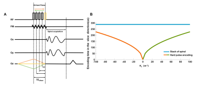 |
50 | A Frequency Modulation HArd Pulse ENcoding Sequence for Ultra-Short Echo Time High-Resolution Three-Dimensional MR Imaging
Jun Zhao1,2, Yupeng Cao1, Weinan Tang3, Quelu Chen4, Wentao Liu1, and Dong Han1
1Key Laboratory of Biological Effects and Safety, National Center for Nanoscience and Technology, beijing, China, 2School of Future Technology, University of Chinese Academy of Sciences, Beijing, China, 3Wandong Medical Inc, Beijing, China, Beijing, China, 4Department of Radiology, Wenzhou Central Hospital, Affiliated Dingli Clinical Institute of Wenzhou Medical University, Wenzhou, China Keywords: Pulse Sequence Design, Lung, hard pulse encoding, variable TE, frequency modulation, spiral Due to its short T2* value and low proton density, MR imaging of the lung is quite challenging. Recently, ultra-short echo time (UTE) techniques, such as stack-of-spiral, were used to conduct lung MRI with good image quality. Here, based on stack-of-spiral, a frequency modulation hard pulse encoding (HAPEN) 3D UTE was proposed to optimize the echo time of different phase encoding steps in the slice direction. As a result, HAPEN can achieve shorter TE and get better SNR in human lung MRI compared to the traditional stack-of-spiral UTE sequence. |
|
2224.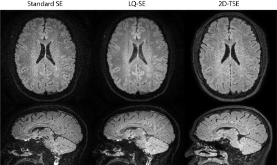 |
51 | Optimized, volumetric, isotropic-resolution T2W 2D FLAIR with high temporal and SNR efficiencies
Dahan Kim1, Tzu Cheng Chao1, Dinghui Wang1, and James G Pipe1
1Department of Radiology, Mayo Clinic, Rochester, MN, United States Keywords: New Trajectories & Spatial Encoding Methods, New Trajectories & Spatial Encoding Methods, Spiral, FLAIR, LQ We describe our novel T2W 2D FLAIR sequence whose temporal and SNR efficiencies are maximized by (1) an efficient IR acquisition that minimizes sequence deadtime and (2) localized quadratic encoding which eliminates SNR-inefficiencies of multi-pass 2D acquisitions. These improvements allowed our FLAIR scan to shorten the scan time while achieving higher SNR than standard SE and 2D-TSE scans of identical/equivalent scan time. |
|
2225.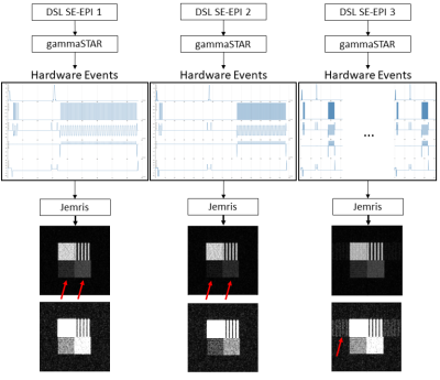 |
52 | High-level and modular description of MRI sequences using domain-specific language
Jörn Huber1, Daniel Christopher Hoinkiss1, Christina Plump2,3, Christoph Lüth2,3, Rolf Drechsler2,3, and Matthias Günther1,4,5
1Fraunhofer Institute for Digital Medicine MEVIS, Bremen, Germany, 2German Research Center for Artificial Intelligence DFKI, Bremen, Germany, 3University of Bremen, Bremen, Germany, 4Faculty 1 (Physics/Electrical Engineering), University of Bremen, Bremen, Germany, 5mediri GmbH, Heidelberg, Germany Keywords: Pulse Sequence Design, Software Tools This work demonstrates the formulation of MRI sequences in a high-level Domain Specific Language (DSL). The DSL approach reduces the complexity of MR sequence programming and enables optimization of preconfigured DSL parameters using MR simulations. Finally, using the gammaSTAR framework, DSL sequences can be directly run on real MR scanners. |
|
2226.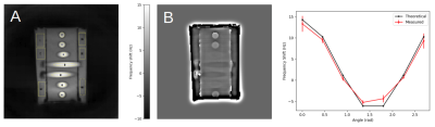 |
53 | Cardiac-Gated Rosette Pulse Sequence Development for Off-Resonance Frequency Imaging
Julian Bertini1, Mira Liu1, Chisondi Simba Warioba1, Yu Fen Chen2, and Timothy Carroll1
1University of Chicago, Chicago, IL, United States, 2Department of Radiology, Northwestern Feinberg School of Medicine, Chicago, IL, United States Keywords: Pulse Sequence Design, Phantoms Intracranial atherosclerotic disease (ICAD) is a leading cause of preventable ischemic stroke. Long-term management of chronic ICAD would benefit from directly quantifying key risk factors, such as the oxygen extraction fraction (OEF) and cerebrovascular reserve (CVR). The accurate measurement of the phase signal is vital to measuring OEF using MRI and seeing these effects through the cardiac cycle promises to elucidate CVR. A cardiac-gated, multi-shot, multi-echo rosette pulse sequence is developed and validated with simulation and a phantom study. The proposed pulse sequence and reconstruction pipeline produces strong off-resonance frequency measurements with a scan time under two minutes. |
|
2227. |
54 | Modified non-contrast enhanced spatially-selective time-resolved vessel imaging by using cylinder-shaped pre-saturation pulse train
Masahiro Takizawa1, Takashi Nishihara1, and Chikako Moriwake1
1FUJIFILM Healthcare Corporation, 2-1, Shintoyofuta, Japan Keywords: Pulse Sequence Design, Lung Cylinder-shaped pre-saturation pulse train is modified to achieve non subtract scheme for non-contrast enhanced spatially-selective and time-resolved vessel imaging. The target vessel is selected by cylinder-shaped pre-saturation, and the dynamics of blood flow in the target vessel is observed by changing the number of applied pre-saturation pulses. The developed pulse train was demonstrated to visualize dynamics of a target pulmonary vessel in the lung. |
|
2228.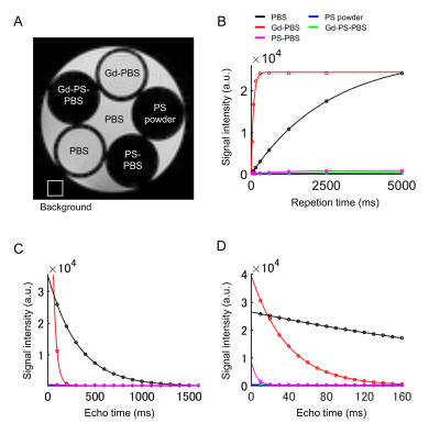 |
55 | Reduction of artifacts and background signals in ex vivo mouse embryo MRI by potato starch suspension
Tomokazu Tsurugizawa1,2,3, Takuma Kumamoto4, and Yoshichika Yoshioka3,5
1Human Informatics and Interaction Research Institute, National Institute of Advanced Industrial Science and Technology (AIST), Tsukuba, Japan, 2Faculty of Engineering, University of Tsukuba, Tsukuba, Japan, 3Center for Information and Neural Networks (CiNet), Osaka University and National Institute of Information and Communications Technology (NICT), Osaka, Japan, 4Developmental Neuroscience Project, Department of Brain & Neurosciences, Tokyo Metropolitan Institute of Medical Science, Tokyo, Japan, 5Graduate School of Frontier Biosciences, Osaka University, Osaka, Japan Keywords: Data Acquisition, Ex-Vivo Applications The high-field MRI enables to investigate the microstructure in the mouse embryo. The proton-free fluid is used for the surrounding liquid around the specimen in MR-microimaging, but the potential issue of the image quality remains due to the air bubbles on the edge of the specimen and the motion artifact. Here, we demonstrated that the potato starch suspension with phosphate-buffered saline showed a low T1 and T2 signal intensity and strongly prevent the motion of the embryo during the scanning. These results indicate the utility of potato starch suspension for MR-microimaging of mouse embryos. |
|
2229.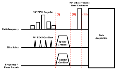 |
56 | Design of a Two-dimensional Ultrashort Echo Time Simultaneous Multi-slice Pulse Sequence
Jason Andrew Reich1, Erin MacMillan2,3,4, and Rebecca Feldman1,5
1Computer Science, Mathematics, Physics and Statistics, University of British Columbia, Kelowna, BC, Canada, 2UBC MRI Research Centre, Department of Radiology, Faculty of Medicine, University of British Columbia, Vancouver, BC, Canada, 3SFU ImageTech Lab, Simon Fraser University, Surrey, BC, Canada, 4Philips Canada, Mississauga, ON, Canada, 5Biomedical Engineering and Imaging Institute, Icahn School of Medicine at Mount Sinai, New York, NY, United States Keywords: Pulse Sequence Design, New Signal Preparation Schemes Simultaneous multi-slice (SMS) pulse sequences have allowed for reductions in scan time with minimal signal-to-noise ratio loss. However, when ultrashort echo times (UTEs) are desired, SMS pulse sequences have been challenging to implement. In this work, we explore a UTE SMS pulse sequence that makes use of a power independent of number of slices prepulse to shape the transverse magnetization profile and a whole volume hard excitation to excite the remaining longitudinal magnetization. The novel pulse sequence is estimated to reduce scan times by a factor of approximately 7.6 when compared to UTE pulse sequences with three-dimensional acquisitions. |
|
2230.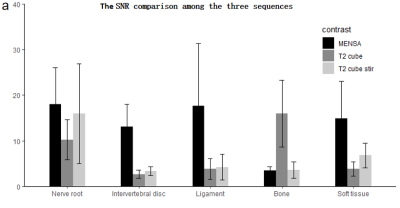 |
57 | Application of 3D MR MENSA in preoperative evaluation of lumbar disc herniation: a prospective study
Xuelin Pan1, Yuting Wen1, Zhenlin Li1, Xin Rong2, and Miaoqi Zhang3
1Department of Radiology, West China Hospital of Sichuan University, Chengdu, Sichuan, China, 2Department of Orthopedics, West China Hospital of Sichuan University, Chengdu, Sichuan, China, 3MR Research, GE Healthcare, Beijing, China Keywords: Data Acquisition, Neuro In this work, we propose a preoperative magnetic resonance examination of patients with lumbar disc herniation using the 3D MENSA sequence. This sequence was superior to cube, cube stir sequence in subjective and objective evaluation.The preoperative 3D MRI MENSA sequence is able to clearly depict the nerve roots and offer desirable contrast between the nerve roots, ligamentum flavum, bone, and intervertebral discs. Patients with lumbar degeneration can effectively benefit from the MENSA sequence since it provides informative imaging information to help understand disc herniation and compression of adjacent tissues when developing preoperative surgical strategies. |
|
2231. |
58 | R2* Estimation by Multispectral Fat-Water Models for GRE and UTE Acquisitions Using Virtual Liver Iron Overload Model and Monte Carlo Simulations
Prasiddhi Neupane1, Utsav Shrestha1, and Aaryani Tipirneni-Sajja1,2
1Biomedical Engineering, The University of Memphis, Memphis, TN, United States, 2St. Jude Children's Research Hospital, Memphis, TN, United States Keywords: Data Analysis, Data Analysis Multispectral fat-water-R2* models are used for the confounder-free assessment of hepatic iron overload. In this study, Monte Carlo-based virtual liver iron overload models were created, MRI signals were synthesized for GRE and UTE acquisitions, and the R2* values estimated using the monoexponential and the multispectral fat-water models were analyzed. Our results demonstrate that both multispectral models exhibit high accuracy and precision for UTE acquisition at both 1.5T and 3T. |
|
2232. |
59 | Model-Based Dynamic B0 Compensation of a 0.35T MRI-Linac using Inclinometer Data
Austen Curcuru1, Taeho Kim2, Deshan Yang3, and H. Michael Gach1,2,4
1Biomedical Engineering, Washington University in Saint Louis, Saint Louis, MO, United States, 2Radiation Oncology, Washington University School of Medicine, Saint Louis, MO, United States, 3Duke University, Durham, NC, United States, 4Radiology, Washington University School of Medicine, Saint Louis, MO, United States Keywords: Artifacts, MR-Guided Interventions Balanced steady state free procession (bSSFP) sequences are commonly used for real-time imaging during MRI guided radiotherapy treatments (MR-IGRT). bSSFP sequences offer high temporal resolution and SNR but are sensitive to B0 fluctuations which lead to imaging artifacts during radiotherapy (RT) gantry rotatation.1 Previous work demonstrated that RT gantry rotation induced artifacts could be significantly reduced by compensating for B0 fluctuations using data from a free induction decay (FID) navigator prior to each bSSFP image.2 A model-based approach to B0 compensation using the gantry inclinometer was evaluated to reduce the SNR loss and temporal resolution associated with the navigator approach. |
|
2233.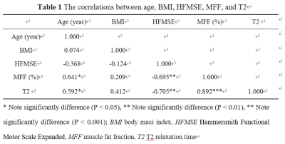 |
60 | Effectiveness of T2-mapping imaging in assessing disease severity in Spinal muscular atrophy: preliminary study
Yingyi Hu1,2, Yang Huang2,3, Taiya Chen1,2, Xinguo Lu4, Diangang Fang2, Kan Deng5, and Zhiyong Li2
1China Medical University, Shenyang, China, 2Department of Radiology, Shenzhen Children's Hospital, Shenzhen, China, 3Shantou University Medical College, Shantou, China, 4Internal Medicine-Neurology, Shenzhen Children's Hospital, Shenzhen, China, 5Philips Healthcare, Guangzhou, China Keywords: Data Analysis, Quantitative Imaging, T2-mapping To evaluate the potential of T2-mapping imaging as a qMRI marker for disease severity in Spinal muscular atrophy (SMA), we compared the T2 with muscle fat fraction (MFF), and clinical assessment of 13 muscles in the pelvis and thighs of 20 patients with SMA. A significant correlation was found between the mean T2 of all muscles and the patient’s clinical evaluation and MFF. Moreover, the highest mean T2 was found in the gluteus maximus, while the lowest in the adductor longus. Therefore, T2 mapping can be used as a quantitative and objective MRI technique to assess disease severity in SMA. |
|
 Back to Meeting Home
 Back to the Program-at-a-Glance
The International Society for Magnetic Resonance in Medicine is accredited by the Accreditation Council for Continuing Medical Education to provide continuing medical education for physicians.
 Back to Meeting Home
 Back to the Program-at-a-Glance
 View Presentation Video